Introduction
Entomopathogenic nematodes (EPNs) of the genera Steinernema and Heterorhabditis of the order Rhabditida, in the families Steinernematidae and Heterorhabditidae respectively, are used to control a wide range of pest insects (Grewal et al., Reference Grewal, Ehlers and Shapiro-Ilan2005), as they have been proven to be safe and effective biological control agents (Ehlers & Hokkanen, Reference Ehlers and Hokkanen1996). The control of insects is based on the interactions between the nematodes and their symbiotic bacteria (Poinar & Thomas, Reference Poinar and Thomas1966; Poinar et al., Reference Poinar, Thomas and Hess1977). The association is an obligate mutualism, in which each entity (i.e. nematode and symbiotic bacteria) requires the other to proliferate (Poinar & Thomas, Reference Poinar and Thomas1966; Poinar et al., Reference Poinar, Thomas and Hess1977). The bacteria protect the insect cadaver against colonization by other micro-organisms, by producing broad-spectrum antimicrobial agents and acting as a substrate for growth and reproduction of the nematodes (Frost & Clarke, Reference Frost, Clarke and Gaugler2002).
The bacteria are delivered into the insect haemocoel through the nematode, which serves as the vector. The nematode also enables the bacteria to persist outside an insect host and to be transferred to a new insect host. When nematodes lack the association with symbiotic bacteria, they are not effective biopesticides or, if mortality does takes place, the reproduction of nematodes is greatly impaired (Poinar & Thomas, Reference Poinar and Thomas1966; Poinar et al., Reference Poinar, Thomas and Hess1977; Han & Ehlers, Reference Han and Ehlers1998). The bacterial symbionts can be isolated from the nematodes, and cultivated separately.
The bacterial symbionts have the ability to persist within the free-living infective juveniles (IJs), until the bacteria are again released within an insect haemocoel (Martens et al., Reference Martens, Vivas, Heungens, Cowles and Goodrich-Blair2004). Symbiotic bacterial cells are stored in the intestine of the IJ and, during the recovery stage of the IJ from non-feeding to feeding stage, such cells are released (Han & Ehlers, Reference Han and Ehlers2000). When IJ recovery to the feeding stage takes place, the IJ develop from the arrested stage to the J4 stage (Strauch & Ehlers, Reference Strauch and Ehlers1998; Hirao & Ehlers, Reference Hirao and Ehlers2009a). The recovered juveniles develop into adults by means of feeding on bacterial cells and degraded host tissue. When the nutritious conditions are optimal, the nematodes produce offspring, which develop into another reproductive adult generation. In steinernematids, such adults are, in general, amphimictic, which enables them to copulate in liquid conditions. Termination of egg-laying by the adults coincides with the depletion of nutrients and bacteria. In place of the former, endotokia matricida (Johnigk & Ehlers, Reference Johnigk and Ehlers1999) then takes place, whereby the first larval stages (J1s) hatch from eggs inside the uterus, and develop into IJs. It was demonstrated that, for Steinernema feltiae (Filipjev, 1934) Wouts, Mráček, Gerdin & Bedding, 1982, a particular level of ammonia produced by nematode excretions mediates emergence of IJs from the insect cadaver (San-Blas et al., Reference San-Blas, Gowen and Pembroke2008). The IJ is adapted to be durable and to survive in the soil environment until it can find a suitable insect host (Lewis, Reference Lewis and Gaugler2002; Torr et al., Reference Torr, Heritage and Wilson2004).
In Europe and the USA, the first steps towards an outdoor commercial biopesticide application have been taken using EPNs. Various EPN formulations against a wide range of insect pests are currently commercially available in both the aforementioned regions of the world. However, to obtain sufficient quantities of EPNs to satisfy the need for commercial field application against pest insects, the former have to be mass cultured, using in vitro liquid culturing techniques (Ehlers, Reference Ehlers2001). In South Africa, locally occurring EPNs are not yet available as a product on the market (Ferreira & Malan, Reference Ferreira and Malan2014). Therefore, Steinernema yirgalemense Tesfamariam, Gozel, Gaugler & Adams, 2005, which is native to South Africa, requires mass culturing for future commercialization.
The liquid culture process is extremely vulnerable to contamination, as a result of the long process time involved, and the even distribution of fluids and organisms that is required during the mixing in the bioreactor (Grewal et al., Reference Grewal, Ehlers and Shapiro-Ilan2005). The presence of non-symbiotic micro-organisms results in the reduction of nematode yields and, since the liquid culture process can last up to 3 weeks, it presents quite a challenge to maintain sterile conditions throughout the period concerned. It is of importance that the monoxenicity of the cultures is ensured from the onset of inoculum production (Grewal et al., Reference Grewal, Ehlers and Shapiro-Ilan2005). Symbiotic bacteria can be isolated from nematode-infected insect larvae, and stock cultures are obtained by mixing the bacteria with 15% (v/v) glycerol and by subsequently freezing at − 80°C (Boemare & Akhurst, Reference Boemare and Akhurst1988). Establishing bacteria-free nematodes from eggs is a more laborious process (Lunau et al., Reference Lunau, Stoessel, Schmidt-Peisker and Ehlers1993; Han & Ehlers, Reference Han and Ehlers1998).
Steinernema yirgalemense was found in a single sample in the Mpumalanga province during a survey for EPNs that was conducted in citrus orchards (Malan et al., Reference Malan, Knoetze and Moore2011). This nematode was first described by Nguyen et al. (Reference Nguyen, Tesfamariam, Gozel, Gaugler and Adams2004) from Yiglemen in Ethiopia, where it was found to be the dominant species, and later from the Central Rift Valley of Kenya (Mekete et al., Reference Mekete, Gaugler, Nguyen, Mandefro and Tessera2005; Mwaniki et al., Reference Mwaniki, Nderitu, Olubayo, Kimenju and Nguyen2008). The nematode in question belongs to the bicornutum-group of eight described species, of which the unsheathed IJ has two horn-like structures in the cephalic region (Nguyen et al., Reference Nguyen, Tesfamariam, Gozel, Gaugler and Adams2004). Currently, the nematode has not been reported outside of the African continent. In South Africa, S. yirgalemense has shown promise as a biological control agent in laboratory bioassays against larvae, pupae and emerging moths of Thaumatotibia leucotreta Meyrick (Lepidoptera: Tortricidae), the false codling moth (Malan et al., Reference Malan, Knoetze and Moore2011). This nematode species has also been found to be effective in controlling codling moth, Cydia pomonella (L.) (Lepidoptera: Tortricidae) (De Waal et al., Reference De Waal, Malan and Addison2011), mealybug, Planococcus citri (Risso) (Hemiptera: Pseudococcidae) (Van Niekerk & Malan, Reference Van Niekerk and Malan2012) and Planococcus ficus (Signoret) (Le Vieux & Malan, Reference Le Vieux and Malan2013).
The aim of the current study was to evaluate the mass production potential of S. yirgalemense, using liquid culture technology. During the study, the population dynamics of S. yirgalamense in liquid culture were monitored, and the growth characteristics of the associated symbiotic bacterium, Xenorhabdus indica Somvanshi, Lang, Ganguly, Swiderski, Saxena & Stakebrant, 2006, were described.
Materials and methods
Source of insects and nematodes
Galleria mellonella (L.) (Lepidoptera: Pyralidae) larvae were reared on a diet containing the following ingredients: five parts brown bread flour; five parts baby Cerelac NestléTM regular cereal; two parts wheat germ; two parts yeast; two parts glycerine; and one part honey. All the ingredients were mixed together, with a beeswax comb being added to the mixture (Woodring & Kaya, Reference Woodring and Kaya1988).
Steinernema yirgalemense (EU625295) was obtained from the collection of EPNs held by Stellenbosch University's Department of Conservation Ecology and Entomology from previous surveys (Malan et al., Reference Malan, Knoetze and Moore2011). In vivo-produced IJs were reared on last-instar Galleria and harvested at room temperature, according to the procedures devised by Kaya & Stock (Reference Kaya, Stock and Lacey1997). The IJs were stored in vented 500-ml culture flasks, placed horizontally at 14°C in filtered tap water. The flasks, each containing 150 ml nematode suspension, were shaken weekly for aeration. Nematodes were used within 1 month of harvesting.
Isolation of symbiotic bacteria and axenization of nematodes
The primary form of X. indica was isolated from last-instar G. mellonella infected with S. yirgalemense and the identity verified using molecular techniques (Ferreira et al., Reference Ferreira, Van Reenen, Tailliez, Pagé, Malan and Dicks2014b). Agar plates (diameter 9.5 cm) were incubated at 25 ± 2°C for 48 h (Akhurst, Reference Akhurst1980; Kaya & Stock, Reference Kaya, Stock and Lacey1997), propagated in trypticase soy broth (TSB), and stored in 15% glycerol at − 80°C. When required, the glycerol stocks were melted at room temperature, propagated in TSB for 2 days at 30°C, and subsequently used.
To obtain nematode eggs, last-instar G. mellonella larvae were inoculated with S. yirgalemense, left in a growth chamber at 25°C and dissected 4 days after inoculation. Gravid females were hand-picked and transferred to a 10-ml test tube. Pieces of razor blades were added to 8 ml of Ringer's solution, vortexed and poured through a 38-μm sieve. The filtrate was transferred to 1-ml Eppendorf tubes and centrifuged (Eppendorf 5415R, AEC-Amersham Ltd, Kyalami, South Africa) for 1 min at 2000 rpm, the supernatant discarded and the tube again filled with 1 ml fresh Ringer's solution. This process was repeated until the supernatant remained clear. A mixture of 0.5 ml sodium hypochlorite, 1.5 ml NaOH (4 n) and 10 ml distilled water was added to the egg pellet and centrifuged for 2 min at 2000 rpm; the supernatant was discarded and the tube filled with 1 ml TSB. The eggs were transferred to a sterile 12-multiwell plate (flat bottom, Sigma-Aldrich, Aston Manor, South Africa), the wells of which were filled with 300 μl TSB, and incubated for 72 h (Ehlers et al., Reference Ehlers, Lunau, Krasomile-Osterfeld and Osterfeld1998).
An Erlenmeyer flask was filled with 30 ml TSB and inoculated with X. indica. The culture was incubated on an orbital platform shaker (OrbiShake 30 kg, Labotek, Cape Town, South Africa) at 180 rpm at 30°C in the dark. Wout's agar plates (diameter 5.4 cm) were inoculated with two drops of the X. indica suspension, after which 50–100 J1 larvae from the sterile cell-wells were added to the plates (Wouts, Reference Wouts1981). The plates were then sealed with Parafilm and incubated in the dark at 25°C.
Monoxenic cultures
In order to provide a homogeneous Xenorhabdus inoculum, the symbiotic bacteria were produced in one batch culture, and then distributed over three Erlenmeyer flasks (Hirao et al., Reference Hirao, Ehlers and Strauch2010). When the bacterial pre-cultures had reached a cell density of 107 colony-forming units (cfu) ml− 1, 300 μl was transferred to sterile 30-ml Erlenmeyer flasks, and then incubated on an orbital platform shaker, at 180 rpm at 30°C in the dark for 42 h. The IJs were subsequently inoculated into the liquid medium in 250-ml Erlenmeyer flasks, at a density of 4000 IJs ml− 1, and incubated on an orbital platform shaker at 180 rpm at 25°C in the dark for 15 days. IJs were taken from monoxenic pre-cultures (Lunau et al., Reference Lunau, Stoessel, Schmidt-Peisker and Ehlers1993).
The liquid medium (complex medium) in which the nematodes were propagated consisted of 15.0 g l− 1 yeast extract (Merck Pty, Modderfontein, South Africa), 20.0 g l− 1 soy powder (Nature's Choice, Meyerton, South Africa), 4 g NaCl, 0.35 g KCl, 0.15 g CaCl2, 0.1 g MgSO4 (Merck) and 46 ml of vegetable oil per litre (Ehlers et al., Reference Ehlers, Lunau, Krasomile-Osterfeld and Osterfeld1998). The 250-ml Erlenmeyer flasks containing 30 ml of nematode culture medium were inoculated with 1% of the bacterial culture, and pre-cultured at 30°C for 48 h before inoculating IJs from the monoxenic cultures. The flasks were incubated in a growth chamber at 25°C for 15 days. One-millilitre samples were taken daily under sterile conditions and washed with Ringer's solution through a 25-μm sieve. Nematodes were counted using a stereomicroscope (Leica MZ75, Leica Microsystems, Wetzlar, Germany) and the concentration was determined (Ferreira et al., Reference Ferreira, Addison and Malan2014a). Three flask cultures were used for each experiment, and the experiments were repeated on different test dates.
Bacterial growth curve
The bacterial growth curve was determined in order to investigate the cell density dynamics of the bacterial symbiont over a period of 48 h. This experiment was conducted at a temperature of 30°C, using TSB as a medium. Forty microlitres of the bacterial stock solution was inoculated into 10 ml of TSB, and incubated on an orbital platform shaker at 180 rpm, for 2 days in the dark, at 30°C. Thereafter, 400 μl of the bacterial culture was inoculated into 400 ml of TSB, with a sample of 1 ml being taken every 3 h for 48 h. A dilution series was conducted for each sample and streaked out in triplicate and replicated twice, using plates of nutrient agar (Biolab, Biolab Diagnostics, Midrand, South Africa) supplemented with 0.004% (w/v) triphenyltetrazolium chloride and 0.025% (w/v) bromothymol blue (NBTA plates). Plates were incubated at 30°C for 2 days, and the cfu were counted (Atlas, Reference Atlas1988). The optical density was also measured every 3 h using a spectrophotometer at 600 nm (OD600).
Bacterial cfu were also determined once the IJs had been inoculated into the liquid (complex medium) (Hirao & Ehlers, Reference Hirao and Ehlers2009b; Hirao et al., Reference Hirao, Ehlers and Strauch2010). Samples were taken every day for 15 days, streaked out in quadruplicate and replicated twice, using NBTA plates. Plates were incubated at 30°C in the dark for 2 days, after which colonies were counted using a dilution series.
Virulence studies
The in vitro-produced IJs were harvested by washing them with Ringer's solution through a 25-μm sieve (Ferreira et al., Reference Ferreira, Addison and Malan2014a). Galleria larvae were infected with in vitro-produced S. yirgalemense and their mortality was compared to that of Galleria larvae infected with IJs harvested from Galleria-cultured nematodes (in vivo).
Filter paper discs (diameter 13 mm) were added to 10 wells of a 24-well bioassay plate (flat bottom, NuncTM, Cat. No. 144530, Thermo Fisher Scientific (Pty) Ltd, Johannesburg, South Africa). A concentration of 200 IJ per 50 μl was inoculated on to the filter paper discs, and Galleria larvae added. Control plates with Galleria larvae were inoculated with water only. The plates were incubated at 25°C, in the dark. Insect mortality was assessed after 2 days, and infection was confirmed by means of dissection in Ringer's solution. The experiment was repeated with a second batch of in vitro- and in vivo-cultured nematodes.
Data analysis
All statistical analyses were performed using STATISTICA 10 software (Statsoft Inc., 2011). Analysis of variance (ANOVA) was used to compare the results of the virulence studies, followed by Bonferroni multiple comparisons. If residuals were not normally distributed, bootstrap multiple comparisons were used (Efron & Tibshirani, Reference Efron and Tibshirani1993). For the rest of the study, if there were no significant differences between the main effects (time and treatment), the data from the two test dates were pooled. Results were depicted by using descriptive statistics.
Results
Population dynamics of S. yirgalemense
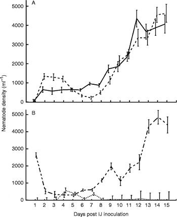
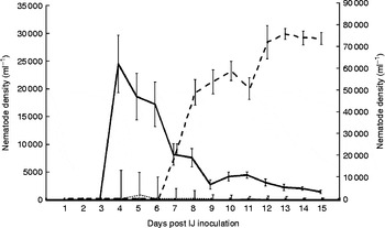
Analyses of data obtained for the life cycle development on each day (1–15) showed no significant differences between the main effects of treatment and date, and data from the two experiments were pooled. The population development for S. yirgalemense at 25°C, for males, females, IJs, J1, J2, pre-adult juveniles (J3 and J4) and endotokia matricida females in monoxenic liquid culture, is presented in figs 1 and 2.
The first males and females occurred on day 2. The highest concentration of males was recorded on day 12, and the highest concentration of females on days 14 and 15 (fig. 1A). During the process time concerned (15 days), the sex ratio was biased towards females from day 2 to day 4. From days 5 to 9, and then on days 12 and 13, the sex ratio was in favour of the males. On days 14 and 15, the number of females exceeded that of the males. The second and third adult (male and female) generations of S. yirgalemense were observed to increase from day 7 onwards, with the maximum density being observed on day 13 (fig. 1A).
(A) Mean population density of Steinernema yirgalemense in monoxenic liquid culture at 25°C, over a period of 15 days. The density of (A) males (——) and females (- - -) and (B) pre-adult stage juveniles (J3 and J4 stages) (– - -) and endotokia matricida females ( ) is indicated. Each plot represents daily counts of each of the different stages in three Erlenmeyer flasks. Error bars represent the standard error.

The pre-adult juvenile stages (J3 and J4) reached their highest concentration on day 14 (fig. 1B). Endotokia matricida female numbers were very low throughout the 15 days, except for an increase that took place on day 4, and then again on day 7 (fig. 1B). Newly hatched J1 and J2 offspring occurred simultaneously on day 4 (fig. 2). The J2 concentration was low, except for a slight increase that occurred on day 5 (fig. 2).
Daily mean population density (95% confidence interval) of J1 ( ), J2 (——) and infective juveniles (IJ) and pre-infective juveniles (- - -) of Steinernema yirgalemense, in monoxenic liquid culture at 25°C, in three Erlenmeyer flasks, over a period of 15 days. Each plot represents the mean daily counts of three Erlenmeyer flasks. Error bars represent the standard error.

The recovery of the IJs was observed as 67 ± 10.15% (fig. 1) on the second day post IJ inoculation, with newly formed IJs being observed on day 7 (fig. 2). The first offspring of the second generation occurred on day 7. The highest concentration of IJs and pre-infective juveniles occurred from day 13 onwards (fig. 2).
Bacterial growth curve
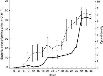
Analyses of data obtained for the cell densities and optical densities showed no significant differences between the main effects of treatment and date, and the results from the two experiments were pooled. The cfu of X. indica, measured every 3 h up to 48 h at 30°C, with the optical density, is shown in fig. 3. The lag phase of X. indica was recorded as lasting from 0 to 15 h. The exponential phase started after 15 h, being followed, after 42 h, by the stationary phase, with an average of 51 × 107cfu ml− 1 (fig. 3).
The mean colony-forming units (cfu) ml− 1 (95% confidence interval) ( ) and the mean optical density (measured at 595 nm) (—) of Xenorhabdus indica in tryptic soy broth, over a period of 48 h, at 30°C in the dark. Each plot represents daily cfu counts. Error bars represent the standard error.

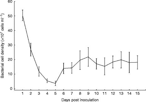
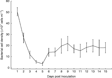
The cell density of X. indica, measured over a period of 15 days, at 25°C, after inoculation of the IJs, is indicated in fig. 4. A marked reduction in bacterial cell density was observed until day 5, after the nematodes were added. The reduction can be ascribed to the recovery of the nematodes, and to their feeding on the bacteria, which caused the former's cell density to decrease dramatically. After the bacteria had been grown for 42 h, the cell density was found to be 50 × 107 ml− 1. From day 5 onwards, the bacterial cell density increased until day 9. On day 10, a slight decrease in bacterial cell density was observed, after which the results of the cell counts seemed to remain constant until day 15 (fig. 4).
The cell density (95% confidence interval) of Xenorhabdus indica in liquid culture over a period of 15 days, at 25°C. Error bars represent the standard error.
Virulence studies
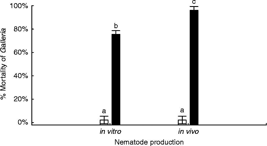
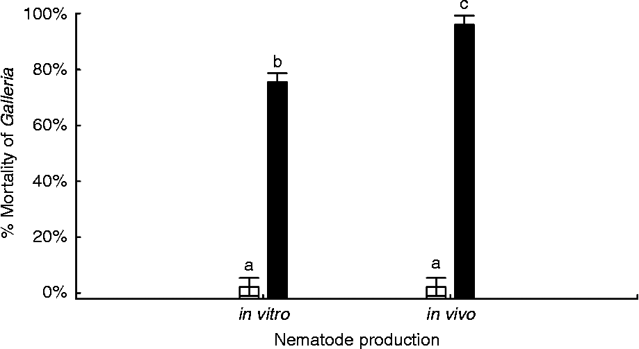
A high percentage mortality rate of G. mellonella larvae was obtained for both in vitro- and in vivo-produced IJs of S. yirgalemense (fig. 5). However, the in vivo-produced nematodes were found to cause a higher percentage mortality (96 ± 2.07%) of G. mellonella larvae in comparison to in vitro-produced larvae (75 ± 1.08%). Results were analysed using a one-way ANOVA (F (1,68)= 41.783; P < 0.001), indicating that the percentage mortality of G. mellonella larvae differs significantly (P < 0.005) between infection with the in vitro- and the in vivo-produced nematodes.
The mean percentage mortality (95% confidence interval) of Galleria mellonella larvae 2 days after inoculation with 200 IJs/insect of Steinernema yirgalemense (black bars; one-way ANOVA: F (1,68)= 41.783; P< 0.001). White bars indicate controls. Different letters above bars indicate significant differences.
Discussion
For nematode population development in liquid culture to be successful, IJ recovery is essential because it is the primary factor determining the number of adults in the parental generation, as it affects the size of the F1 generation (Hirao & Ehlers, Reference Hirao and Ehlers2010). The recovery of IJs varies unpredictably among cultures, making it difficult to adjust the number of nematodes feeding on the bacteria. As a consequence, food depletion and subsequent IJ formation does not always take place after a single generation. Likewise, non-synchronous recovery also exposes early offspring to conditions of sufficient food, for the starting of another generation. Hence, maximum population density is sometimes reached only after a prolonged processing time (Ehlers, Reference Ehlers2001).
Results obtained by Hirao & Ehlers (Reference Hirao and Ehlers2010) showed IJ recovery of >90% for S. feltiae. In the current study, the recovery of IJs was recorded as being 67%. Such a low percentage recovery for the IJs could be ascribed to the low bacterial cell density present in the medium. In order to assist with nematode development in liquid culture, the symbiotic bacteria were inoculated into the liquid medium at least 1 day prior to the IJ inoculation. The volume of the bacterial culture inoculated was between 0.5 and 1% of the culture volume (Ehlers, Reference Ehlers2001). The addition of the pre-cultured symbiotic bacteria triggered the recovery of the IJs, by providing appropriate food signals (Aumann & Ehlers, Reference Aumann and Ehlers2001; Hirao & Ehlers, Reference Hirao and Ehlers2009a). The inoculation was essential, otherwise the artificial media employed did not tend to produce sufficient food signals to allow for IJ recovery (Strauch & Ehlers, Reference Strauch and Ehlers1998).
When IJs of S. yirgalemense were inoculated, the cell density of X. indica was 5 × 10⁸cfu ml− 1. In an earlier study, when S. feltiae IJs were inoculated, the cell density of X. bovienii (Akhurst, Reference Akhurst1980) Akhurst & Boemare, 1993 was 10 × 109ml− 1 (Hirao & Ehlers, Reference Hirao and Ehlers2010), which was substantially higher than that of the bacterial symbiont in the current study. Results from the research conducted by Hirao & Ehlers (Reference Hirao and Ehlers2010) indicate that bacterial cell density is the key factor for IJ recovery, and not IJ inoculum density, as was thought previously.
The method used for determining cell density in the present study differed from the method used by Hirao & Ehlers (Reference Hirao and Ehlers2010), who assessed bacterial cell density by counting cells in a Thoma chamber. In the current study, dilution series were made of each sample and cfu were determined by streaking out on to NBTA plates (Atlas, Reference Atlas1988). Bacteria were grown initially in TSB to determine their general growth characteristics, including their growth rate and their optimum growth temperature, as no work with X. indica associated with S. yirgalemense had been undertaken. Thereafter, the bacteria were grown in the complex medium, prior to it being inoculated with S. yirgalemense IJs. During both growth experiments, on determining the cfu, no difference was found between the bacteria grown in either the TSB, or the complex medium. The different methods used could explain the difference in bacterial cell density between the current study and that of Hirao & Ehlers (Reference Hirao and Ehlers2010). It was concluded that direct cell counts would probably have been a better option for determining cell density and should, consequently, be used in future.
When asynchronous (when not all the IJs start developing simultaneously) recovery occurs in heterorhabditid cultures, the subsequent development of a second generation can cause a complete loss of the liquid culture process, due to the inability of F1 amphimictic adults to copulate in liquid cultures (Strauch et al., Reference Strauch, Stoessel and Ehlers1994). In comparison, Steinernema are generally less vulnerable to a low recovery rate of IJs, because the adults are able to copulate in liquid (Strauch et al., Reference Strauch, Stoessel and Ehlers1994). In this study, the amount of IJ recovery continued to increase after newly hatched juveniles were observed, and no new-generation IJs were observed 4 days after IJ inoculation. This indicates that asynchronous development took place in the first generation. The research of Hirao & Ehlers (Reference Hirao and Ehlers2010) showed that the optimum inoculum density for Steinernema carpocapsae was between 3 × 10³ and 6 × 10³IJs ml− 1, which would, consequently, result in 1 × 10³ and 3 × 10³ml− 1 parental females being required for successful production. For S. feltiae, the optimum inoculum density is >5 × 10³IJs ml− 1, which would result in >2 × 10³ parental females being present per millilitre (Hirao & Ehlers, Reference Hirao and Ehlers2010). In the current study, the inoculum density for S. yirgalemense was 4 × 10³IJs ml− 1, which resulted in a parental female density of only 1 × 10³ml− 1 on day 9 and an increased density of 4 × 10³IJ ml− 1 on day 15. The increase in adult density after day 8 indicates that the progeny of the late-recovered IJs, as well as those of the second and third generations, were induced to develop into adults, instead of into IJs. The cell density of X. indica reached the lowest density on day 5, after which the cell density increased again. The increase prevented the starvation conditions necessary for inducing development into IJs, rather than into adults. The cell density reached two peaks, one of which occurred on day 1 and the other which occurred on day 9, during the 15-day developmental period. Control of the bacterial cell density is critical at the moment at which the progeny of the F1 generation hatches, as achieving high numbers of IJs within a short period of process time is required.
In the current study, the sex ratio was not constantly biased towards the females, as was recorded in previous research on Steinernema (Selvan et al., Reference Selvan, Campbell and Gaugler1993; Alsaiyah et al., Reference Alsaiyah, Ebsa, Zenner, O'Callaghan and Griffin2009; Hirao & Ehlers, Reference Hirao and Ehlers2010). The ratio concerned varied between the different days of the study, and whether this was caused by the low initial recovery of the IJs, by the low bacterial cell density or by both, still requires investigation. Inoculum density has a strong effect on the fertility of the parental females (Hirao & Ehlers, Reference Hirao and Ehlers2010) and in the present study, the bacterial concentration was relatively constant from day 9 onwards. Therefore, fewer nutritional resources were available for the females, with increasing densities (Hirao & Ehlers, Reference Hirao and Ehlers2010). This was shown to have the same effect on S. carpocapsae parental females as it did on those of S. feltiae in the research that was undertaken by Hirao & Ehlers (Reference Hirao and Ehlers2010). Reproduction of Steinernema is amphimictic, with male and female copulation occurring, and with the females producing eggs. When food is abundant in the liquid culture, the offspring develop further into reproductive adults. In comparison, the response to depleting food resources tends to be the encouragement of the development of IJs (Strauch et al., Reference Strauch, Stoessel and Ehlers1994).
The development of a large number of offspring into adult males and females might cause problems when downstreaming of the process is required. As all non-IJs died off rapidly during formulation, the amount of contaminants increased, resulting in the nematode product being spoiled. Therefore, these stages should be removed from the IJ suspension before formulation. Second- and third-generation adults should also be avoided, as the increase concerned is caused by an increase in the bacterial density. This results in a switching to adult development, instead of IJ formation, as the attainment of a one-generation process is the objective, as was achieved for S. carpocapsae in the past (Hirao & Ehlers, Reference Hirao and Ehlers2010).
The virulence of IJs produced from employing in vivo methods was more effective compared to that of IJs produced from the use of in vitro methods, with a significant difference being observed between the two. Similar results were obtained from research done by Gaugler & Georgis (Reference Gaugler and Georgis1991), in which Heterorhabditis bacteriophora Poinar, 1976, produced by liquid culture, achieved significantly lower mortality against the Japanese beetle, Popillia japoica Newman (Coleoptera: Scarabaeoidea), than did those that were reared in vivo (Gaugler & Georgis, Reference Gaugler and Georgis1991). A concentration of 200 IJ 50 μl− 1 was used in this study, which was high for G. mellonella larvae, as they are already highly susceptible to entomopathogenic nematodes. Even with the relatively high concentration, there was still a significant difference between the in vivo- and in vitro-produced IJs. Such differences could have been the result of the in vivo-produced IJs containing higher amounts of lipids (Molyneux, Reference Molyneux1985). Nematodes that have been cultured in vitro appear to be more transparent when compared to their in vivo-produced counterparts. This condition is generally associated with the reduced presence of lipids, and is related to a reduction in pathogenicity against Tenebrio molitor L. (Coleoptera: Tenebrionidae) larvae, according to Vanninen (Reference Vanninen1990). As the amount of time between production and application increases, the reduced pathogenicity also tends to increase (Vanninen, Reference Vanninen1990). This has important consequences for S. yirgalemense, as the desired end result is commercial production and application in the field.
The successful culturing of S. yirgalemense in vitro led to valuable information being gained in the current study. The maximum population density of 75,000 IJs ml− 1 on day 13 after inoculation in the Erlenmeyer flasks was relatively low with regard to the potential of 250,000 IJs ml− 1 obtained using internal-loop airlift bioreactors (Chavarría-Hernández et al., Reference Chavarría-Hernández, Maciel-Vergara, Chavarría-Hernández, Castro-Rosas, Rodríguez-Pastrana, De la Torre-Martínez and Rodríques-Hernández2011). However, the liquid culture production of S. yirgalemense requires further investigation, especially regarding the production of IJs within a single generation. The bacterial growth should be further analysed, as the nutrients provided by the medium might not yet be fully utilized by the bacteria. Improving the culture medium, as well as increasing the IJ inoculum, might result in an improved final number of IJs of S. yirgalemense, in a shorter process time.
Acknowledgements
The authors would like to thank R.-U. Ehlers for his valuable contributions.
Financial support
The authors would like to thank the South African Apple and Pear Producers’ Association (SAAPPA), Citrus Research International (CRI), and the National Research Foundation of South Africa (NRF-THRIP TP2011060100026) for funding the project.
Conflict of interest
None.