Introduction
The internal carotid artery (ICA) has been traditionally described as running a straight course to the skull base, without branching. However, variations such as kinking and coiling do exist. These phenomena may be under-reported as they are asymptomatic in most cases. We report an uncommon case of an ICA variant presenting as an oropharyngeal mass, and we discuss the implications of inadvertent surgical trauma.
Case report
A 71-year-old woman presented with a visible mass on the posterior pharyngeal wall, left of the midline. This was an incidental finding following routine examination by her general practitioner, who urgently referred the case to our department as a suspected cancer of the upper aero-digestive tract. The patient was otherwise asymptomatic, and did not report dysphagia or throat soreness on direct questioning.
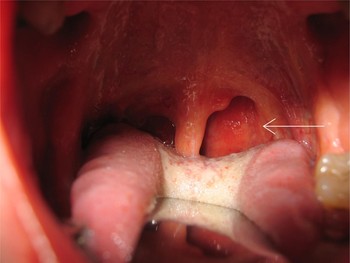
Nasendoscopy revealed a well defined, smooth, submucosal mass which was limited to the oropharynx (Figure 1). The mass was hard and non-pulsatile, and there were no palpable cervical lymph nodes.
Large, smooth mass on the left posterior pharyngeal wall (arrow).

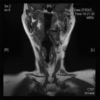
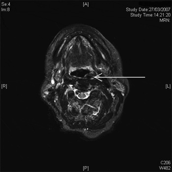
Magnetic resonance imaging revealed a large internal carotid artery tonsillar kink indenting into the pharyngeal wall (Figures 2 and 3). Having ruled out neoplasia and vascular malformation, the patient was reassured and discharged.
Coronal magnetic resonance imaging scan showing kinking of the left internal carotid artery and indenting into the pharynx.

Axial magnetic resonance imaging scan showing anatomical asymmetry of the internal carotid artery (arrow).

Discussion
According to the descriptions of most anatomy textbooks, the cervical part of the internal carotid artery (ICA) runs a straight course to the base of the skull, without branching. However, this only occurs in 65–70 per cent of cases. Variations of the ICA do occur, such as a curved course (25 per cent) or kinking or coiling (5 per cent).Reference Fix, Daffner and Deeb1 Such anatomical variation occurs in approximately 6 per cent of the general population, and it is of great clinical importance in view of the large number of surgical procedures performed in the oropharynx.Reference Paulsen, Tillman, Christofides, Richter and Koebke2, Reference Palacios, Kirsch and Rojas3 The development of these variations has been attributed to arteriosclerotic changes in the carotid artery, malignant processes, or incomplete straightening of the ICA during embryological development.Reference Palacios, Kirsch and Rojas3 Such factors result in congenital tortuosity or an aberrant ICA in the retropharyngeal space. The non-pulsatile nature of the mass in this case may have been due to long-standing arteriosclerosis.
Inadvertent trauma from intubation, rigid endoscopy or a Boyles–Davis gag may result in torrential intra-operative haemorrhage.Reference Shanley4 Management of such haemorrhage may be difficult due to restricted access to the oral orifice, compounded by the presence of an endotracheal tube should the patient be intubated. Furthermore, control cannot be achieved by packing alone and may only be possible by ligating or embolising the common carotid, which carries significant implications such as cerebral infarction.
Anatomical variations of the ICA are usually asymptomatic, but they may present as dysphagia, throat pain, hoarseness or increasing sensations of a foreign body in the pharyngeal area.Reference Avitia, Hamilton and Osborne5, Reference Sergi, Alberti, Paludetti and Snider6 A new onset of upper digestive tract symptoms may be the presenting sign of carotid artery aneurysm presenting as a pharyngeal mass.Reference Sergi, Alberti, Paludetti and Snider6
As there is no bony structure protecting the pharyngeal wall, injury to the ICA can easily occur from diagnostic puncture during biopsy. Since retropharyngeal masses in most instances arouse suspicion of malignant disease, the ENT surgeon should defer any attempt at oropharyngeal biopsy until appropriate imaging has been performed. Certainly, this should not be adopted as dogma, but there is a compelling argument for being prudent in any patient presenting with a smooth, submucosal mass in the pharyngeal wall.