Introduction
Obesity, especially abdominal adiposity, is associated with cardiovascular disease (CVD).Reference Mathieu, Pibarot and Larose 1 , Reference Canoy 2 Abdominal adiposity functions as an active endocrine organ and may lead to adverse metabolic consequences that increase cardiovascular risk factors (e.g. insulin resistance, dyslipidemia, high-blood pressure).Reference Ordovas and Corella 3 – Reference Gower, Nagy and Goran 5 Unlike subcutaneous adipose tissue, visceral adipose cells also produce significant amounts of proinflammatory cytokines that are associated with atherosclerosis process.Reference Funahashi, Nakamura and Shimomura 6 , Reference Ohman, Wright, Wickenheiser, Luo and Eitzman 7
Abdominal circumference (AC) has been widely used to assess abdominal adiposity and has been shown to be more closely linked to CVD risk factors than is body mass index (BMI).Reference Lofgren, Herron and Zern 8 However, AC can not distinguish subcutaneous fat accumulation in the abdominal region from visceral fat accumulation.Reference Klein, Allison and Heymsfield 9 To overcome this limitation the use of ultrasonography to evaluate abdominal fat has been suggested as an alternative technique.Reference van der Kooy and Seidell 10 Ultrasound scan is a non-invasive and low-cost method, which can be used to image body fat without radiation exposure and has a high correlation with radiographic computed tomography. Suzuki et al.Reference Suzuki, Watanabe and Hirai 11 in 1993 introduced the Abdominal wall Fat Index (AFI), to indicate the ratio between the thickness of preperitoneal and subcutaneous fat evaluated by Ultrasound. This method reflects the amount of visceral fat and some studies have shown a positive correlation between AFI and CVD risk factors.Reference Kawamoto, Oka, Tomita, Kodama and Ootsuka 12 , Reference Soyama, Nishikawa and Ishizuka 13
Obesity track from childhood to adulthoodReference Clarke and Lauer 14 and epidemiological studies suggest that fat distribution could be programmed in fetal life.Reference Oken and Gillman 15 , Reference Sayer and Cooper 16 Children who are born small for gestational age have a predisposition to accumulate visceral fat in childhood and adulthood.Reference Sayer and Cooper 16 , Reference González, Nazmi and Victora 17 However, it is not clear if visceral adiposity is present at birth or if it is accumulated during the rapid postnatal catch-up growth. To the best of our knowledge there are no studies assessing the visceral adiposity in newborns with ultrasound scan. On the basis of this evidence, the aim of our exploratory study was to measure the visceral adiposity using the AFI in a sample of newborns and to assess its correlation with birth size, that is, weight, length and BMI.
Methods
We studied mothers and their newborns, all singleton births and healthy from the Neonatology Department of the Instituto de Medicina Integral Professor Fernando Figueira (IMIP), Brazil, during the period 10 March to 10 May 2009; IMIP cares for the needy population and provides assistance to nearly 600 deliveries per month. These patients have seen at IMIP came from the 85% of Brazilians that use the public health system. All had gestational age ⩾ 37 weeks and Apgar score ⩾ 7. The study was approved by IMIP’s Research Ethics Committee and all the mothers gave informed written consent.
All newborns underwent ultrasound examination of abdomen within the first 24 h of life. An ATL HDI 5000 (CA, Bothell) with CL 4–7 MHz or CL 2–5 MHz curvilinear transducer was used for the study. The thickness of subcutaneous fat, that is, the minimum distance in millimeter from the subcutaneous tissue to the linea alba and that of preperitoneal fat, that is, the maximum distance from linea alba to the surface of the left lobe of the liver, were measured directly from the screen with electronic caliper. AFI was calculated as preperitoneal fat to subcutaneous fat ratio. All measurements were carried out with the neonates laying in a supine position and without compressing the abdominal wall with the transducer. The linear-array probe was kept perpendicular to the skin on the upper median abdomen and longitudinal scanning was done under the xiphoid process.
All neonates were weighed and had their height (crown-heel lengths) measured within the first 24 h of life by using a digital scale (Seca, Germany) to the nearest 0.1 kg and a Harpenden neonatal stadiometer to the nearest 1 mm, respectively. BMI was calculated as weight/lengthReference Canoy 2 (kg/m2). AC was measured at the narrowest circumference between xiphisternum and umbilicus.
A subgroup of 10 neonates with weight, length and BMI comparable with the whole group, had the measurements of preperitoneal fat and subcutaneous fat determined twice by two experienced sonographers who did not have knowledge of the other operator’s scanning results. The inter-operator and intra-operator reliability was assessed.
The statistical analysis was performed by using the SPSS software package. Data are expressed as means ± s.e. (range). The correlation between parameters was assessed by using the Pearson correlation analysis, and P values <0.05 (two tailed) were considered statistically significant.
Results
A total of 118 mothers and their respective offspring were studied. The mothers’ age ranged from 15 to 45 years (25.3 ± 6.9), gestational age ranged from 37 to 41 weeks (38.4 ± 0.5), 62 mothers (53%) were single, 18 mothers (15.2%) had an individual income below US $1.00 ‘per day’ and 19 mothers (16.1%) had less than 4 years of schooling. The anthropometric characteristics of the study population are showed in Table 1; males were taller and heavier than female offspring (P < 0.005) and had a bigger AC at birth (P < 0.001). Twelve offspring (10.1%) had low birth weight and 12 offspring (10.1%) were large for gestational age.
Anthropometric characteristics of the 118 newborns studied

BMI, body mass index.
Data are the mean ± s.d.
*Student’s t-test.
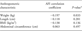
The sonographic measurements showed good inter-rater and intra-rater reliability, with intra-class correlation coefficients of subcutaneous and preperitoneal fat thickness, respectively: 0.96 (95% CI: 0.86–0.99) and 0.95 (95% CI: 0.83–0.99). There was a weak but statistically significant negative correlation between AFI and birth weight (−0.197; P = 0.033), but not with length, BMI or AC (Table 2).
Correlation between AFI and weight, length, BMI and abdominal circumference

AFI, Abdominal Wall Fat Index; BMI, body mass index.
*Pearson’s correlation test.
Discussion
Our results indicate a significant negative weak correlation between AFI and birth weight, but not with height, BMI or AC. Birth weight is an indicator of the conditions experienced prenatally and low birth weight usually represents an intra-uterine growth restriction (IUGR). Ten percent of our sample was low birth weight and we did not assess IUGR but our results may generalize to the very low end of the birth-weight spectrum.
Yajnik et al.Reference Yajnik, Lubree and Rege 18 found that Indian neonates, though lighter and shorter, had significantly greater absolute visceral adiposity than white European babies. These results are in agreement with our findings that lighter neonates may have visceral adiposity in early life. It could be a protective mechanism developed during fetal life to provide survival advantage under conditions of nutrient deficiency.
The importance of fat distribution in early life is not well-known and few studies have investigated visceral adiposity in neonates. As first hypothesized by Sparks,Reference Sparks 19 fetal fat-free mass may represent growth regulated primarily by intrinsic/genetic factors, whereas fat mass may be more readily affected by environmental factors. Animal studies demonstrate that IUGR decrease the muscle mass concomitant with increased adiposity that is correlated with insulin resistance.Reference Miles, Kathryn, Caroline, Jeffrey and Julie 20 , Reference Greenwood, Hunt, Hermanson and Bell 21 Furthermore, stress may play an essential role in body fat accumulation by activation of the hypothalamic-pituitary–adrenal axis, which increases glucocorticoids production and exerts hyperphagic and anti-thermogenic effects.Reference Drapeau, Therrien, Richard and Tremblay 22 Abdominal adipose tissue has more fat cells, higher blood flow and more glucocorticoid receptors. Besides, glucocorticoids affect abdominal fat to a greater extent than subcutaneous adipose tissue.Reference Ljung, Ottosson and Ahlberg 23
To date, ultrasound measure of subcutaneous and preperitoneal fat thickness has been used only in adults and there are a few studies in children.Reference Suzuki, Watanabe and Hirai 11 – Reference Clarke and Lauer 14 AFI is a practical approach for evaluating the distribution of abdominal adipose tissue in adults and has a strong association with cardiovascular risk factors. Given the relatively cheap, non-invasive, technically less demanding nature of ultrasound scan together with its good reproducibility, measurement of preperito neal and subcutaneous fat thickness may potentially become a useful imaging tool in the investigation of the early origins of obesity. To the best of our knowledge this is the first time that this method has been used in newborns and our results need to be confirmed.
Our study has some limitations. At first fat thickness in neonates is lower than in adults or children, making it more difficult to evaluate by ultrasound. This means that ultrasound measurement of fat thickness in neonates is more difficult. However, our results had a good reliability. Secondly our population includes few low birth weight and large for gestational age newborns.
In conclusion, we suggest that AFI is a useful parameter for evaluating the fat distribution in newborns. We found that visceral adiposity has a weak negative correlation with birth weight. Our findings need to be confirmed and further studies are needed to assess the fat distribution through the early postnatal life.
Acknowledgements
Renata Maria Gazineu was sponsored by Programa Institucional de Bolsas de Iniciação Científica (PIBIC), Conselho Nacional de Desenvolvimento Científico e Tecnológico (CNPq).
Statement of Interest
None.