Background
Premenstrual dysphoric disorder (PMDD) is a disabling affective disorder, characterized by negative emotional and physical symptoms that recur up to 2 weeks before the onset of each menstrual period (American Psychiatric Association, 2013). These symptoms include difficulty regulating emotions (Petersen et al. Reference Petersen, London, Liang, Ghahremani, Gerards, Goldman and Rapkin2016; Reuveni et al. Reference Reuveni, Dan, Segman, Evron, Laufer, Goelman, Bonne and Canetti2016), and emotional problems are featured heavily in the diagnostic criteria for PMDD (Bloch et al. Reference Bloch, Schmidt and Rubinow1997; Wittchen et al. Reference Wittchen, Becker, Lieb and Krause2002; Dickerson et al. Reference Dickerson, Mazyck and Hunter2003; Epperson et al. Reference Epperson, Steiner, Hartlage, Eriksson, Schmidt, Jones and Yonkers2012). PMDD can seriously diminish quality of life (Rapkin & Winer, Reference Rapkin and Winer2009), and some evidence suggests that women with PMDD are at higher risk of suicidal ideation and attempts compared with unaffected women (Hong et al. Reference Hong, Park, Wang, Chang, Sohn, Jeon, Lee, Cho, Kim, Bae and Cho2012; Pilver et al. Reference Pilver, Libby and Hoff2013). Nonetheless, PMDD has been relatively understudied compared with other affective disorders, and was moved from the appendix to the main text of the Diagnostic and Statistical Manual of Mental Disorders (DSM) only in the most recent edition, the DSM-5, released in 2013 (Hantsoo & Epperson, Reference Hantsoo and Epperson2015).
Because the core affective symptoms of PMDD involve difficulty regulating negative emotions, it is reasonable to suggest that women with PMDD may have transient, menstrual phase-related deficits in brain functions that are involved in emotion regulation during the symptomatic period. In healthy research participants, emotion regulation is associated with top–down control of amygdala activity by the prefrontal cortex (Ochsner et al. Reference Ochsner, Ray, Cooper, Robertson, Chopra, Gabrieli and Gross2004; Ochsner & Gross, Reference Ochsner and Gross2005; Banks et al. Reference Banks, Eddy, Angstadt, Nathan and Phan2007; McRae et al. Reference Mcrae, Misra, Prasad, Pereira and Gross2012), and patients with major depressive disorder exhibit less activation in the right dorsolateral prefrontal cortex (dlPFC) (Erk et al. Reference Erk, Mikschl, Stier, Ciaramidaro, Gapp, Weber and Walter2010) during regulation than healthy controls (Johnstone et al. Reference Johnstone, Van Reekum, Urry, Kalin and Davidson2007). Dysfunction in the bilateral dlPFC of women with PMDD has previously been documented (Baller et al. Reference Baller, Wei, Kohn, Rubinow, Alarcon, Schmidt and Berman2013), but has not yet been explicitly linked to symptoms of the disorder, or to difficulties in emotion regulation.
To address this problem, we performed a functional magnetic resonance imaging (fMRI) study, implementing an emotion-regulation paradigm, in 18 women with PMDD and 18 healthy controls. The paradigm selected is similar or identical to tasks used in previous investigations of emotion regulation that generated robust and reliable regulation-related brain activation and behavioral responses (Koenigsberg et al. Reference Koenigsberg, Fan, Ochsner, Liu, Guise, Pizzarello, Dorantes, Guerreri, Tecuta, Goodman, New and Siever2009, Reference Koenigsberg, Fan, Ochsner, Liu, Guise, Pizzarello, Dorantes, Tecuta, Guerreri, Goodman, New, Flory and Siever2010; Silvers et al. Reference Silvers, Shu, Hubbard, Weber and Ochsner2015, Reference Silvers, Insel, Powers, Franz, Helion, Martin, Weber, Mischel, Casey and Ochsner2016). Each woman completed the experiment once during the follicular phase, and once during the late luteal phase. Behavioral and neural measurements of emotion regulation were then compared between the two groups and menstrual phases. We predicted that during the luteal phase, while symptomatic, women with PMDD would experience difficulty regulating negative emotions, and that this difficulty would be accompanied by less task-related activation in the right dlPFC and greater activation in the amygdala in response to negative emotional stimuli relative to measurements in healthy controls and in the same participants during the follicular phase. The right dlPFC was selected for study because it is a primary prefrontal region that shows activation during emotion regulation (Banks et al. Reference Banks, Eddy, Angstadt, Nathan and Phan2007; Goldin et al. Reference Goldin, Mcrae, Ramel and Gross2008; Buhle et al. Reference Buhle, Silvers, Wager, Lopez, Onyemekwu, Kober, Weber and Ochsner2014), and because women with PMDD exhibit differences in dlPFC function bilaterally as compared with control women, albeit not during emotion regulation (Baller et al. Reference Baller, Wei, Kohn, Rubinow, Alarcon, Schmidt and Berman2013; Gingnell et al. Reference Gingnell, Bannbers, Wikstrom, Fredrikson and Sundstrom-Poromaa2013). The left and right amygdala were selected for similar reasons – i.e. the amygdala has been implicated as the target of top–down control by the prefrontal cortex during emotion regulation (Ochsner et al. Reference Ochsner, Silvers and Buhle2012; Buhle et al. Reference Buhle, Silvers, Wager, Lopez, Onyemekwu, Kober, Weber and Ochsner2014) and also has been linked to abnormal function in women with PMDD (Gingnell et al. Reference Gingnell, Morell, Bannbers, Wikstrom and Sundstrom Poromaa2012).
Methods
Participants and recruitment
The authors assert that all procedures contributing to this work comply with the ethical standards of the relevant national and institutional committees on human experimentation and with the Helsinki Declaration of 1975, as revised in 2008. All methods were approved by the UCLA Institutional Review Board. Participants were recruited via Internet advertisements and flyers. They were required to be 18–44 years old non-smokers, fluent in English, right-handed [verified by the Edinburgh Handedness Inventory (Oldfield, Reference Oldfield1971)], generally healthy as determined through a medical history and physical examination by a nurse practitioner (see online Supplemental Information for additional detail), willing to use non-hormonal contraception for the duration of the study [because hormonal contraception may affect PMDD symptoms (Lopez et al. Reference Lopez, Kaptein and Helmerhorst2012) and amygdala reactivity (Petersen & Cahill, Reference Petersen and Cahill2015)], and to have regular, 24–32-day menstrual cycles. All participants provided written informed consent prior to enrolling in the study.
Exclusion criteria were: any current or lifetime diagnosis of a psychiatric illness other than unipolar mood disorders (these were allowed if occurred >2 years before assessment) identified using the Structured Clinical Interview for DSM-IV Axis I Disorders, Patient Edition (First et al. Reference First, Spitzer, Gibbon and Williams2002); central nervous system, cardiovascular, hepatic, renal, endocrine, or autoimmune disease; substance use disorder(s); hormonal contraception within 1 month of entering the study [normal ovulatory cycles are expected within 1 month of discontinuation; see (Duijkers et al. Reference Duijkers, Engels and Klipping2005; Birtch et al. Reference Birtch, Olatunbosun and Pierson2006; Davis et al. Reference Davis, Kroll, Soltes, Zhang, Grubb and Constantine2008)]; use of medications that affect cerebral perfusion or function; pregnancy; breastfeeding; or metal in the body that could compromise MRI safety or data fidelity. PMDD-group participants were also excluded for using any medication or herbal product to treat PMDD within the 1 month before participating in the study.
Potentially eligible participants completed the Daily Record of Severity of Problems [DRSP; (Endicott et al. Reference Endicott, Nee and Harrison2006)], entering their responses in a secure online database for ⩾2 months; the range of possible scores was 1 (not at all) to 6 (extreme).
To meet criteria for the PMDD group, participants were required to meet four criteria:
1. During days 5–12 after the onset of menstruation, have mean scores <3 for each of the 14 DRSP items.
2. During the premenstrual phase (6 days before menstruation + day 1 of menstruation), on DRSP items 1 through 4 (measuring depression, anxiety, mood swings, and anger/irritability), report scores ⩾3 for at least 4 days, and also report scores ⩾4 for at least 2 of those days for one or more symptoms.
3. During the premenstrual phase, on DRSP items 1 through 11, report scores ⩾3 for at least 2 days on at least five symptoms, and also report scores ⩾4 for at least 2 days.
4. During the premenstrual phase, on DRSP items 12 through 14 (measuring presence and degree of impairment), report scores ⩾3 for at least 2 days, and also report scores ⩾4 for at least 2 days.
To describe the severity of PMDD in this sample, percent change in emotional symptoms from the follicular to premenstrual phase was also calculated. Emotional symptoms were defined as DRSP items 1–4 (describing depression, anxiety, mood swings, and anger/irritability). The follicular and premenstrual phases were defined as above. Percent change was calculated as: [(follicular score–premenstrual score)/premenstrual score] × 100.
To meet criteria for the healthy control group, participants were required to report scores <3 on all DRSP items during the follicular phase, and <2 during the premenstrual phase.
Approximately half of the participants in each group (PMDD, 61%; controls, 50%) entered the study during the follicular phase; the rest began during the late luteal phase. Ovulation was estimated with at-home luteinizing hormone urinalysis (Clearblue® Digital Ovulation kit, SPD Swiss Precision Diagnostics GmbH, Geneva).
Participants were asked to refrain from using marijuana for ⩾48 h, alcohol for ⩾24 h, and caffeine for ⩾2 h before the two fMRI sessions (one during the follicular phase, one during the late luteal phase). Urine tests excluded pregnancy, and breath tests verified abstinence from alcohol and smoking. To the extent possible, fMRI was conducted at similar times on the two testing days to avoid diurnal effects (Cunningham-Bussel et al. Reference Cunningham-Bussel, Root, Butler, Tuescher, Pan, Epstein, Weisholtz, Pavony, Silverman, Goldstein, Altemus, Cloitre, Ledoux, Mcewen, Stern and Silbersweig2009); 86% of repeat scans were performed within 2 h of initial scan times, and 100% were performed within 4.5 h.
Emotion-regulation task
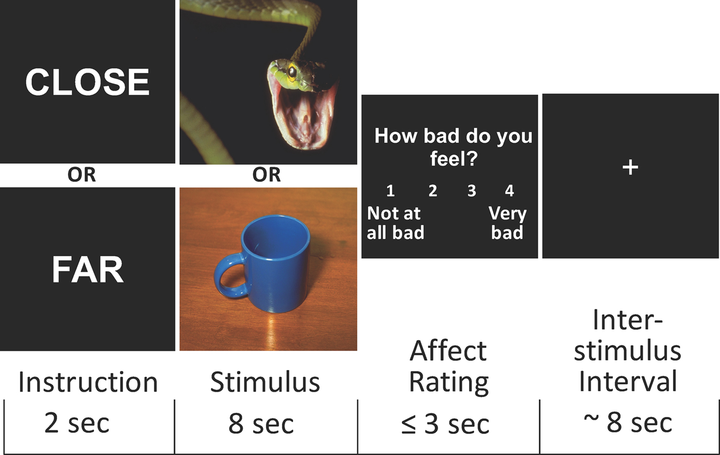
Proximal/distal perspective taking (‘distancing’) was used as a strategy to regulate affect (Silvers et al. Reference Silvers, Shu, Hubbard, Weber and Ochsner2015). Each trial had three components: instruction (2 s), image presentation (8 s), and response interval (max 3 s) (Fig. 1). Negative and neutral images were presented in two conditions. During the ‘close’ condition, participants were instructed, ‘Imagine yourself standing close to the scene depicted in the picture, and allow yourself to experience any emotions that come’; during ‘far’ trials, the instruction was, ‘Imagine yourself standing further away from the scene and focus more on the facts of the photograph than on its emotional details, in the same way that a news reporter might’. Next, participants responded to the question, ‘How bad do you feel?’, on a scale from 1 (‘not at all bad’) to 4 (‘very bad’), using an MR-compatible, four-button box.
Schematic of event sequence during trials of the emotion regulation task. Participants viewed the instruction ‘close’ or ‘far’ (2 s), followed by a negative or neutral image (8 s), and then rated their degree of negative affect (⩽3 s). An inter-stimulus interval of ~3 s preceded the next trial (see Methods section).

The stimuli presented were negatively or neutrally valenced pictures from the International Affective Picture System (Lang et al. Reference Lang, Bradley and Cuthbert2008) and images used by Silvers et al. (Reference Silvers, Shu, Hubbard, Weber and Ochsner2015). Participants completed 80 trials of the task in an event-related fMRI design separated across four runs, generating a total of 20 negative, close trials; 20 negative, far trials; 20 neutral, close trials; and 20 neutral, far trials in each session (see online Supplementary Methods for details on task presentation). In both behavioral and fMRI analyses, emotion regulation was assessed by the difference of data obtained during the negative, far condition from those during the negative, close condition. We also examined data from the negative, far condition alone to assess response to this condition independent of a contrast (i.e. how do ‘negative, far’ ratings differ irrespective of ‘negative, close’ ratings).
fMRI data acquisition
MRI scans were performed using a 3-T Magnetom Trio scanner (Siemens Medical Solutions USA Inc., Malvern, PA, USA) with a 12-channel bird-cage coil. Four runs of 167 functional T2*-weighted echoplanar images (EPIs) were acquired [slice thickness, 4 mm; 34 slices; repetition time (TR), 2 s; echo time (TE), 30 ms; flip angle, 90°; matrix, 64 × 64; field of view (FOV), 192 mm]. The orientation of slices was oblique axial to maximize brain coverage and to optimize signal from ventral prefrontal regions (see online Supplementary Methods for additional information).
Analysis of fMRI data
The fMRI data were analyzed using The Oxford Centre for Functional Magnetic Resonance Imaging of the Brain (FMRIB)’s Software Library (FSL), version 5.0.9. Images were motion-corrected, spatially smoothed (6-mm Gaussian kernel), and temporally filtered. Time-series analysis was performed using FMRIB's Improved Linear Model (FILM) with local auto-correlation correction. Each event was modeled as an impulse, convolved with a canonical hemodynamic response function (double-Ɣ) with a width equaling the event duration, with its temporal derivative. Instruction, image, and response periods were modeled separately. Six motion parameters (three rotational and translational directions) were modeled as nuisance covariates. EPIs were registered to the MBW image, then to the MPRAGE structural image, and finally into standard space (Montreal Neurological Institute avg152 template), using 12-parameter affine transformations (Jenkinson & Smith, Reference Jenkinson and Smith2001). Registration from MPRAGE structural images to standard space was refined using FNIRT non-linear registration (Andersson et al. Reference Andersson, Jenkinson and Smith2007). Contrast and parameter estimate maps (i.e. modeled condition v. the implicit baseline of all unmodeled activation) were registered to standard space.
Regions of interest (ROIs) were selected a priori based on prior literature (Buhle et al. Reference Buhle, Silvers, Wager, Lopez, Onyemekwu, Kober, Weber and Ochsner2014; Silvers et al. Reference Silvers, Shu, Hubbard, Weber and Ochsner2015): one in the right dlPFC (defined by a 10-mm radius sphere centered at MNI coordinates: 33, 24, 51), and others in the left and right amygdala (defined according to the Harvard–Oxford Atlas). Average contrast parameter estimates for each participant from each ROI were extracted using FSL tools (i.e. fslmeants).
Planned comparisons were carried out to assess effects of group and phase in each ROI. First, a manipulation check was performed to test whether the emotion-regulation task produced expected activation in the selected ROIs. In the right dlPFC, activation during the negative, far condition v. the negative, close condition was compared to establish that dlPFC activation was greater during the far v. close trials. Subsequently, full factorial models, including main effects of group, phase, and the group-by-phase interaction, were used to test right dlPFC activation during negative, far trials and the difference between right dlPFC activation during negative, far–negative, close trials. As a negative control, a full factorial model was used to rule out effects of group and phase during negative, close trials. To test the relationship between brain activation during emotion regulation and symptom severity in women with PMDD (excluding healthy controls), an analysis of covariance (ANCOVA) was performed with parameter estimates of dlPFC activation during negative, far trials and a composite mood summary score [i.e. the sum of depression, anxiety, mood swings, and irritability ratings from the DRSP (Rapkin et al. Reference Rapkin, Berman, Mandelkern, Silverman, Morgan and London2011)] entered into the model.
In each amygdala ROI, activation during negative v. neutral trials was compared to establish sensitivity of amygdala activation to negatively valenced stimuli. Subsequently, full factorial models, including main effects of group, phase, and the group-by-phase interaction, were used to test for differences in amygdala activation during negative, far trials; negative, close trials; and the difference between the two.
In addition, exploratory whole-brain voxel-wise group analyses were performed using mixed-effects analysis in FSL, with fixed-effects for within-subject analyses to combine runs and compare across menstrual phases and random-effects analysis for between-subjects analyses, using FSL's Local Analysis of Mixed Effects (FLAME1) tool, which uses hierarchical linear modeling correcting for variance heterogeneity and non-sphericity. Z (Gaussianized T/F) statistic images were thresholded using a cluster-forming threshold of Z > 2.3 and a cluster extent significance threshold of p < 0.05. To isolate activity associated with emotion regulation, activity in ‘negative, far’ trials was contrasted against ‘negative, close’ trials.
Hormone analysis
Serum progesterone levels at each visit were measured to confirm ovulation; they were analyzed by electrochemiluminescence (Roche Elecsys Immunoassay system, F. Hoffman-LaRoche, Basel, Switzerland). The measuring range in this system is 0.03–60 ng/mL (detection threshold of 0.03 ng/mL).
Statistics
Statistical analyses were performed in JMP(R) Pro 11.0.0 (SAS Institute Inc., Cary, NC, USA) on behavioral data and ROI summary data (Bonferroni corrected at α = 0.05/3 = 0.02). FSL 5.0.9 was used for all other fMRI analyses (Z > 2.3, p < 0.05, cluster-corrected). The whole-brain analysis used a cluster detection threshold of Z = 2.3. A 2 × 2 mixed-model ANOVA was used to evaluate group and phase effects, (fixed effects: group, phase) (random effect: participant). ‘Regulation success’ was calculated by subtracting each participant's average ratings on ‘negative, far’ trials from her average ratings on ‘negative, close’ trials for each session separately.
Results
Participants
There were 18 participants in each of the two groups. They ranged in age from 18 to 41 years (controls, M = 25.4, s.d. = 6.99; PMDD, M = 29.2, s.d. = 7.24), with no significant group difference [t(34) = 1.59; p = 0.12]. The groups did not differ significantly in years of education (controls, M = 15.6, s.d. = 2.70; PMDD, M = 16.7, s.d. = 3.61; [t(34) = 1.04, p = 0.30]), maternal education [t(33) = 1.52, p = 0.14], or self-reported income (one participant declined to answer) [t(33) = 0.99, p = 0.33], and reported similar relationship statuses (controls: 16.7% married, 55.6% single, 5.6% divorced, 22.2% in a committed relationship; PMDD: 16.7% married, 55.6% single, 5.6% divorced, 22.2% in a committed relationship). The groups differed somewhat in ethnicity (see online Supplementary Table S1; χ2 test not performed due to small cell sizes).
Hormone levels
Progesterone levels were significantly higher during the luteal phase than the follicular phase for both groups (p < 0.05), with no difference between groups (p = 0.38) (online Supplementary Table S2).
DRSP scores
As expected, PMDD-group women had higher DRSP scores during the luteal phase than the follicular phase [t(34) = 21.07, p < 0.05], and higher scores than luteal controls [t(34) = 19.86, p < 0.05], producing a significant group by menstrual phase interaction [F(1,34) = 219.53, p < 0.05] (see online Supplementary Table S3 for detailed symptom reports). On the four core emotional symptoms (DRSP items 1–4, describing depression, anxiety, mood swings, and anger/irritability), participants reported a 182% change from the follicular to premenstrual phase (range = 54.9–343.4%).
Emotion-regulation task performance
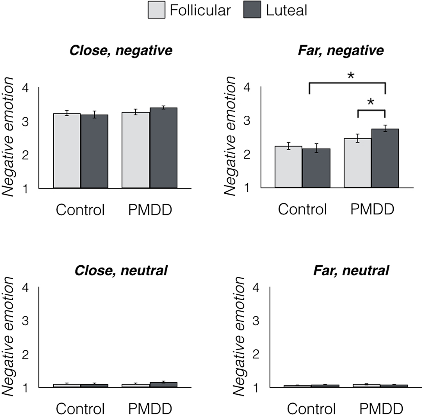
Robust main and interacting effects of instruction (close/far) and stimulus type (negative/neutral) were found, all p < 0.05 (see online Supplementary Fig. S1). The groups had similar performance on neutral trials of the emotion-regulation task (Fig. 2), no main effect of group on self-reported negative emotion in ‘neutral, close’ [F(1,34) = 0.39, p = 0.54] or ‘neutral, far’ trials [F(1,34) = 0.39, p = 0.54], no main effect of menstrual phase on performance in ‘close’ [F(1,34) = 1.22, p = 0.28] or ‘far’ [F(1,34) = 0.06, p = 0.81] trials, and no group-by-phase interaction on performance in either condition [close: F(1,34) = 0.84, p = 0.37; far: F(1,34) = 1.43, p = 0.24].
Negative emotion after each trial condition during the emotion regulation task. Women with PMDD gave reports of stronger negative emotion in the luteal phase than in the follicular phase, and compared with healthy controls during the luteal phase. No group or phase differences were detected in the other task conditions, n = 18/group. *p < 0.05. Error bars indicate ± 1 s.e.m.

There were no significant main or interacting effects of group or phase in negatively valenced trials with a ‘close’ instruction [main effect of group: F(1,34) = 1.28, p = 0.27; main effect of phase: F(1,34) = 0.29, p = 0.60; group-by-phase interaction: F(1,34) = 1.54, p = 0.22]. However, there were significant group and group-by-phase differences in negatively valenced trials with the ‘far’ instruction, indicating significant differences in the ability of women with PMDD to regulate emotions during the luteal phase (Fig. 2). Women with PMDD reported stronger negative emotions during these trials than did controls [main effect of group: F(1,34) = 6.17, p = 0.02]. The group-by-phase interaction (on ratings made after ‘negative, far’ trials) was significant [F(1,34) = 4.85, p = 0.03]. Post hoc t tests comparing scores of each experimental group and each session (i.e. control follicular, control luteal, PMDD follicular, PMDD luteal) after negative, far trials indicated that this interaction was driven by higher ratings by PMDD-group women during the luteal v. follicular phase [t(35) = 2.36, p = 0.02] or v. controls in the luteal phase [t(35) = 3.19, p < 0.01]. Despite the significant group and phase differences in negative, far mean scores, the emotion-regulation difference score (negative, close–negative, far) did not differ significantly between the groups and/or phases of the cycle (main and interacting effects all p > 0.05).
fMRI results: activation related to emotion regulation
As a manipulation check to test whether the task produced activation consistent with what was observed in previous investigations of brain function related to the regulation of negative emotions, activity during negative, far trials was contrasted against negative, close trials in the entire sample (both groups, both sessions). In a whole-brain analysis, five clusters showed significant effects, and they were found in regions that were activated in previous studies of emotion regulation: lingual gyrus (Goldin et al. Reference Goldin, Mcrae, Ramel and Gross2008), angular gyrus (Wager et al. Reference Wager, Davidson, Hughes, Lindquist and Ochsner2008; Kohn et al. Reference Kohn, Eickhoff, Scheller, Laird, Fox and Habel2014), middle frontal gyrus (Wager et al. Reference Wager, Davidson, Hughes, Lindquist and Ochsner2008; Buhle et al. Reference Buhle, Silvers, Wager, Lopez, Onyemekwu, Kober, Weber and Ochsner2014; Kohn et al. Reference Kohn, Eickhoff, Scheller, Laird, Fox and Habel2014; Silvers et al. Reference Silvers, Shu, Hubbard, Weber and Ochsner2015), precuneus (Goldin et al. Reference Goldin, Mcrae, Ramel and Gross2008; Wager et al. Reference Wager, Davidson, Hughes, Lindquist and Ochsner2008; Buhle et al. Reference Buhle, Silvers, Wager, Lopez, Onyemekwu, Kober, Weber and Ochsner2014; Silvers et al. Reference Silvers, Shu, Hubbard, Weber and Ochsner2015), and frontal pole (Wager et al. Reference Wager, Davidson, Hughes, Lindquist and Ochsner2008) (see online Supplementary Fig. S2 and Table S4).
ROI analyses
Right dlPFC: The validity of the selected, independently defined ROI as an index of regulation-related activation was tested via linear mixed model, assessing dlPFC activation in both groups, both sessions. This analysis confirmed significantly greater activation in the right dlPFC during ‘negative, far’ trials than ‘negative, close’ trials [F(1,34) = 6.42, p = 0.01] (i.e. greater BOLD signal while participants were viewing negative stimuli and engaged in the emotion regulation condition than when viewing negative stimuli without regulating). To confirm that regulation-related activation was lateralized to the right dlPFC, greater activation in ‘negative, far’ v. ‘negative, close’ trials from a left dlPFC ROI (MNI – 33, 24, 51) was extracted and submitted for analysis. No significant difference was found [F(1,34) = 0.08, p = 0.77].
To test whether right dlPFC activation was associated with emotion regulation, regulation success (self-reported negative, far–negative, close scores) was tested for a relationship with right dlPFC activation related to emotion regulation (activation during negative, far trials–activation during negative, close trials) in a mixed model with participant as a random effect including both groups, both sessions. A significant relationship between neural and behavioral emotion regulation was found [F(1,34) = 1.94, p = 0.03] (online Supplementary Fig. S3).
Because the two groups differed in ethnicity, a one-way ANOVA was used to confirm that ethnicity was not significantly related to overall right dlPFC activation during all trials [F(1,34) = 0.57, p = 0.68] or specifically to ratings in ‘negative, close’ trials [F(1,34) = 1.34, p = 0.26] or ratings in ‘negative, far’ trials [F(1,34) = 1.05, p = 0.39].
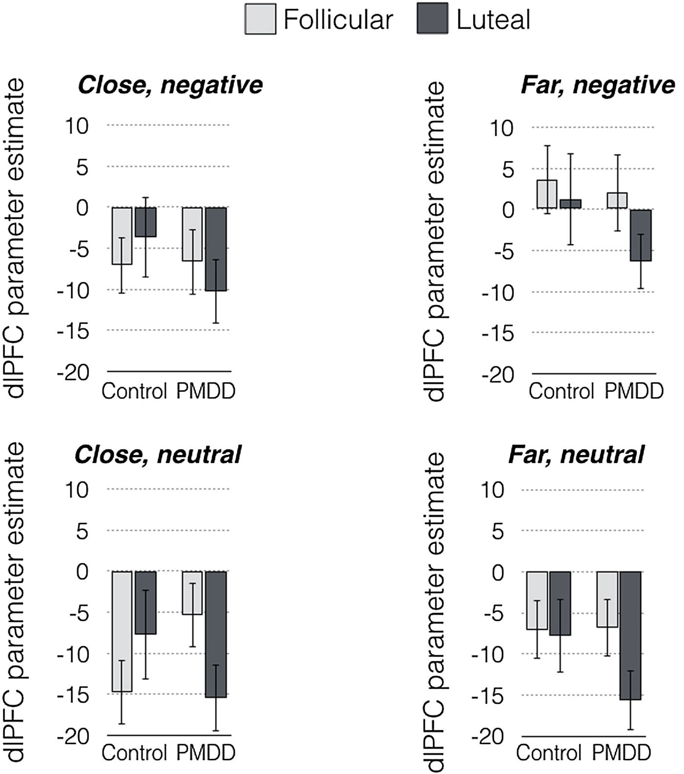
Contrary to the a priori hypotheses, a subsequent exploratory analysis showed that the effect of lower right dlPFC activity in PMDD-group women during the luteal phase was not restricted to any particular task condition (Fig. 3). No main or interacting effects of group or phase on right dlPFC activity were found in any of the individual conditions (all p > 0.05). Similarly, the emotion-regulation difference score (greater activation during negative, far trials than during negative, close trials) showed no significant effect of group or phase on right dlPFC activation (p > 0.05).
Right dlPFC activation for each group and each task condition during the emotion regulation task. Parameter estimates of right dlPFC activation during each task condition were not significantly different between groups or phases. N = 18/group. Error bars indicate ± 1 s.e.m.

A significant group-by-phase interaction on right dlPFC activity during the emotion regulation task [F(1,34) = 5.98, p = 0.02] was obtained when data were collapsed across all task conditions (neutral, negative, close, and far). No significant main effects of group or phase were found (p > 0.10). Post hoc t tests showed that right dlPFC activation was significantly lower in the PMDD group during the luteal phase than the follicular phase [t(35) = 2.79, p < 0.02], and marginally lower in the PMDD group compared with controls during the luteal phase [t(35) = 2.01, p = 0.05] (Fig. 4).
Women with PMDD had significantly less activation in the right dlPFC while performing the emotion regulation task during the late luteal phase, when they are symptomatic. Statistical parameter estimates of right dlPFC activity during all trials of the emotion regulation task combined (compared with the intertrial-baseline condition) were significantly lower for women with PMDD during the luteal phase than during the follicular phase, and compared with healthy women during the luteal phase. N = 18/group. *p < 0.05. Error bars indicate ± 1 s.e.m.

Activation in right dlPFC during ‘negative, far’ trials correlated significantly with the severity of emotional PMDD symptoms in women with PMDD. Severity of symptoms was indexed by a composite mood summary score [the sum of core ratings of depression, anxiety, mood swings, and irritability from the DRSP (Rapkin et al. Reference Rapkin, Berman, Mandelkern, Silverman, Morgan and London2011)] and was significantly related to right dlPFC activation during emotion regulation (negative, far trials) in a one-way ANCOVA, controlling for ethnicity [F(1,14) = 4.22, p = 0.03].
Amygdala: In an analysis with the groups combined, the BOLD response was significantly greater in the left [t(35) = 3.66, p < 0.01] and right [t(35) = 2.79, p < 0.01] amygdala during the presentation of negative v. neutral images, indicating amygdala activation when emotional stimuli were presented. No main effect of instruction was found [left amygdala: F(1286) = 2.80, p = 0.09; right amygdala: F(1286) = 0.84, p = 0.36], nor was a significant instruction-by-valence interaction [left amygdala: F(1286) = 1.56, p = 0.21; right amygdala: F(1286) = 0.94, p = 0.33]. There were no significant effects of group or phase on amygdala activation (all p > 0.05) during any task condition or with all conditions combined (online Supplementary Fig. S4).
Whole-brain results
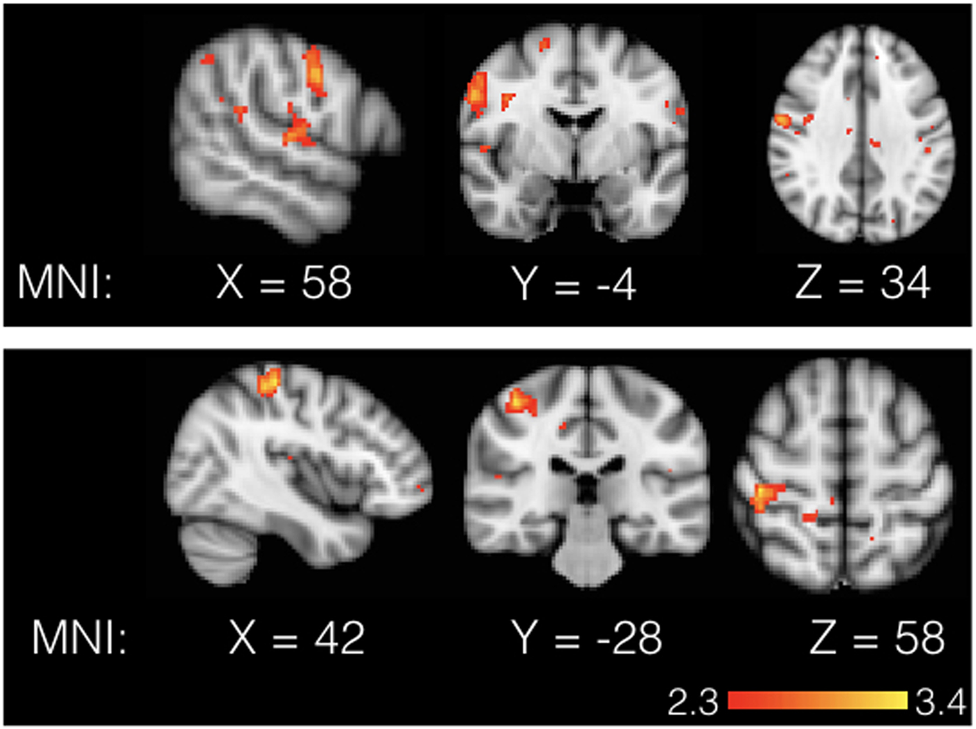
To measure brain activation during emotion regulation using whole-brain voxel-wise analyses, ‘negative, far’ trials were contrasted against ‘negative, close’ trials (far, negative greater than close, negative). Women in the PMDD group showed greater activation in the right precentral gyrus in the follicular phase than in the luteal phase (peak voxel x, y, z = 58, −4, 34; Z = 3.49; cluster size = 523; p < 0.05). Activation in the right postcentral gyrus was also lower during the luteal phase (peak voxel x, y, z = 42, −28, 58; Z = 3.79; cluster size = 403; p = 0.02; see Fig. 5) when compared with control women.
Whole-brain voxel-wise analysis results. Top: Group comparison of regulation-related activity (far, negative greater than close, negative) in the luteal phase v. the follicular phase, showing significantly greater activation in the right precentral gyrus during the follicular phase in PMDD women, N = 18/group, FWE cluster corrected, Z > 2.3, p < 0.05, centered on peak voxel at 58, −4, 34. Bottom: A similar analysis showed significantly greater regulation-related activity in the right postcentral gyrus of healthy controls compared with women with PMDD, both tested in the luteal phase, FWE cluster-corrected, Z > 2.3, p < 0.05, centered on peak voxel at 42, −28, 58.

Conclusions
These findings indicate that women with PMDD experience emotion-regulation difficulty as measured with a behavioral task that engages cognitive reappraisal of emotion using a distancing strategy, as well as transient hypoactivity in the right dlPFC during the luteal phase of the menstrual cycle, when they are symptomatic, and that this hypoactivity is related to the severity of emotional symptoms during the late luteal phase. The data suggest that the observed right dlPFC hypoactivation underlies negative affect in general rather than a specific emotion-regulation deficit in symptomatic women with PMDD.
Previous findings have pointed to the dlPFC as a potential neural locus of dysfunction in women with PMDD. Women with PMDD differed from controls in bilateral dlPFC activation when they performed a working memory task, as indicated by fMRI and positron emission tomography (Baller et al. Reference Baller, Wei, Kohn, Rubinow, Alarcon, Schmidt and Berman2013). In that case, dlPFC activity was greater in women with PMDD, but the participants were subjected to gonadal hormone suppression with leuprolide acetate alone, then leuprolide acetate with add-back estrogen, and separately, add-back progesterone. Activation in dlPFC was greater than in controls through all hormonal conditions, even when participants were asymptomatic. Notably, perfusion in the dlPFC is sensitive to leuprolide acetate treatment with or without add-back estrogen and progesterone (Berman et al. Reference Berman, Schmidt, Rubinow, Danaceau, Van Horn, Esposito, Ostrem and Weinberger1997). Another fMRI investigation found greater right dlPFC activation during the luteal phase in women with PMDD compared with controls when they were anticipating negative stimuli (Gingnell et al. Reference Gingnell, Morell, Bannbers, Wikstrom and Sundstrom Poromaa2012). One possible way to reconcile this result with the finding of right dlPFC hypoactivity throughout the emotion-regulation task is that the right dlPFC may be generally hypoactive in women with PMDD, but uniquely shows increases in activity during the anticipation of negative stimuli. Thus, whereas a variety of evidence points to the dlPFC dysfunction in women with PMDD, this dysfunction may depend on task demands, hormonal status, or another variable not yet identified.
The finding of hypoactivity in the right pre- and/or postcentral gyri in women with PMDD is convergent with literature on the function of these brain regions. The left and right precentral gyri show activation during emotion regulation (Domes et al. Reference Domes, Schulze, Bottger, Grossmann, Hauenstein, Wirtz, Heinrichs and Herpertz2010), as does the postcentral gyrus (Ochsner et al. Reference Ochsner, Ray, Cooper, Robertson, Chopra, Gabrieli and Gross2004; Domes et al. Reference Domes, Schulze, Bottger, Grossmann, Hauenstein, Wirtz, Heinrichs and Herpertz2010), although the postcentral gyrus has been implicated in increasing rather than suppressing emotion, more so in men than in women (Domes et al. Reference Domes, Schulze, Bottger, Grossmann, Hauenstein, Wirtz, Heinrichs and Herpertz2010). Specific to PMDD, women with PMDD showed less activation in the postcentral gyrus than controls during a Go/NoGo response inhibition task independent of cycle phase (Bannbers et al. Reference Bannbers, Gingnell, Engman, Morell, Comasco, Kask, Garavan, Wikstrom and Sundstrom Poromaa2012). Here, we report less activation in the right precentral gyrus in women with PMDD during the luteal compared with the follicular phase, and less activation in the right postcentral gyrus in the symptomatic women compared with controls. Additional studies are warranted to understand the significance of these effects in the context of PMDD.
Although ample evidence supports a role for the amygdala in emotion regulation [for meta-analysis, see (Buhle et al. Reference Buhle, Silvers, Wager, Lopez, Onyemekwu, Kober, Weber and Ochsner2014)], the current investigation shows no differences in amygdala activation in PMDD across menstrual phases, consistent with one previous study in which amygdala reactivity did not differ in women with PMDD between the luteal phase and follicular phase, or in women with PMDD compared with controls in the luteal phase (Gingnell et al. Reference Gingnell, Morell, Bannbers, Wikstrom and Sundstrom Poromaa2012).
While these findings advance understanding of the neural circuitry underlying PMDD, some limitations constrain their interpretation. First, the sample size, although commensurate with other PMDD imaging studies, is small, and the study may have been underpowered to detect some effects. For instance, a single task condition may be driving the right dlPFC hypoactivity that was observed across all conditions in the emotion-regulation task, but the statistical power may be insufficient to detect differences between conditions. Low statistical power may also underlie the lack of convergence between the two related measures of emotion regulation (i.e. negative, far trials alone v. the difference between negative, far and negative, close trials). With a larger sample, the two ways of assessing emotion regulation may have produced more similar results. Similarly, an effect of instruction (‘close’ v. ‘far’) on amygdala activation was not observed, although a trend in the expected direction (lower activation during ‘far’ trials) was observed in the left amygdala (p = 0.09). With more statistical power, a significant relationship between instruction and amygdala activation might be observed.
A second concern is the negative direction of the right dlPFC activity relative to the implicit baseline. Because the dlPFC is generally regarded as belonging to a task-positive network (Greicius et al. Reference Greicius, Krasnow, Reiss and Menon2003), positive parameter estimates for each task condition were expected. However, the parameter estimates were only positive during negative, far trials – the most cognitively demanding condition – and even then, they were still negative for women with PMDD during the luteal phase. This finding could be attributed to less right dlPFC activation during task performance, or to more activation during the implicit baseline or ‘null’ condition (inter-stimulus interval) than predicted. Nonetheless, the regions of peak activation during emotion regulation were the same as those activated in other reports of emotion regulation (Goldin et al. Reference Goldin, Mcrae, Ramel and Gross2008; Wager et al. Reference Wager, Davidson, Hughes, Lindquist and Ochsner2008; Buhle et al. Reference Buhle, Silvers, Wager, Lopez, Onyemekwu, Kober, Weber and Ochsner2014; Kohn et al. Reference Kohn, Eickhoff, Scheller, Laird, Fox and Habel2014; Silvers et al. Reference Silvers, Shu, Hubbard, Weber and Ochsner2015), and right dlPFC activation was greatest when the task called for the most cognitive control (i.e. far, negative trials).
A third concern involves the selection of the control group and therefore generalizability of the findings. Healthy controls in this investigation were required to report almost no premenstrual discomfort, which does not represent the entire population. Many women [approximately 10–11% of the population; (Halbreich et al. Reference Halbreich, Borenstein, Pearlstein and Kahn2003)] experience menstrual-related distress that does not meet criteria for PMDD, and it is not clear from this investigation what brain function may be observed during emotion regulation in that portion of the population.
In summary, this investigation extends existing evidence that women with PMDD are less able to regulate their experience of negative emotion during the premenstrual phase compared with the follicular phase, or compared with controls in the premenstrual phase. This finding expands on a previous report (on the same participants) of trait-like difficulties with emotion regulation in women with PMDD (Petersen et al. Reference Petersen, London, Liang, Ghahremani, Gerards, Goldman and Rapkin2016), and strengthens the evidence that dysfunction in the right dlPFC and the right pre- and postcentral gyri may be linked with affective symptoms in PMDD. These findings support the use of existing therapies, and may be helpful in guiding the development of new therapies, and personalizing treatment for PMDD. Notably, dlPFC function can predict symptom improvement as a result of cognitive–behavioral therapy (CBT) in patients with major depressive disorder (Ritchey et al. Reference Ritchey, Dolcos, Eddington, Strauman and Cabeza2011); if this is also true in PMDD, then fMRI measurements of dlPFC activity may be used to identify patients who are likely to respond to CBT. Finally, therapies such as transcranial magnetic stimulation or transcranial direct current stimulation rely on localization of cortical dysfunction. Although more research is needed, the findings identify the right dlPFC and right pre- and postcentral gyri as potential candidate regions for such therapies, which can relatively easily target superficial cortical structures.
Supplementary Material
The supplementary material for this article can be found at https://doi.org/10.1017/S0033291717003270.
Acknowledgements
The authors are grateful for the dedicated, conscientious work of the research assistants who helped collect and manage data for this project: Aqsah Choudhary, Rachel Gerards, and Chelsea Cox. They also thank Linda Goldman, NP, for her clinical contributions and Dr Jennifer Silvers for providing some of the images used as stimuli in the emotion regulation task. The authors report no conflicts of interest, financial or otherwise. This work was supported by a National Institute of Mental Health Grant (R21MH098668) to EDL and AJR, and endowments from the Thomas P. and Katherine K. Pike Chair in Addiction Studies, and the Marjorie M. Greene Trust. These funding sources provided financial support but had no role in study design, data collection, data analysis, or publication decisions.
Declaration of Interest
None.