
Introduction
There is an increased interest toward the application of low-dimensional nanostructured materials for the past about 10 years.[Reference Augustine, Singh, Srivastava, Sharma, Das and Malhotra1–Reference Hahm3] Many efforts have been made toward the preparation and application of one-dimensional (1D) nanomaterials based on transition metals. In this context, the 1D morphologies (e.g., nanotubes, nanowires, and nanorods)[Reference Hahm3] have been predicted to show enhanced characteristics making them suitable for a wide range of applications including biosensors, electrochromic devices, light-emitting diodes, field emitters, super-capacitors, drug delivery, bioimaging, nanogenerators, biofuel cells, and nanobioelectronics. Among these, applications of 1D nanostructured metal oxides toward the development of biosensors for cancer diagnostics have recently aroused much interest.[Reference Bagchi, Maji, Sardar, Lemmens, Bhattacharya, Karmakar and Pal2, Reference Liu, Guo and Qiu4]
Breast cancer is currently one of the most prevalent cancers.[Reference Stewart and Wild5, Reference Adair, Berg, Joubert and Robbins6] Several factors such as hormone replacement therapy, ionizing radiation, smoking, alcohol consumption, late or no child birth, and genetic susceptibility, etc., may lead to the onset of breast cancer.[Reference Sankaranarayanan, Ramadas, Thara, Muwonge, Prabhakar, Augustine, Venugopal, Anju and Mathew7–Reference Chiarelli, Majpruz, Brown, Thériault, Shumak and Mai9] The conventional techniques used for breast cancer detection include immunohistochemistry, fluorescence in situ hybridization (FISH), and biopsy.[Reference Slamon, Clark, Wong, Levin, Ullrich and McGuire10] These techniques are complex, time consuming, expensive, and require specially trained personnel for analysis. Biosensors are being seen as an alternative technique for breast cancer detection as they offer high sensitivity, reduced cost, minimal sample volume requirement, and point-of-care diagnostics.[Reference Bleyer and Welch11] For the development of biosensors as a point of care devices, biomarkers have been known to play a prominent role as they are correlated with the diagnosis and prognosis of a particular disease. Many biomarkers such as CA 27.29, estrogen receptor,[Reference Bohunicky and Mousa12, Reference Ali, Srivastava, Mondal, Chavhan, Agrawal, John, Sharma and Malhotra13] progesterone receptor, CA-15-3, RS/DJ-1,[Reference Goode, Rushworth and Millner14] and human epidermal growth factor receptor-2 (HER-2), etc., can be associated with the presence of the breast cancer.[Reference Le Naour, Misek, Krause, Deneux, Giordano, Scholl and Hanash15] The early-stage screening, monitoring, and recurrence of breast cancer via electrochemical techniques has recently attracted the attention of researchers.[Reference Ravalli, da Rocha, Yamanaka and Marrazza16] Among these, HER-2 is a promising biomarker for breast cancer diagnosis.[Reference Chandra, Suman, Mukherjee and Kumar17] Moreover, the HER-2 gene is amplified in 30% of the breast cancer patients leading to increased expression of HER-2 receptor protein on to a cell surface.[Reference Gohring, Dale and Fan18] HER-2 is a tyrosine kinase receptor that encodes for a 180 KDa protein located on chromosome 17 and is composed of cytoplasmic domain, a transmembrane domain and extra cellular domain (ECD). It releases its ECD into the serum fraction on over expression during the presence of breast cancer. The cut-off concentration of HER-2 in serum sample of a breast cancer patient can be >15 ng/mL.[Reference Gohring, Dale and Fan18] Thus monitoring HER-2 in serum may yield important information pertaining to tumor growth of a breast cancer patient.[Reference Tchou, Lam, Li, Edwards, Ky and Zhang19, Reference Fehm, Becker, Duerr-Stoerzer, Sotlar, Mueller, Wallwiener, Lane, Solomayer and Uhr20] Gohring et al. developed an optical-based biosensor based on optofluidic ring resonator for HER-2 detection.[Reference Gohring, Dale and Fan18] The detection range for this biosensor was found to be as 13–100 ng/mL. In another approach, Khafaji et al. demonstrated an electrochemical immunoassay based on labeled sandwich assay comprising of antibody-functionalized via magnetic beads coupled to screen-printed cells in the range 0–30 ng/mL.[Reference Al-Khafaji, Harris, Tombelli, Laschi, Turner, Mascini and Marrazza21] Ravalli et al. showed that gold nanostructured based screen-printed graphite platform can be used for HER-2 detection in the range 0–40 µg/mL.[Reference Ravalli, da Rocha, Yamanaka and Marrazza16]
The dimensionality has been predicted to play a promising role in tuning the properties of a 1D nanomaterial for biosensing applications. The 1D nanomaterials may perhaps provide an electrical conduction path between the immobilized proteins and the electrode resulting in enhanced sensitivity of biosensing electrodes due to fast ion or electron transport rates.[Reference Kolhar, Anselmo, Gupta, Pant, Prabhakarpandian, Ruoslahti and Mitragotri22] Unlike spherical nanoparticles (0D), the 1D nanostructured metal oxide can be coupled to a biomolecular surface to obtain increased exposed surface area for receptor binding.[Reference Kolhar, Anselmo, Gupta, Pant, Prabhakarpandian, Ruoslahti and Mitragotri22, Reference Barua, Yoo, Kolhar, Wakankar, Gokarn and Mitragotri23] Thus, 1D nanostructured metal oxides may perhaps facilitate faster electron transfer resulting in enhanced characteristics of a biosensing device. In addition, aligned 1D nanorods have immense potential in providing improved performance of an electrochemical biosensor due to improved electron transfer properties, electrocatalytic activity leading to increased diffusion of the redox species.[Reference Chen, Cheah, Madhavi and Lou24] The application of nanostructured metal oxide[Reference Solanki, Singh, Rupavali, Tiwari and Malhotra25] and nanostructured transition metal oxides (nTMOs) toward the development of an efficient biosensing platform has stimulated much interest.[Reference Pumera and Loo26] The nTMOs[Reference Tadi, Narayanan, Arepalli, Banerjee, Viswanathan, Liepmann, Ajayan and Renugopalakrishnan27] are known to have electron-filled s-shells of positive metallic ions, whereas the d-shells remain partially filled resulting in good electrical characteristics, semiconducting behavior, and high dielectric constants.[Reference Rao and Raveau28]
Among the different transition metal oxides, nanostructured molybdenum trioxide (nMoO3) has been predicted to have excellent characteristics including electrochemical activity, optical transparency, photochemical stability, excellent electrical, and surface charge properties.[Reference Shakir, Shahid, Yang, Cherevko, Chung and Kang29, Reference Sui, Zhang, Cheng, Wang, Xu, Gao, Zhao and Huo30] The nMoO3 belongs to the family of transition metal dichalcogenides and comprises of a layered structure in a hexagonal lattice held by Van der Waals forces. Moreover, the polar nature of MoO3 facilitates better binding and increased charge adhesion leading to the formation of electrical double layer with improved immobilization of desired protein.[Reference Zhou, Yang, Yuan, Zou, Wu and Yu31] Besides this, the variable oxidation states of nMoO3 facilitate the movement of electrons with ease. The nMoO3 with different morphologies including nanoparticles, nanofibers, nanoflakes, nanospheres, and nanowires has been predicted to have a large number of applications including biosensors.[Reference Radhakrishnan, Reed, Oyama, Seman, Kondo, Domen, Ohminami and Asakura32, Reference Sreedhara, Matte, Govindaraj and Rao33] Balendhran et al. demonstrated the use of a 2D MoO3-based FET biosensor using bovine serum albumin (BSA) as model protein.[Reference Balendhran, Walia, Alsaif, Nguyen, Ou, Zhuiykov, Sriram, Bhaskaran and Kalantar-zadeh34] Shakir et al. fabricated enzymatic biosensor based on α-MoO3 toward the detection of L-lactate.[Reference Shakir, Shahid, Yang, Cherevko, Chung and Kang29] The morphology of nMoO3 may play an important role toward the performance of a biosensor. For instance, in case of nanorods, the electronic conduction in nMoO3 can occur due to the tunneling mechanism, enhanced surface scattering, bulk conduction, and higher density of electronic states.[Reference Balendhran, Walia, Alsaif, Nguyen, Ou, Zhuiykov, Sriram, Bhaskaran and Kalantar-zadeh34] Further, the concentration of surface atoms in 1D nanomaterials has been reported to increase with decrease in diameter resulting in higher conductivity.[Reference Chen, Cheah, Madhavi and Lou24] Moreover, the 1D nanorods of nMoO3 promote electrical charge throughout its length due to binding of the biomolecules.
We demonstrate for the first time the application of 1D nMoO3-based immunosensing platform for breast cancer detection. We have investigated the physical, electrochemical, and biocompatible properties of the synthesized 1D nMoO3 nanomaterial for breast cancer detection. Further we have evaluated the performance of nMoO3 and spherical nMoO3-based biosensors for HER-2 detection.
Experimental section
Materials and reagents
Sodium tetra molybdate dihydrate (Na2MoO4.2H2O), 3-aminopropyl triethoxysilane [APTES (C9H23NO3Si)] were purchased from Alfa-Aesar, India. Sodium monophosphate (NaH2PO4), sodium diphosphate dihydrate (Na2HPO4·2H2O), N-hydroxysuccinimide [NHS (C4H5NO3)], sodium chloride (NaCl), potassium ferricyanide [K3{Fe(CN)6}], and potassium ferrocyanide [K4{Fe(CN)6}3H2O] were procured from Fisher Scientific, India. Nitric acid (HNO3) was purchased from Rankem, India. The cetyl trimethylammoniumbromide,1-(3-(dimethylamino)-propyl)-3-ethylcarbodiimide hydro-chloride [EDC (C8H17N3)] were procured from Sigma Aldrich, USA. The analytical grade chemicals were used for all the experiments without purification. Fresh phosphate buffer saline (PBS) solution using Na2HPO4·2H2O (0.05 mol/L) and NaH2PO4 (0.05 mol/L) was prepared using Milli-Q water (resistivity of 18 MΩ cm). The ELISA kit (HER-2) was purchased from Promocell GmbH, Germany. The monoclonal antibodies (anti-HER-2) and antigen (HER-2) were obtained from Biorbyt Ltd, UK. Further the biomolecules were diluted in PBS (pH 7.0) and stored at 4 °C till further use.
Fabrication of BSA/anti-HER-2/APTES/nMoO3/ITO biosensing platform
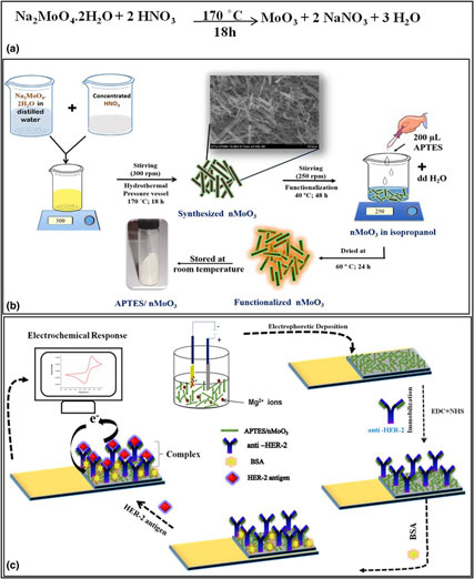
For the fabrication of the biosensing platform, indium tin oxide (ITO)-coated glass electrode (0.5 cm × 2.5 cm) was used. The ITO electrode was hydrolyzed with a solution of NH3:H2O2:H2O in a ratio of 1:1:5 to introduce –OH functional groups on the surface to promote the formation of nMoO3 film.[Reference Kumar, Kumar, Tiwari, Srivastava, Srivastava, Yadav, Kumar, Tran, Dewan and Mulchandani35, Reference Srivastava, Kumar, Ali, Solanki, Srivastava, Sumana, Saxena, Joshi and Malhotra36] For this purpose, a dispersed colloidal solution (1 mg/mL) of functionalized nMoO3 (APTES/1D nMoO3) was prepared in acetonitrile. The electrophoretic deposition (EPD) technique (50 V for 2 min) was used to obtain uniform thin film on hydrolyzed ITO electrodes. The uniform film formation was further confirmed by AFM studies of APTES/1D nMoO3/ITO electrode (Fig. S6). The AFM results indicated a uniform deposition of film onto the ITO substrate with an average roughness of ~2.93 nm. For the EPD technique, platinum was used as the anode and the ITO electrode was used as the cathode and these were separated at a distance of 1 cm.
Anti-HER-2 were immobilized onto the APTES/1D nMoO3/ITO electrode in a ratio 2:1 via EDC-NHS coupling chemistry. In brief, 15 µL of monoclonal anti-HER-2 (50 µg/mL) was mixed with 7.5 µL of EDC (0.4 M) and 7.5 µL NHS (0.1 M) and stored at room temperature (25 °C) for 20 min to activate the –COOH groups at F c region of anti-HER-2. Further, this mixed solution (30 µL) was drop cast uniformly onto the APTES/1DnMoO3/ITO electrode surface and stored in a humid chamber (2 h). The interaction resulted in the formation of covalent bond between –NH2 groups present on APTES/1D nMoO3/ITO electrode and the activated –COOH groups of anti-HER-2. To remove the unbound biomolecules, the electrode was washed with PBS (pH 7.0). For blocking of the non-specific binding sites, 20 µL of BSA (0.1 mg/dL) was used as it did not interact with the specific binding sites. These BSA-incorporated immunoelectrodes were kept for 2 h at room temperature (25 °C) and were subsequently washed with PBS. The fabricated BSA/anti-HER-2/APTES/1D nMoO3/ITO immunoelectrodes were stored at 4 °C until further use. Scheme S1(c) represents the various steps relating to the fabrication of a BSA/anti-HER-2/APTES/1D nMoO3/ITO immunoelectrode.
Results and discussion
Morphological, structural, Raman studies
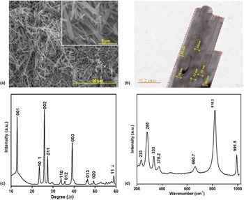
The SEM image [Fig. 1(a)] obtained for nMoO3 indicated the formation of needle-like structure.[Reference Bleyer and Welch11] The magnified image [inset in Fig. 1(a)] revealed the structure of nMoO3 to be as rods. The diameter of nMoO3 was found to lie between 60 and 80 nm with a few micrometers in length [Fig. 1(b)]. The XRD spectra [Fig. 1(c)] of the nMoO3 obtained in the 20–60° diffraction angle (2θ) range with a scan rate of 0.2/min revealed crystallographic (hkl) planes: (001), (101), (002), (011), (110), (012), (003), (013), (020), (−114), and (023) were well indexed with JCPDS No. 47-1320 indicating the presence of the monoclinic phase of 1D nMoO3. The (002) plane seen at 2θ = 25.83° had the highest intensity revealing that the 1D nMoO3 crystallites were more orderly along the (002) plane. Figure 1(d) shows the Raman spectra obtained for the nMoO3 nanorods. The high-intensity characteristic peak found at 818.2/cm was attributed to the stretching mode of the doubly connected bridge oxygen atom in Mo–O–Mo, present in the MoO6 octahedrons. The peak obtained at 991.5/cm was due to asymmetric stretching modes of terminal oxygen in Mo=O.[Reference Radhakrishnan, Reed, Oyama, Seman, Kondo, Domen, Ohminami and Asakura32] The peak at 280/cm was a doublet comprising of wagging modes of the terminal oxygen atoms. The peak at 660.7/cm was attributed to the triply coordinated oxygen atoms linked to the Mo–O stretching modes of three octahedrons.[Reference Radhakrishnan, Reed, Oyama, Seman, Kondo, Domen, Ohminami and Asakura32] The peaks found at 333 and 375.2/cm were assigned to the bending mode of O=Mo=O and scissoring modes of O–Mo–O, respectively.
SEM image (a), TEM image (b), XRD pattern (c), Raman spectra (d) of the synthesized 1D nMoO3.

BET studies
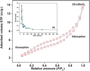
The specific surface area and porosity of the synthesized 1D nMoO3 were determined via BET studies. For sampling the specific surface area, the sample was degassed in a vacuum at 200 °C for 2 h and then the nitrogen adsorption and desorption isotherms were recorded. The pore size distribution and nitrogen adsorption–desorption isotherm for the 1D nMoO3 are shown in Fig. 2. The results of the BET studies obtained 1D revealed the total pore volume of 1D nMoO3 to be 0.029 cc/g. The specific surface area of the 1D nMoO3 calculated by BJH method was found to be 17.19 m2/g. The pore size distribution curve indicated that the 1D nMoO3 was dominated by mesoporous with a minor fraction of microporous [Fig. 2 inset (a)]. The mean pore diameter was found to be 6.8 nm. The obtained results revealed the highly mesoporous behavior of the synthesized 1D nMoO3 facilitate increased loading of anti-HER-2.[Reference Ali, Srivastava, Mondal, Chavhan, Agrawal, John, Sharma and Malhotra13]
BET studies for 1D nMoO3 (inset a: pore size distribution).

Fourier transform infrared spectroscopy studies
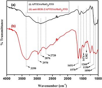
Figure 3 shows Fourier transform infrared spectroscopy (FTIR) spectra obtained for (i) APTES/1D nMoO3/ITO and (ii) anti-HER-2/APTES/1D nMoO3/ITO electrodes, respectively. In both the spectra, bands appearing in the range 1350–1000/cm indicated the presence of C–N (amines) group onto the nMoO3-bound APTES molecules. Bands found at 1474, 2880/cm and in the 1640–1550/cm range were due to C–H bending vibration of alkane (back bone of APTES), –C–H bond and N–H bending for primary amine.[Reference Srivastava, Kumar, Ali, Solanki, Srivastava, Sumana, Saxena, Joshi and Malhotra36, Reference Singh37] The band found at 1382/cm was due to stretching vibration of C–N bond between –NH2 group of APTES functionalized nMoO3 and F c region of anti-HER-2.[Reference Pavia, Lampman, Kriz and Vyvyan38–Reference Liu, Liu, Mernaugh and Zeng40] The singlet band observed at 1651/cm was due to bending vibration of C=O group of amide bond (O=C–N–H). The broad band found at 3350/cm was assigned to stretching vibration of monosubstituted N–H group of amide (R–CO–NH–R) bond. The bands seen at 2720 and 2970/cm were attributed to –CH-band of alkane of the biomolecules and APTES present in the BSA/anti-HER-2/APTES/1D nMoO3/ITO.[Reference Chen, Wang and Wu41, Reference Pavia42] These FTIR results confirmed successful covalent immobilization of anti-HER-2 biomolecules onto APTES/1D nMoO3/ITO electrode.[Reference Liu, Liu, Mernaugh and Zeng40]
FTIR spectra of (a) APTES/1D nMoO3, (b) anti-HER-2/APTES/1D nMoO3/ITO electrode.

Cytotoxicity studies
The biocompatible behavior was evaluated by conducting cytotoxicity studies. In brief, the cytotoxicity of the functionalized nMoO3 (APTES/1D nMoO3) was determined in the concentration range 0–30 µg/mL by MTT assay on HEK 293T cells. The level of toxicity of the 1D nMoO3 was found to vary as a function of concentration. The APTES/1D nMoO3 did not exhibit any significant cytotoxicity on HEK 293 cell lines up to 15 µg/mL. This was adequate for the development of the biosensing platform. Figures S2(a)–S2(d) show morphology of the HEK 293 cell lines on exposure to different concentrations. The % cell viability of APTES/1D nMoO3 with respect to control was found to be about 75% for 15 µg/mL (Fig. S3).
X-ray photoelectron spectroscopic studies
The survey scan X-ray photoelectron spectroscopic (XPS) spectra of APTES/1D nMoO3 and anti-HER-2/APTES/1D nMoO3 onto the ITO electrode is shown in Fig. S4. The peaks pertaining to Mo 3d, Si 2p, O 1s and N 1s were found at 232, 102, 530.5, and 399.6 eV, respectively. During the process of anti-HER-2 immobilization, the APTES/1D nMoO3/ITO electrode was washed with PBS (pH 7.0). The peaks corresponding to Na and Cl can be seen in the survey scan spectra. The Na (KLL), O (KLL), and C (KLL) Auger transitions were observed. The P transition was attributed to anti-HER-2 molecules. It was found that the intensity of Mo (3d) and Si (2p) peaks decreased, and the N (1s) peak intensity increased after the anti-HER-2 immobilization on APTES/1D nMoO3/ITO electrode.
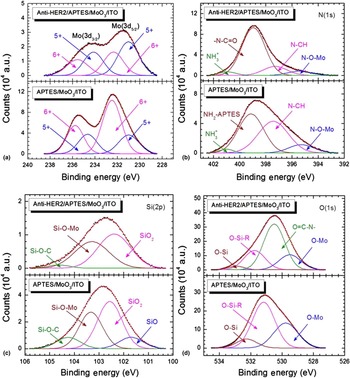
The XPS core-level spectra of Mo (3d), Si (2p), N (1s), and O (1s) were deconvoluted into corresponding binding energy peaks [Figs. 4(a)–4(d)]. The two distinct states Mo (3d 5/2) and Mo (3d 3/2) separated by 3.15 eV arising due to spin-orbit splitting were visible [Fig. 4(a)]. The presence of two distinct valance states of Mo 5+ and 6+ in the deconvoluted spectra revealed the Mo valence state. The sharp doublet at Mo(3d 5/2) = 231 eV and Mo(3d 3/2) = 234 eV corresponded to Mo+5 state while the doublet at Mo(3d 5/2) = 232.02 eV and Mo(3d 3/2) = 235.76 eV pertained to Mo+6 state.[Reference Choi and Thompson43] The atomic percentage area of +6 states in anti-HER-2/APTES/1D nMoO3/ITO decreased from 66.9% to 41.2%. This could be due to immobilization of antibodies onto the APTES/1D nMoO3/ITO electrode. The N(1s) peak observed for APTES/1D nMoO3/ITO and anti-HER-2/APTES/1D nMoO3/ITO electrodes deconvoluted to four peaks at 395.3, 397.4, 399.1, and 401.0 Ev corresponded to the N–O–Mo, N–CH, –NH2, and NH3+, respectively [Fig. 4(b)]. It may be noted that the contribution of the highest energy peak of –NH2 (399.1 eV) in APTES/1D nMoO3/ITO was perhaps due to –N–C=O (398.9) in anti-HER-2/APTES/1D nMoO3/ITO.[Reference Choi and Thompson43–Reference Song, Hildebrand and Schmuki45] This was assigned to the formation of amide bond (–N–C=O) between the carboxyl terminal of anti-HER-2 and the free –NH2 groups of APTES molecules. The deconvolution of Si (2p) yielded four peaks at 101.7, 102.5, 103.3, and 104.4 eV corresponding to Si in Si–O, SiO2, Si–O–Mo, Si–O–C, respectively [Fig. 4(c)],[Reference Baltrusaitis, Mendoza-Sanchez, Fernandez, Veenstra, Dukstiene, Roberts and Fairley46] confirmed the functionalization of 1D nMoO3 with the APTES molecules. The O (1s) deconvolution was carried out in the range 528–534 eV [Fig. 4(d)]. The three deconvoluted peaks were ascribed to the oxygen linked to the O–Si–, O–Si–R wherein R was attributed to the presence of CH2/NH2 groups in APTES and the O–Mo.[Reference Martin, Schulz, Bumgardner and Walters44] After immobilization of anti-HER-2, an additional peak found at 530.5 eV was due to oxygen atom of the amide bond in O=C–N–.[Reference Ali, Srivastava, Mondal, Chavhan, Agrawal, John, Sharma and Malhotra13, Reference Kumar, Sharma, Maji and Malhotra47] These results confirmed decrease in N–O–Mo and O–Mo bonding in N 1s and O 1s core-level spectra after the anti-HER-2 immobilization [Figs. 4(b) and 4(d)], respectively.
Deconvoluted core-level XPS spectra of (a) Mo (3d), (b) N (1s), (c) Si (2p), and (d) O (1s) and of APTES/1D nMoO3/ITO and anti-HER-2/APTES/1D nMoO3/ITO.

Electrochemical studies
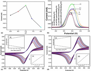
The effect of pH (6.0–8.0) on BSA/anti-HER-2/APTES/1D nMoO3/ITO immunoelectrode was investigated using DPV in PBS buffer (50 mM, 0.9% NaCl) containing 5 mM of [(Fe (CN)6)]3−/4− ions. The maximum current response was recorded at pH 7.0 [Fig. 5(a)]. This could perhaps be due to the fact that at pH 7.0, biomolecules (e.g., antibodies, antigens, enzymes, DNA, mRNA, etc.) existed in their original form and had the highest bio-molecular activity. However, in the acidic or basic medium, the biomolecules perhaps got denatured due to the interaction of H+ or OH− ions in the amino acid sequence of the antibodies.[Reference Solanki, Singh, Rupavali, Tiwari and Malhotra25] In further electrochemical studies, PBS (pH 7.0) were used.
pH study of (a) BSA/anti-HER-2/APTES/1D nMoO3/ITO with respect to pH (ranging from 6.0 to 8.0) in PBS (50 mM, 0.9% NaCl) containing 5 mM of [Fe (CN) 6]−3/4− (b) DPV studies at each step of electrode fabrication. Scan rate studies of (c) APTES/1D nMoO3/ITO and BSA/anti-HER-2/APTES/1D nMoO3/ITO (d) with square root of scan rate versus current (inset i) and peak potential versus square of scan rate (inset ii).

To investigate the electrochemical behavior of ITO, APTES/1D nMoO3/ITO, anti-HER-2/APTES/1D nMoO3/ITO, and BSA/anti-HER-2/APTES/1D nMoO3/ITO electrodes, the DPV studies were conducted in the potential range −0.2 to 0.6 V [Fig. 5(b)]. The peak current obtained for the APTES/1D nMoO3/ITO (0.31 mA) was found to be higher as compared to that of ITO (0.20 mA) due to high electrocatalytic efficiency and large surface area of the nMoO3 nanorods leading to more efficient electron transfer between the electrolyte and the electrode surface.[Reference Malik, Srivastava, Verma, Kumar, Kumar and Singh48] The value of current decreased to 0.24 and 0.23 mA after immobilization of anti-HER-2 antibodies. This decrease was attributed to the insulating nature of the anti-HER-2 and BSA that acted as a barrier to free electron movement to the immunoelectrode.[Reference Malik, Srivastava, Verma, Kumar, Kumar and Singh48]
Interfacial kinetics studies on the APTES/1D nMoO3/ITO and BSA/anti-HER-2/APTES/1D nMoO3/ITO electrodes were carried out as a function of scan rate (40–140 mV/s) via CV. The magnitude of peak currents obtained for both the electrodes exhibited a linear relationship with square root of scan rate [inset (i) of Figs. 5(c) and 5(d)] indicating that the electrochemical reaction was a diffusion-controlled process and followed Eqs. (1)–(4).
 $$\eqalign{&I_{{\rm pa}({\rm APTES}/{\rm 1D}\,{\rm nMoO3}/{\rm ITO})} \cr &= - [12.7 {\rm \mu}{\rm A}({\rm s}/{\rm mV}) \times (\hbox{scan rate}\,[{\rm mV}/{\rm s}])^{1/2}] \cr & \quad\hskip 1pt - {10.3}\,{\rm \mu}{\rm A},\;R^{2} = 0.996,\;{\rm SD} = 24.8 \times 10^{ - 4},} $$
 $$\eqalign{I_{{\rm pc}({\rm APTES}/{\rm 1D\,nMoO3}/{\rm ITO}))}R^2 = 0.996,\;{\rm SD} = 34.5 \times 10^ - 4,} $$
 $$\eqalign{& I_{{\rm pc}({\rm BSA}/{\rm anti} - {\rm HER} - {2}/{\rm APTES}/{\rm 1D\,nMoO3}/{\rm ITO} )} \cr & = [14.9\,{\rm \mu} {\rm A}({\rm s}/{\rm mV}) \times (\hbox{scan rate}\,[{\rm mV}/{\rm s}])^{1/2}] \cr &\quad \, + 15.3\,{\rm \mu} {\rm A},\;R^{2} = 0.997,\;{\rm SD} = 13.809 \times 10^{ - {4}},} $$
 $$\eqalign{& I_{{\rm pa}({\rm BSA}/{\rm anti} - {\rm HER} - 2/{\rm APTES}/{\rm 1D}\,{\rm nMoO3}/{\rm ITO})}R^2 \cr& = 0.997,\;{\rm SD} = 20.9 \times 10^{ - 4}.} $$
With increasing scan rate, the oxidation and the reduction peaks shifted linearly toward more positive and negative potential, respectively. The linear variation was found between the magnitude of difference peak potentials calculated as (ΔE p = E pc–E pa, where E pa is anodic peak potential and E pc is cathodic peak potential) and function of the square root of scan rate [inset (ii) of Figs. 5(c) and 5(d)]. The observed linear variation indicated efficient and more facile electron transfer kinetics between the medium to electrode and followed Eqs. (5) and (6).
 $$\eqalign{& \Delta E_{{\rm pa}({\rm APTES}/{\rm 1D\,nMoO3}/{\rm ITO} )} \cr & = [0.41\,{\rm V}({\rm s}/{\rm mV}) \times (\hbox{scan rate}[{\rm mV}/{\rm s}])^{1/2}] \cr &\quad\hskip 1pt + 0.11\,{\rm V},\;R^{2} = 0.995,\;{\rm SD} = 2.77 \times 10^{ - 4},} $$
 $$\eqalign{& \Delta E_{{\rm pc}({\rm BSA}/{\rm anti} - {\rm HER} - {2}/{\rm APTES}/{\rm 1D\,nMoO3}/{\rm ITO})} \cr & = [0.30\,{\rm V}({\rm s}/{\rm mV}) \times (\hbox{scan rate}[{\rm mV}/{\rm s}])^{1/2}] \cr & \quad\hskip 1pt+ 0.079\,{\rm V},\;R^{2} = 0.997,\;{\rm SD} = 4.77 \times 10^{ - 4},} $$
where R is the correlation coefficient and SD is the standard deviation. The diffusion coefficient (D) for the BSA/anti-HER-2/APTES/1D nMoO3/ITO immunoelectrode was calculated to be 5.9 × 10⁻⁵ cm2/s by using Randle Sevick equation that is based on the assumption that mass transport occurred only by diffusion:
 $$I_{\rm p} = (\hbox{2}.\hbox{69} \times \hbox{1}0^{\rm 5}){\rm} n^{{\rm 3}/{\rm 2}}\hbox{A}\hbox{D}^{{\rm 1}/{\rm 2}}C\upsilon ^{{\rm 1}/{\rm 2}},$$
where I p is the peak current obtained for the immunoelectrode, n is the number of electrons transferred (1), A is the active surface area of the immunoelectrode (0.25 cm2), D is the diffusion coefficient, C is the concentration of redox species (5 × 10⁻³ mol/cm−2) and υ is the scan rate (50 mV/s). The surface concentration (γ) of the BSA/anti-HER-2/APTES/1D nMoO3/ITO immunoelectrode was calculated to be 1.049 × 10⁻⁷ mol/cm2 using Brown–Anson Eq. (8)
 $$I_{\rm p} = n^{\rm 2}F^{\rm 2}\gamma A\upsilon (\hbox{4RT})^{ - {\rm 1}},$$
where I p is the peak current of immunoelectrode, n is the number of electrons transferred (1), F is the Faraday constant (96485 C/mol), γ is the surface concentration of the absorbed electro-active species, A is the surface area of the electrode (0.25 cm2), υ is the scan rate (V/s), R is the gas constant (8.314 J/mol/K) and T is the temperature (25 °C).
Incubation time studies
To determine the time required for interaction between the epitope of HER-2 antigen (2.5 ng/mL) and paratope of the immobilized anti-HER-2 onto BSA/anti-HER-2/APTES/1D nMoO3/ITO immunoelectrode, incubation time studies were performed via differential pulse voltammetry (DPV) every 5 min up to 30 min (Fig. S5).[Reference Srivastava, Ali, Solanki, Chavhan, Pandey, Mulchandani, Srivastava and Malhotra49] It was observed that the peak current response decreased gradually from 0 to 20 min after which it became nearly constant. This result showed that 20 min were required for complete binding of HER-2 onto BSA/APTES/1D nMoO3/ITO immunoelectrode.
Electrochemical detection of the cancer biomarker
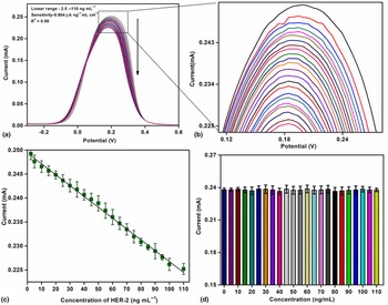
The electrochemical response studies of the BSA/anti-HER-2/APTES/1D nMoO3/ITO immunoelectrode were conducted as a function of HER-2 concentration (0–110 ng/mL) in PBS (pH 7.0) containing 5 mM [Fe (CN)6]3−/4− via DPV technique in the potential range −0.2 to +0.6 V [Figs. 6(a) and 6(b)]. Each measurement was repeated three times for a given concentration of HER-2 (n = 3). It was observed that the anodic peak current was found to decrease proportionally with increasing HER-2 concentration. The decrease in the response current was attributed to the formation of insulating antigen–antibody complex molecules resulting due to the interaction of the specific binding sites of the HER-2 with anti-HER-2 at the transducer surface. This perhaps obstructed the electron transfer of Fe(CN)6]3−/4− from the bulk solution to the electrode resulting in reduced current.[Reference Srivastava, Kumar, Ali, Solanki, Srivastava, Sumana, Saxena, Joshi and Malhotra36] The calibration curve [Fig. 6(c)] obtained by plotting change in the magnitude of the anodic peak current of BSA/anti-HER-2/APTES/1D nMoO3/ITO immunoelectrode as a function of HER-2 concentration in the detection range 2.5–110 ng/mL followed Eq. (9):
 $$\eqalign{I_{\rm p} =& \,[0.\hbox{9}0\hbox{4}\,{\rm \mu} /\hbox{Ang/mLc}\hbox{m}^{\rm 2} \times {\rm} (\hbox{conc}.\hbox{of HER - 2 in ng/mL}) \cr & + 0.0\hbox{954}\,{\rm \mu A}],\;R^{\rm 2} = 0.\hbox{997},\;\hbox{SD} = \hbox{1}.\hbox{83} \times \hbox{1}0^{ - {\rm 7}}.} $$
Electrochemical response of the (a) BSA/anti-HER-2/APTES/1D nMoO3/ITO immunoelectrode as a function of HER-2 concentration using DPV, (b) the magnified view of oxidation peak current, (c) calibration curve plot between magnitude of peak current and concentration of HER-2 (2.5–110 ng/mL) and (d) control experiment of APTES/1D nMoO3 against HER-2.

The lower detection limit calculated using the standard equation Eq. (10) was obtained as 2.47 ng/mL
 $$\hbox{Lower detection limit} = \hbox{3}\sigma /m{\rm,} $$
where σ is the standard deviation (SD) of the calibration graph and m is the sensitivity. The linear detection range was obtained as 2.5–110 ng/mL (S/N = 3) and the sensitivity was calculated from the slope/surface area (0.25 cm2), which was found to be as 0.904 µAng/mL/cm2.[Reference Tabasi, Noorbakhsh and Sharifi50]
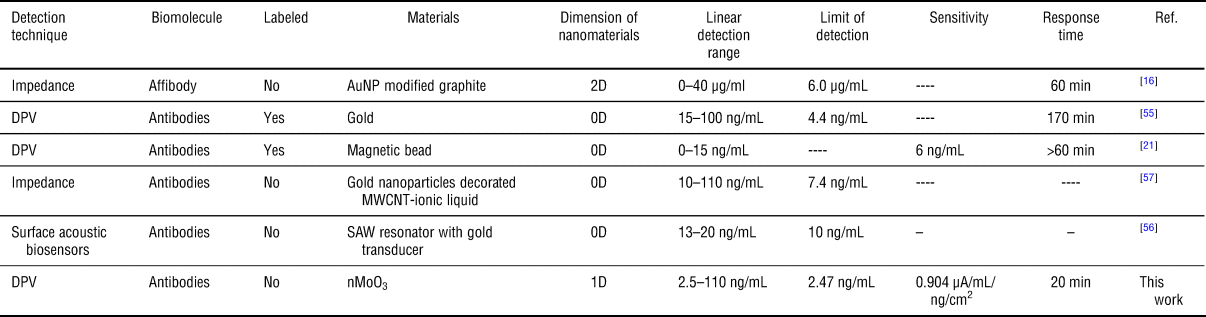
To determine the effect of morphology onto the efficiency of biosensor, the electrochemical sensing studies of spherical nanoparticles of spherical MoO3 were simultaneously carried out against HER-2 biomarker. Figure S7 is the SEM image of the as-synthesized spherical nanoparticles. The linear detection range was obtained as 5–60 ng/mL with sensitivity 0.52 µ/Ang/mL/cm2. The poor sensitivity and narrow detection range of the spherical nanoparticle with respect to 1D nMoO3 immunoelectrode can be ascribed to the fact that the exposed surface area available for binding of antibodies is far less as compared with that of the MoO3 nanorods.[Reference Barua, Yoo, Kolhar, Wakankar, Gokarn and Mitragotri23] The higher sensitivity and the wide linear range (2.5–110 ng/mL) of the 1D MoO3-based immunoelectrode (with respect to those reported in Table 1 and spherical MoO3) can be attributed to the high electro activity of 1D nMoO3.[Reference Chen, Cheah, Madhavi and Lou24]
Characteristics of the BSA/anti-HER2/APTES/1D MoO3/ITO immunosensor along with those reported in the literature.

The association constant (K a) is known to determine the affinity of antigen toward antibodies. The higher the K a value, the stronger is the affinity of HER-2 toward BSA/anti-HER-2/APTES/1D nMoO3/ITO bio electrode. The affinity between the HER-2 and anti-HER-2 obtained using Hanes–Wolf plot (the plot between substrate concentration verses substrate concentration/current) (Fig. S8)[Reference Jagadeesan, Kumar and Sumana51, Reference Tiwari, Gupta, Bagbi, Sarkar and Solanki52] was estimated to be 4.91 × 10¹² mol/L indicating strong affinity.
The electrochemical response of bare APTES/1D nMoO3/ITO electrode toward different concentrations (2.5–110 ng/mL) of HER-2 antigen was determined [Fig. 6(d)] in order to evaluate the current response of the developed sensor. No significant change was observed in the peak current indicating that the APTES/1D nMoO3/ITO electrode did not generate any false electrochemical response. This control experiment revealed that the sensor response was entirely due to the immunoreactions between anti-HER-2 and HER-2 biomolecules. Regeneration studies of the fabricated immunosensor were carried out using the same electrolyte after immersing the BSA/anti-HER-2/APTES/1D nMoO3/ITO in 0.2 M solution of glycine-HCl buffer (pH 2.4 for 2 min). It was found that there was a gradual decrease in peak current with the increased regeneration times (Fig. S9). This could be due to the fact that at low pH, the protein binding sites repelled each other and tended to fold making the molecules move further apart.[Reference Goode, Rushworth and Millner14, Reference Andersson, Areskoug and Hardenborg53] The results demonstrated that the proposed immunosensor could be regenerated and used for at least five times.
Selectivity studies
To determine the selectivity of the BSA/anti-HER-2/APTES/1D nMoO3/ITO immunoelectrode toward various biomolecules present in serum samples of breast cancer patients, interferent studies were conducted via DPV technique (Fig. S10). The serum samples of breast cancer patients are known to contain glucose [7 mg/mL], cardiac troponin [cTn-I, >0.6 ng/mL], CYFRA-21-1 [>3.5 ng/mL],[Reference Kumar, Kumar, Tiwari, Srivastava, Srivastava, Yadav, Kumar, Tran, Dewan and Mulchandani35] CEA [>3 ng/mL], cTn-I [>0.19 ng/mL],[Reference Kumar, Sen, Kumar, Augustine, Yadav, Mishra and Malhotra54] etc. Firstly, we measured the electrochemical response of BSA/anti-HER-2/APTES/1D nMoO3/ITO immunoelectrode toward the HER-2 (2.5 ng/mL). Thereafter we added cTn-I, glucose, and CYFRA-21-1 and ET-1 one by one in the same solution containing HER-2 and measured the electrochemical response. The measured peak current exhibited no significant change. The results obtained indicated that 1D nMoO3 based immunoelectrode was highly selective toward the HER-2 biomarker only. The selectivity coefficient was calculated using Eq. (11):
 $$\hbox{SC} = \displaystyle{{I_{\rm c} + i} \over {I_{\rm c}}},$$
where I c+i is the current response of bioelectrode for interference and I c is the current obtained in absence, of each interferent, respectively. The SC value obtained for each interferent was ~1.
Shelf life studies
The shelf life of the BSA/anti-HER-2/APTES/nMoO3/ITO immunoelectrode was determined at a regular interval of 2 days up to 40 days (Fig. S11). The fabricated biosensor was stored at 4 °C when not in use. The measured current response exhibited 95% of the initial electrochemical current response up to 30 days after which the current gradually decreased. The sensing characteristics of the fabricated immunoelectrode with the reported literature are summarized in Table 1.[Reference Marques, Viswanathan, Nouws, Delerue-Matos and González-García55, Reference Gruhl, Rapp and Länge56]
Patient's serum samples analysis
Quantification of HER-2 in serum samples of breast cancer patients was accomplished via double sandwich ELISA. After determination of the HER-2 concentration, five serum samples of breast cancer patients were utilized to test the accuracy of data obtained using fabricated BSA/anti-HER-2/APTES/1D nMoO3/ITO platform. The reasonable correlation with an acceptable percentage relative standard deviation was obtained between (a) HER-2 concentration in serum samples determined by ELISA and (b) standard concentration of HER-2 (Table S2).
Conclusions
We have demonstrated the fabrication of the label-free immunosensor based on biocompatible 1D nMoO3 biosensing platform for breast cancer biomarker detection. The results of electrochemical studies have shown wider linear detection range (2.5–110 ng/mL) and excellent sensitivity (0.904 µAmL/mg/cm2). Besides this, BSA/anti-HER-2/APTES/1D nMoO3/ITO immunoelectrode can be used to detect HER-2 concentration up to 2.47 ng/mL. The better sensitivity of this platform has been attributed to the mesoporous behavior and high electro catalytic activity of 1D MoO3 that provides high aspect ratio for improved bimolecular loading. The observed specific surface area of the 1D nMoO3 (17.19 m2/g) calculated by BET analysis and the pore size distribution have revealed the presence of mesoporous compared with micro and macropores. It should be interesting to utilize this emerging 1D nMoO3 toward the application in biomolecular electronics, biofuel cells, therapeutics, and detection of other biomarkers released in communicable as well as other non-communicable diseases.
Supplementary material
The supplementary material for this article can be found at https://doi.org/10.1557/mrc.2018.182.
Acknowledgments
The authors thank Prof. Yogesh Singh, Vice Chancellor, Delhi Technological University (DTU), Delhi, India for providing the facilities. Shine Augustine is thankful to UGC-MANF [2015-16/MANF-2015-77-DEL-55426] and CSIR-SRF [08/133(0019)/2018-EMR-I] for the award of research fellowship. The authors thank Dr. Prabhakar Rai and Prof. Ashutosh Sharma, IIT Kanpur for the BET analysis.