Percutaneous pulmonary valve implantation is a less invasive treatment for right ventricular outflow tract dysfunction and has shown to be feasible and safe, with a low complication rate.Reference Khambadkone, Nordmeyer and Bonhoeffer 1 , Reference Eicken, Ewert and Hager 2 Most common complications are stent fractures or migration, complete atrioventricular block, endocarditis, homograft rupture,Reference Khambadkone, Nordmeyer and Bonhoeffer 1 , Reference Nordmeyer, Khambadkone and Coats 3 and coronary extrinsic compression, which is usually avoided by performing balloon testing of the right ventricular outflow tract.Reference Eicken, Ewert and Hager 2 , Reference Kostolny, Tsang and Nordmeyer 4 Here we report on a case of an acute antero-septal non-ST myocardial infarction secondary to partial extrinsic compression of the left coronary artery after percutaneous pulmonary valve implantation despite a negative balloon testing.
Case report
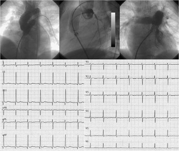
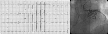
A 14-year-old boy after neonatal surgical coarctation repair had a Ross operation for progressive aortic valve regurgitation – bicuspid valve – at the age of 11 years. An 18-millimetre homograft conduit was implanted in the pulmonary position. He presented to our centre with severe right ventricular outflow tract obstruction for percutaneous pulmonary valve implantation. Under general anaesthesia, percutaneous femoral access was obtained – artery and vein; a complete haemodynamic study was performed followed by a selective left coronary angiogram combined with balloon BALT 20 × 40-millimetre (BALT, Montmorency, France) inflation in the conduit to exclude potential coronary arterial compression (Fig 1 left upper). After this, an AndraStent XXL 39-millimetre (Andramed, Reutlingen, Germany) fixed on a BALT 20 × 45-millimetre balloon was implanted into the conduit. A Melody valve (Medtronic, Inc., Minneapolis, Minnesota, United States of America) mounted on a 22-millimetre Ensemble was then delivered. Owing to a residual gradient of 20 millimetres of mercury, the valve was re-dilated using a MULLINS 22 × 45-millimetre balloon (Fig 1, right upper). At the end of the procedure, there were no signs of dissection, normal electrocardiogram (Fig 1, bottom), and stable haemodynamic conditions (right ventricular systolic pressure passed from 90 to 35 millimetres of mercury and right ventricular to pulmonary artery peak systolic gradient from 65 to 10 millimetres of mercury). After 1 hour of the procedure, the patient experienced chest pain. A 12-lead electrocardiogram showed ST segment depression in V2–V6 (Fig 2, left). Coronary angiography showed a partial obstruction of the proximal-mid tract of the left anterior descendent coronary artery due to extrinsic compression near the site of Melody valve implantation (Fig 2, right). He was immediately sent to the operating room where surgeons removed the Melody valve and found a laceration of the lateral-posterior left wall of the homograft: a thrombus was evident between the tissue and the stents, compressing the left coronary artery passing exactly beneath it. It was repaired with a pericardial patch, and a biological prosthetic pulmonary ASPIRE no. 23 valve was implanted.
Left upper: balloon test performed in the anteroposterior view and ascending aortogram showing no signs of coronary compression; Middle: selective left coronary angiography during balloon test showing no signs of coronary compression; Right upper: angiography in the anteroposterior view of Melody valve after re-dilation with Mullins balloon; Bottom: electrocardiography 12-leads at the end of procedure 124 × 127 millimetres (300 × 300 dots per inch).

Left: electrocardiography 12-leads with signs of anterior acute myocardial ischaemia; Right: coronary angiography in the anteroposterior view showing partial obstruction of proximal-mid tract of left anterior descending 137 × 43 millimetres (300 × 300 dots per inch).

Comment
Very few cases of extrinsic compression of the left coronary artery after percutaneous pulmonary valve implantation are reported in the literature.Reference Eicken, Ewert and Hager 2 , Reference Kostolny, Tsang and Nordmeyer 4 Performing an aortogram – or, in case of unfavourable anatomy detected at cardiac computed tomography or remote magnetic navigation, a selective coronary angiography – during balloon inflation in the right ventricular outflow tract has become a mandatory preliminary step to the procedure.Reference Eicken, Ewert and Hager 2 In our case, this event occurred in a different way. A cardiac magnetic resonance imaging did not clearly show unfavourable coronary anatomy. Balloon testing was negative. We needed to post-dilate the valve with a larger diameter balloon (20 to 22 millimetres) because of a residual gradientReference Nordmeyer, Khambadkone and Coats 3 and this caused the laceration of the homograft, which led to the thrombus formation and the subsequent coronary compression.
Probably the use of a high-pressure balloon for testing could have avoided false-positive results; however, having some degree of right outflow tract stenosis can be useful to obtain a better valve anchoring, reducing risk of embolisation. In addition, aggressive high-pressure balloon dilation may cause homograft fractures in a moment of the procedure when there is no protection by the coverage of Melody valve. On the other hand, the presence of a residual gradient and need for Melody valve post-dilatation cannot be predicted in advance. Moreover, in our case, the delay in clinical and electrocardiographic presentation, probably related to the time needed for thrombus formation between the stent and the tissue, represented a real trap. In conclusion, our case underlines the need for continuous and careful monitoring and the tight collaboration between interventional cardiologists and surgeons.