




No CrossRef data available.
Article contents
- Extract
- Pre-operative anatomy
- Post-operative anatomy
- Functionally univentricular physiology
- Optimizing systemic oxygen delivery in neonates with functionally univentricular physiology
- Optimizing systemic oxygen delivery by manipulation of pulmonary vascular resistance
- Optimizing systemic oxygen delivery by manipulation of systemic resistance
- Optimizing total cardiac output
- Optimizing pulmonary recruitment to avoid pulmonary venous desaturation
- Summary
- References
Neonatal physiology of the functionally univentricular heart
Published online by Cambridge University Press: 21 September 2005
Article contents
- Extract
- Pre-operative anatomy
- Post-operative anatomy
- Functionally univentricular physiology
- Optimizing systemic oxygen delivery in neonates with functionally univentricular physiology
- Optimizing systemic oxygen delivery by manipulation of pulmonary vascular resistance
- Optimizing systemic oxygen delivery by manipulation of systemic resistance
- Optimizing total cardiac output
- Optimizing pulmonary recruitment to avoid pulmonary venous desaturation
- Summary
- References
Extract
The term “functionally single ventricle” includes a variety of congenital cardiac anomalies where there is only one ventricle pumping blood to the systemic and pulmonary circulations. The physiology in this arrangement is a considerable challenge for the cardiac specialist, because the complexity encountered in patients with these lesions necessitates particularly specialized medical and surgical management. Patients with such functionally univentricular physiology often respond to common interventions, such as supplemental oxygen, mechanical ventilation, and vasoactive drugs, differently than patients with conventional circulations.1 Furthermore, these patients tend to be encountered more frequently by pediatricians and cardiologists because they undergo multiple operations, may be more adversely affected by intercurrent illnesses, or have chronic cardiac problems requiring frequent attention. A thorough understanding of the complexities of the physiology encountered is thus imperative for the pediatric cardiologist. In this review, we will address important physiologic and anatomic principles that influence care of neonates with functionally univentricular hearts. Although the anatomy and physiology of each reconstructive stage of palliation are unique, we will focus upon the pre- and post-operative physiology as encountered in the neonate.
Information
- Type
- Norwood Procedure and Staged Palliation
- Information
- Copyright
- © 2004 Cambridge University Press
Pre-operative anatomy
In congenital cardiac malformations, anatomy dictates physiology. Various anatomic lesions can produce a functionally univentricular circulation (Table 1). By definition, newborns with this physiology have mixing of the pulmonary and systemic venous returns, but the relative amounts of each vary substantially depending on the underlying anatomy. The most important anatomic issues affecting planned interventions and management of intensive care are the outflows of blood to and from the functionally single ventricle, and the respective pulmonary and systemic circulations. The newborn may have either pulmonary or aortic obstruction, or bilaterally unobstructed outflows. Either the systemic or pulmonary venous return may also be abnormally connected or obstructed in the newborn with a functionally univentricular circulation.
Table 1. Anatomic diagnoses commonly associated with functionally univentricular physiology in the newborn. Very few malformations have an anatomically single ventricle.

Obstruction to the systemic outflow
Systemic obstruction is present in multiple anatomic variations, including hypoplastic left heart syndrome, tricuspid atresia with discordant ventriculo-arterial connections, and variants of double inlet left ventricle. The physiology encountered with systemic obstruction also applies to newborns with critical aortic stenosis, severe coarctation of the aorta, or interruption of the aortic arch. Important features of these lesions include complete mixing of the systemic and pulmonary venous returns, with the ventricular outflow being directed primarily to the lungs. When systemic obstruction is severe, systemic blood flow is provided by right-to-left ductal shunting, and thus depends upon a non-restrictive arterial duct. With a non-restrictive duct, systemic flow itself is dependent upon the overall output from the functionally single ventricle, coupled with the relative pulmonary and systemic vascular resistances. Severe obstruction to systemic outflow is poorly tolerated in the face of such anatomy, resulting in signs or symptoms of shock concomitant with closure of the arterial duct.
Obstruction to pulmonary outflow
Obstruction to the flow of blood to the lungs occurs, among others, in lesions such as tricuspid atresia, pulmonary atresia, and severe Ebstein's malformation of the tricuspid valve. Salient anatomic features include complete mixing of systemic and pulmonary venous returns, with the ventricular outflow directed predominantly to the aorta. The restricted flow of blood to the lungs imposes an obligatory intracardiac right-to-left shunt, generally at the atrial level, and results in deoxygenated blood reaching the systemic circulation. The clinical finding is one of cyanosis. Clinical consequences of obstructed blood flow to the lungs are variable, and depend on the severity of the lesion. Mild obstruction may permit excessive amounts of the total cardiac output to go to the pulmonary circulation, sometimes at the expense of the systemic cardiac output. Infants with this type of anatomy are only minimally cyanotic, and often develop signs and symptoms of congestive heart failure, so treatment is directed at limiting, rather than increasing the pulmonary flow. At the other end of the spectrum are those with severe obstruction, or even atresia of the pulmonary pathway. These patients are profoundly cyanotic unless an alternate source of flow to the lungs is quickly established. These lesions require ductal patency to maintain pulmonary flow, and are thus referred to as duct-dependent malformations.
The atrial septum
In the functionally univentricular situations, unobstructed pulmonary or systemic venous return often requires a non-restrictive interatrial communication. When an atrioventricular valve is severely stenotic or atretic, as occurs in hypoplastic left heart syndrome, tricuspid atresia, or pulmonary atresia with intact ventricular septum, a large atrial septal defect is necessary to decompress the atrium that exits through the inadequate atrioventricular valve. Restricted systemic venous return causes increased central venous pressures, accumulation of fluid in the third space, and limited cardiac output. Although patency of the oval foramen usually allows right-to-left shunting of blood across the atrial septum, it is occasionally inadequate to permit unobstructed flow of all systemic venous return to the systemic ventricle. Obstructed flow from the pulmonary venous atrium causes elevated pulmonary venous pressure and pulmonary hypertension. Although this can be helpful in the immediate newborn period to limit flow of blood to the lungs, and enhance systemic blood flow, the atrial septum must be opened at the time of the first palliative operation to avoid the long-term consequences of elevated pulmonary vascular resistance. A severely restrictive or intact atrial septum with pulmonary venous hypertension requires creation of a shunt at atrial level as an emergency to alleviate the profound cyanosis. These procedures carry a high risk of morbidity, and may imply a worse prognosis for further palliative surgery.2, 3
Post-operative anatomy
The goal of all initial palliative procedures for the newborn with functionally univentricular anatomy is to establish unobstructed pulmonary and systemic venous return, unobstructed systemic outflow, and to limit the flow of blood to the lungs with normal pulmonary arterial pressures. This is typically accomplished through the first stage Norwood procedure, construction of a modified Blalock-Taussig shunt, or banding of the pulmonary trunk. Although variations on these palliative procedures exist, they represent the spectrum of post-operative anatomy encountered in cardiac intensive care. Since each anatomic arrangement establishes similar physiology, the important differences are the means by which each operation accomplishes the ultimate goal defined above. The Norwood procedure requires cardiopulmonary bypass, cardioplegia, and a period of deep hypothermic circulatory arrest, although newer techniques can limit the period of circulatory arrest.4–8 This maneuver subjects the tissues to a period of ischemia with subsequent reperfusion, which is often followed by transient post-operative dysfunction, especially in the heart, lungs, kidneys, and vascular endothelium.9–11 A Blalock-Taussig shunt, either alone or as part of the first stage Norwood procedure, can lower diastolic arterial pressure, which may compromise coronary arterial perfusion. Unlike the Blalock-Taussig shunt, a band on the pulmonary trunk is not associated with diastolic run-off to the lungs, but some investigators have suggested that banding may increase the risk of subaortic obstruction and ventricular hypertrophy,12 although this has been disputed.13 Shunting and banding procedures both carry the risk of distorting the pulmonary arteries, or producing unilateral pulmonary arterial obstruction, which can contribute to cyanosis after either of these procedures.
Sano and others have proposed a modification of the Norwood procedure which entails supplying flow to the lungs directly from the ventricle by insertion of a Goretex conduit from the morphologically right ventricle to the bifurcation of the pulmonary tank, rather than constructing a conventional Blalock-Taussig shunt. Theoretical benefits of this modification include stabilization of the ratio of pulmonary-to-systemic flows and elimination of the aortic diastolic run-off to the lungs, which may improve coronary arterial perfusion. Proponents report excellent results, and assert that postoperative management following this modified procedure is less complicated than after the conventional first stage Norwood palliation. On the other hand, placement of such a conduit requires a ventriculotomy, and introduces an additional volume load on the already volume-loaded funtionally single ventricle, which could increase the risk for ventricular dysfunction or arrhythmias over the long-term.
Functionally univentricular physiology
The balance of pulmonary and systemic flows
Regardless of underlying anatomy, univentricular physiology entails mixing of systemic and pulmonary venous return, and partitioning the total cardiac output to pulmonary and systemic flows based on the amount of anatomic obstruction and the vascular resistance to flow in the respective circulations. It is generally accepted that the arterial oxygen saturation reflects the ratio of flows in the unoperated or shunted newborn with a functionally single ventricle. This is based on a simplification of the Fick principle, which involves two important assumptions. The Fick equations for calculation of pulmonary and systemic flows are:


where VO2 represents oxygen consumption, CaO2 is the arterial oxygen content, CmvO2 is the mixed venous oxygen content, CpvO2 is the pulmonary venous oxygen content, and CpaO2 is the pulmonary arterial oxygen content. Since systemic and pulmonary arterial oxygen saturations are identical in functionally univentricular physiology, the equation for the ratio of pulmonary to systemic blood flows (Qp:Qs) becomes:

where SmvO2 is the oxygen saturation of mixed venous blood, SaO2 is the oxygen saturation of arterial blood, and SpvO2 is the oxygen saturation of pulmonary venous blood. Since the lungs in most infants with congenital cardiac malformations are relatively healthy, one can assume that the saturation of oxygen in the pulmonary venous blood is normal, at about 95% in room air. Furthermore, if one also assumes the difference between the systemic arterial and venous oxygen saturation is normal, at approximately 25%, the simplified equation for the ratio of pulmonary to systemic blood flow becomes:

This simplified version of the Fick equation allows estimation of the pulmonary to systemic blood flow ratio based on systemic oxygen saturation alone. Given the ease with which this latter value can be measured in clinical practice, the simplified equation has clear advantages. Specifically, it allows the clinician to estimate the ratio of pulmonary to systemic flows, and systemic delivery of oxygen, simply by looking at the systemic arterial saturation. Theoretically, this allows one to assess the effects of interventions designed to alter flows by observing changes in arterial oxygen saturation. It is important to remember, however, that such estimation of the ratio of pulmonary to systemic blood flows is based on assumptions regarding oxygen saturation of mixed venous and pulmonary venous blood. The assumption regarding the difference in systemic arterial-venous oxygen saturation is only accurate if systemic perfusion is normal. In low output states, which are common in pre- and post-operative neonates with functionally univentricular physiology, the oxygen saturation of mixed venous blood can be quite low, so that arterio-venous oxygen saturation differences will be substantially greater than 25%. If the decrease in systemic flow and mixed venous oxygen saturation is offset by an increase in the amount of well-saturated blood returning from the lungs, the arterial oxygen saturation may remain unchanged, or even increase. Clinical data suggest that the oxygen saturation of mixed venous blood varies considerably in patients with functionally univentricular physiology.14–21 Many centers have begun to monitor this value routinely following Norwood palliation for hypoplastic left heart syndrome, using a sample from the superior caval vein as representative of mixed venous blood, since there is no true site of systemic mixed-venous blood in the functionally univentricular circulation.1, 14–19 Adapted from Tweddell et al.,18 Figure 1 shows simultaneous mean arterial blood pressure and the oxygen saturations of arterial and mixed venous blood in a patient early after Norwood palliation. The arterial oxygen saturation remains relatively constant, but mixed venous oxygen saturation plummets at about 17:00, accompanied by a slight increase in blood pressure. The only plausible explanation for this event is that systemic vascular resistance and the ratio of pulmonary to systemic flow increase dramatically. The decrease in mixed venous oxygen saturation and systemic flow is offset by a larger amount of well-saturated blood returning to the heart from the lungs, resulting from the increase in the ratio of flows. In this situation, estimation of the ratio of flows from arterial oxygen saturation alone would lead the clinician to the erroneous conclusion that the circulations are well balanced, when in reality systemic flow is critically low. As predicted by the assumptions inherent in equation 4, clinical data indicates that the difference in arterio-venous oxygen saturation averages 25 to 30% in postoperative Norwood patients,19, 20 but the range of values varies considerably, limiting the applicability of equation 4 for any particular patient. Although the saturation of oxygen in pulmonary venous blood is likely to be normal in the absence of clinical or X-ray evidence of pulmonary parenchymal disease, there are conditions under which this assumption is incorrect. Using catheters placed in the left lower pulmonary vein and superior caval vein in infants undergoing Norwood palliation, Taeed and colleagues19 found unexpected pulmonary venous desaturation is common early after Norwood palliation, particularly with fractional inspired oxygen levels less than 0.3 (Fig. 2). Failure to account for pulmonary venous desaturation results in a falsely low estimate of the ratio of pulmonary to systemic flow, using equation 4. As shown in Figure 3, small errors in such assumptions can result in gross inaccuracy in the estimated ratio.22 The clinical relevance of this principle is that maneuvers that decrease pulmonary venous oxygen saturation, rather than the ratio of flows, result in lower arterial oxygen saturation and reduced systemic oxygen delivery, because systemic flow remains unchanged.

Figure 1. Arterial oxygen saturation (SaO2), mean arterial blood pressure (MAP), and mixed venous oxygen saturation (SmvO2) over time in a patient following Norwood palliation. The mixed venous saturation falls precipitously while pressures and systemic saturation do not change significantly. This can be explained by a rise in systemic vascular resistance, associated with an increase in pulmonary blood flow and a decrease in systemic blood flow. (Used with permission from Tweddell et al.64)

Figure 2. Pulmonary venous oxyhemoglobin saturation (SpvO2) plotted as a function of fractional inspired oxygen (FiO2), demonstrates that pulmonary venous desaturation occurs frequently following the Norwood operation. Pulmonary venous saturations of less than (points below the dashed line) most often occur at fractional inspirations of less than 0.21, but can also occur at values as high as 0.40. (Used with permission from Taeed et al.62)

Figure 3. The proportional error in the ratio of pulmonary to systemic blood flows as a function of pulmonary venous oxyhemoglobin saturation (SpvO2). Arterial oxyhemoglobin saturation (SaO2) is assumed to be 77%, and mixed venous oxyhemoglobin saturation is assumed to be 45%. The relationship is plotted for both a 5% and 10% error in pulmonary venous saturations. (Used with permission from Barnea et al.6)
The importance of accurately estimating the ratio of pulmonary to systemic flow is depicted in Figure 4, which demonstrates the relationships between the ratio of flows, systemic oxygen delivery, and total cardiac output.23 Using mathematical modeling with the oxygen saturation of pulmonary venous blood constant at 96%, Barnea et al.23 generated a series of curves showing systemic delivery of oxygen as a function of the ratio of pulmonary to systemic flow. Since the total cardiac output of the functionally single ventricle is the combined flows, unless total cardiac output increases, an increase in pulmonary flow is accompanied by a decrease in systemic flow, and vice versa. The dashed line in Figure 4 shows that maximum oxygen delivery occurs at ratios between 0.5 and 1, depending upon total cardiac output. The slope of each isobar for a given cardiac output is steepest on either side of maximal systemic oxygen delivery, suggesting that small changes in the ratio of flows can be associated with large changes in oxygen delivery. The key point demonstrated in Figure 4, however, is that systemic oxygen delivery is increased much more effectively by augmenting total cardiac output than by altering the ratio of flows. One limitation to this type of model is the use of systemic oxygen saturation and systemic flow as interchangeable components of systemic oxygen delivery. Although newborns tolerate cyanosis well, the oxyhemoglobin dissociation curve dictates that, once arterial oxygen saturation becomes critically low, further decreases can no longer be compensated for by increases in systemic flow.24 This is validated in clinical practice by the fact that infants with functionally univentricular physiology may develop acidosis, and become quite unstable as the systemic saturation of oxygen falls below a certain level, usually between 60 and 65%, presumably due to myocardial ischemia. Thus, although optimization of total cardiac output is the best way to assure adequate systemic oxygen delivery, manipulation of the ratio of pulmonary to systemic flow is still very important for improvement of marginal oxygen delivery.

Figure 4. Systemic oxygen availability (mlO2/min) as a function of the ratio of pulmonary to systemic blood flows (Qp/Qs)at different levels of cardiac output. Pulmonary venous oxyhemoglobin saturation (SpvO2) is assumed to be 96%, and oxygen consumption (VO2) is assumed to be 18 mlO2/min. (Used with permission from Barnea et al.5)
Total cardiac output
Low total cardiac output in the functionally univentricular situation causes poor tissue perfusion and reduced arterial oxygen saturation, so rapid diagnosis and treatment are essential. In the absence of mixed venous saturation monitoring, poor cardiac function is usually manifest as low arterial saturation coupled with the clinical signs of low cardiac output, such as anuria, poor perfusion, high filling pressure or ongoing metabolic acidosis. The functionally univentricular circulation places the newborn at an increased risk of ventricular contractile dysfunction.25–27 The functionally single ventricle is volume loaded compared to an anatomically normal heart, in which the systemic ventricle needs only to supply systemic flow. Low systemic flows, particularly with low diastolic blood pressure as seen in the newborn with a large shunt or patent arterial duct, or high end-diastolic ventricular pressure as occurs in a volume-loaded heart or after cardiopulmonary bypass, can cause the perfusion pressure in the coronary arteries to become critically low. Additional factors that may exacerbate the balance of myocardial oxygen supply and demand include tachycardia, cyanosis, and inotropic agents. Limited coronary perfusion can compromise systolic ventricular function, further raising end-diastolic pressure and lowering systemic arterial pressure. If not rapidly corrected, this type of situation can result in profound hemodynamic decompensation.
Optimizing systemic oxygen delivery in neonates with functionally univentricular physiology
The goal of management in this setting is not to maximize the systemic arterial saturation, but to ensure adequate delivery of oxygen to all tissues. Optimization of systemic oxygen delivery requires maintenance of cardiac inotropy while balancing flows, and maintaining adequate blood pressure and saturations of oxygen. Although systemic delivery of oxygen is best augmented by optimizing total cardiac output, inherent inter-relationships between the various hemodynamic variables can make this task challenging in the patient with a functionally single ventricle. Furthermore, blood pressure and arterial oxygen saturation may not reflect moment-to-moment alterations in underlying physiology, and may remain constant over a wide range of varies of flows.18, 19 Although management of this situation in neonates has traditionally focused on manipulation of flows via changes in pulmonary vascular resistance, newer data suggest management of total cardiac output and systemic vascular resistance may be more effective.14–20 Defects in pulmonary gas exchange resulting in unrecognized pulmonary venous desaturation have also been shown to compromise systemic oxygen delivery.19 In all instances, maintenance of oxygen-carrying capacity by keeping hemoglobin in the range of 13 to 16 mg/dl is beneficial. In patients with intracardiac shunting, an increased concentration of hemoglobin increases mixed venous and arterial oxygen saturations, and decreases the ratio of pulmonary to systemic flow, by increasing the ratio of pulmonary to systemic resistances.28, 29
Optimizing systemic oxygen delivery by manipulation of pulmonary vascular resistance
Differential manipulation of the resistances in patients with functionally single ventricles has traditionally emphasized the use of oxygen, carbon dioxide, and acid–base status (Table 2).30, 31 In infants with unrestricted pulmonary flow, subatmospheric levels of oxygen, or induction of respiratory acidosis, can effectively raise pulmonary resistance and decrease systemic resistance, and thus decrease the ratio between them.32 Subatmospheric oxygen should be used with caution since it may cause pulmonary venous desaturation and reduce systemic oxygen delivery, especially in the postoperative patient.19 Respiratory acidosis can be induced either through alveolar hypoventilation, or by altering inspired concentrations of carbon dioxide in mechanically ventilated and paralyzed patients. Recent data from both pre-32, 33 and post-operative34 newborns with hypoplastic left heart syndrome suggest that inhaled carbon dioxide may be more effective than subatmospheric oxygen in increasing cerebral and/or systemic perfusion in ventilated and paralyzed patients, although these studies did not differentiate whether inspired carbon dioxide improves systemic or cerebral delivery of oxygen, or both. It is important to recognize that these studies did not assess the effect of inspired carbon dioxide in spontaneously ventilated patients. Although both inspired carbon dioxide and subatmospheric oxygen are often used to balance the ratio of flows in spontaneously breathing patients, data on this are lacking.
Table 2. Effects of respiratory maneuvers on pulmonary and systemic vascular resistance.

In infants with low pulmonary vascular resistance and anatomically restricted pulmonary blood flow, it is less clear that manipulation of pulmonary resistance is useful to alter the ratio of flows. Since all palliative procedures are aimed to restrict blood flow to the lungs by mechanical obstruction, it is likely that pulmonary flow is primarily limited by the size of the systemic-to-pulmonary arterial shunt or the tightness of the pulmonary arterial band, so that further decreases in downstream resistance are of minimal consequence. Taeed et al.19 demonstrated no significant changes in the ratio of flows with subatmospheric oxygen following Norwood palliation. Although the arterial-venous difference in oxygen increased with inspired carbon dioxide in postoperative Norwood patients34, these alterations could also result from direct effects of arterial carbon dioxide and pH on systemic vascular resistance.
Increases in pulmonary resistance can also be induced by increased positive end-expiratory pressure.31 When lung compliance is normal, positive airway pressure increases resistance by compressing inter-alveolar pulmonary arterioles. Since the nadir of pulmonary vascular resistance occurs at functional residual capacity rather than zero pressure, the level of pressure must result in an end-expiratory lung volume greater than functional residual capacity. Initial application of positive pressure results in pulmonary recruitment, and aids vascular recruitment through radial traction forces applied to the pulmonary vasculature, which therefore actually decreases resistance. Further increases in pressure above functional residual capacity results in compression of pulmonary vessels, and thus increases resistance. Increased levels of pressure should be used with caution, however, as this may reduce cardiac output by impeding venous return. Optimal lung recruitment with positive pressure may also prevent pulmonary venous desaturation by optimizing pulmonary gas exchange.19
Not all patients with functionally single ventricles demonstrate pulmonary overcirculation. Elevated pulmonary resistance can easily persist in the newborn with such physiology, and can cause severe cyanosis. When pulmonary flow is very low, with saturations less than 30 Torr, it can effectively increase the pulmonary dead space and impair alveolar ventilation. The occurrence of respiratory acidosis in this setting is of grave concern, because this will further increase pulmonary vascular resistance, limiting the ability to hyperventilate or alkalinize the patient. Treatment of high pulmonary resistance in such patients is much the same as in any other population. Strategies of alveolar recruitment are appropriate when there is atelectasis or pulmonary disease, but airway pressures should be the minimum necessary to maintain the functional residual capacity. High-frequency oscillatory or jet ventilation may be effective to attain hyperventilation at low mean airway pressure,35 a desirable combination in pulmonary hypertension. Use of supplemental inspired oxygen, hyperventilation, and alkalosis may be beneficial. Inhaled nitric oxide and infusion of prostaglandin have also been used in these patients to selectively lower pulmonary vascular resistance.36, 37 Raising systemic blood pressure by vasoconstriction may increase pulmonary flow, and thus increase systemic arterial oxygen saturation.
Optimizing systemic oxygen delivery by manipulation of systemic resistance
Another approach to differential regulation of the vascular resistances is pharmacological manipulation of systemic resistance. Although intravenous vasodilators tend to have similar effects on the pulmonary and systemic vasculature, in the setting of poor systemic perfusion with high systemic and low pulmonary resistance, they will have a greater relative effect on the systemic vasculature. Nitroprusside, phenoxybenzamine, amrinone, and milrinone have been advocated to reduce systemic afterload, and to block the α-adrenergic receptor mediated vasoconstriction that occurs with drugs such as epinephrine and norepinephrine. Phenoxybenzamine lowers systemic resistance, decreases the ratio of pulmonary to systemic flow, and improves systemic delivery of oxygen after the Norwood operation, even though it is associated with reductions in systemic blood pressure18. Beneficial effects reported with use of phenoxybenzamine may also result from increased use of inotropic agents in these patients.18, 20 β-adrenergic stimulation of the myocardium, in conjunction with vasodilation, can increase total cardiac output without associated vasoconstriction. Other vasodilating agents can be used to accomplish this same goal, although they involve different receptor mechanisms and cellular pathways. Systemic vasodilation may be particularly valuable after operations that require deep hypothermic circulatory arrest, which are often associated with systemic vasoconstriction, such as the Norwood operation. Both systemic and pulmonary endothelial dysfunction have been observed following cardiopulmonary bypass with or without circulatory arrest, presumably resulting from ischemia-reperfusion injury to the endothelium.9, 11, 38–40 Systemic vasoconstriction in this setting as a response to low systemic output is a non-adaptive response. Inappropriate increases in systemic resistance increase pulmonary flow at the expense of systemic flow while maintaining blood pressure and systemic arterial saturation, thus masking potential warning signs of low systemic output. It is important to recognize that data supporting manipulation of systemic vascular resistance to balance flows in neonates with functionally single ventricles is limited to postoperative patients. The utility of manipulating systemic resistance in newborns with unrestricted pulmonary flow has not been studied.
Optimizing total cardiac output
Increasing total cardiac output is among the most effective ways to optimize systemic oxygen delivery. Increasing systemic flow by inotropic support or afterload reduction may also increase systemic arterial saturation simply by increasing the mixed venous saturation. Inotropic agents can improve ventricular contractile function and augment stroke volume for a given preload, but it is important to recognize the systemic vasoconstriction associated with certain inotropic agents. Alterations in vascular tone associated with particular inotropic agents may cause alterations in flows in addition to increases in total cardiac output. In a porcine model of functionally univentricular physiology, Riordan et al.41 studied the effects of epinephrine, dobutamine and dopamine on pulmonary and systemic flows. They reported that the ratio of pulmonary to systemic flow was increased by dobutamine, decreased by low-dose epinephrine, and not altered significantly by dopamine. While it is likely that these observations are not fully applicable to the clinical situation since human neonates are more likely to show systemic vasoconstriction to epinephrine or high-dose dopamine, these data illustrate the importance of using vasodilator drugs to accompany inotropic agents with prominent vasoconstrictor properties. Furthermore, since the neonatal heart is particularly sensitive to elevated afterload, vasodilator drugs may also augment total cardiac output through afterload reduction. When systemic vasodilators are used in patients with functionally univentricular physiology, it is important to recognize that cardiac output and systemic arterial pressure are particularly sensitive to a reduction in preload, so the clinician must be certain that adequate preload is maintained. Figure 4 demonstrates the importance of combining inotropic and vasodilator agents in patients with univentricular physiology. Systemic oxygen delivery can be improved dramatically by increasing total cardiac output, and further optimized by adjusting the ratio flows, thus the combination of inotropic support and decreasing systemic resistance is often the optimal strategy to maximize systemic oxygen delivery.
Optimizing pulmonary recruitment to avoid pulmonary venous desaturation
The data of Taeed and colleagues19 suggest that postoperative Norwood patients may have abnormalities in pulmonary gas exchange resulting from cardiopulmonary bypass, circulatory arrest, and the extensive surgical procedure. Ventilatory maneuvers used to manipulate pulmonary vascular resistance in these patients, such as controlled hypoventilation, or inhalation of low concentrations of oxygen, may also predispose patients to microatelectasis and ventilation-perfusion mismatch. The presence of unrecognized pulmonary venous desaturation will compromise systemic oxygen delivery by lowering the systemic arterial oxygen saturation. In each of the patients studied by Taeed et al.,19 pulmonary venous desaturation normalized with higher inspired oxygen or increased positive end expiratory pressure, which suggests that judicious use of inspired oxygen and/or positive end expiratory pressure may be beneficial in selected patients early after Norwood palliation. These data suggest that optimal alveolar recruitment may be the best way to preserve pulmonary gas exchange and prevent pulmonary venous desaturation in the patient with a functionally single ventricle.19
Summary
The neonate with functionally univentricular physiology presents unique challenges to the cardiac team. An integrated approach that applies working knowledge of cardiac anatomy, cardiopulmonary physiology, and basic principles of intensive care is essential to guide management of each individual patient. This requires cooperative and constructive involvement of a surgical, medical, nursing and respiratory care team experienced in the management of such patients. In the neonate with this physiology, systemic oxygen delivery is optimized by manipulating pulmonary and systemic resistances, augmenting total cardiac output, and utilizing strategies for ventilation that preserve optimal pulmonary recruitment.
References
Schwartz SM, Taeed R, Nelson DP. Intensive Care of Infants and Children with Hypoplastic Left Heart Syndrome. In: Hennein HA, Bove EL (eds). Hypoplastic Left Heart Syndrome. Armonk, NY: Futura 2002.
Atz AM, Feinstein JA, Jonas RA, Perry SB, Wessel DL. Preoperative management of pulmonary venous hypertension in hypoplastic left heart syndrome with restrictive atrial septal defect. Am J Cardiol 1999; 83: 1224–1228.Google Scholar
Rychik J, Rome JJ, Collins MH, DeCampli WM, Spray TL. The hypoplastic left heart syndrome with intact atrial septum: atrial morphology, pulmonary vascular histopathology and outcome. J Am Coll Cardiol 1999; 34: 554–560.Google Scholar
Imoto Y, Kado H, Shiokawa Y, Fukae K, Yasui H. Norwood procedure without circulatory arrest. Ann Thorac Surg 1999; 68: 559–561.Google Scholar
Pearl JM, Nelson DP, Schwartz SM, Manning PB. First-stage palliation for hypoplastic left heart syndrome in the twenty-first century. Ann Thorac Surg 2002; 73: 331–339; discussion 339–340.Google Scholar
Pigula FA, Siewers RD, Nemoto EM. Regional perfusion of the brain during neonatal aortic arch reconstruction. J Thorac Cardiovasc Surg 1999; 117: 1023–1024.Google Scholar
Pigula FA, Nemoto EM, Griffith BP, Siewers RD. Regional low-flow perfusion provides cerebral circulatory support during neonatal aortic arch reconstruction. J Thorac Cardiovasc Surg 2000; 119: 331–339.Google Scholar
Pigula FA. Arch reconstruction without circulatory arrest: Scientific basis for continued use and application to patients with arch anomalies. Semin Thorac Cardiovasc Surg Pediatr Card Surg Annu 2002; 5: 104–115.Google Scholar
Schermerhorn ML, Tofukuji M, Khoury PR, Phillips L, Hickey PR, Sellke FW, Mayer JE, Nelson DP. Sialyl Lewisx Oligosaccharide Preserves Cardiopulmonary and Endothelial Function after Hypothermic Circulatory Arrest in Lambs. J Thorac Cardiovasc Surg 2000; 120: 230–237.Google Scholar
Paparella D, Yau TM, Young E. Cardiopulmonary bypass induced inflammation: pathophysiology and treatment. An update. Eur J Cardiothorac Surg 2002; 21: 232–244.Google Scholar
Wessel DL, Adatia I, Giglia TM, Thompson JE, Kulik TJ. Use of inhaled nitric oxide and acetylcholine in the evaluation of pulmonary hypertension and endothelial function after cardiopulmonary bypass. Circulation 1993; 88: 2128–2138.Google Scholar
Freedom RM, Sondheimer H, Sische R, Rowe RD. Development of “subaortic stenosis” after pulmonary arterial banding for common ventricle. Am J Cardiol 1977; 39: 78–83.Google Scholar
Webber SA, LeBlanc JG, Keeton BR, Salmon AP, Sandor GG, Lamb RK, Monro JL. Pulmonary artery banding is not contraindicated in double inlet left ventricle with transposition and aortic arch obstruction. Eur J Cardiothorac Surg 1995; 9: 515–520.Google Scholar
Rossi AF, Sommer RJ, Lotvin A, Gross RP, Steinberg LG, Kipel G, Golinko RJ, Griepp RB. Usefulness of intermittent monitoring of mixed venous oxygen saturation after stage I palliation for hypoplastic left heart syndrome. Am J Cardiol 1994; 73: 1118–1123.Google Scholar
Tweddell JS, Hoffman GM, Fedderly RT, Ghanayem NS, Kampine JM, Berger S, Mussatto KA, Litwin SB. Patients at risk for low systemic oxygen delivery after the Norwood procedure. Ann Thorac Surg 2000; 69: 1893–1899.Google Scholar
Tweddell JS, Hoffman GM, Mussatto KA, Fedderly RT, Berger S, Jaquiss RD, Ghanayem NS, Frisbee SJ, Litwin SB. Improved survival of patients undergoing palliation of hypoplastic left heart syndrome: lessons learned from 115 consecutive patients. Circulation 2002; 106: I82–89.Google Scholar
Tweddell JS, Hoffman GM. Postoperative management in patients with complex congenital heart disease. Semin Thorac Cardiovasc Surg Pediatr Card Surg Annu 2002; 5: 187–205.Google Scholar
Tweddell JS, Hoffman GM, Fedderly RT, Berger S, Thomas JP, Jr, Ghanayem NS, Kessel MW, Litwin SB. Phenoxybenzamine improves systemic oxygen delivery after the Norwood procedure. Ann Thorac Surg 1999; 67: 161–167; discussion 167–168.Google Scholar
Taeed R, Schwartz SM, Pearl JM, Raake JL, Beekman RH 3rd, Manning PB, Nelson DP. Unrecognized pulmonary venous desaturation early after Norwood palliation confounds Gp:Gs assessment and compromises oxygen delivery. Circulation 2001; 103: 2699–2704.Google Scholar
Hoffman GM, Ghanayem NS, Kampine JM, Berger S, Mussatto KA, Litwin SB, Tweddell JS. Venous saturation and the anaerobic threshold in neonates after the Norwood procedure for hypoplastic left heart syndrome. Ann Thorac Surg 2000; 70: 1515–1520; discussion 1521.Google Scholar
Riordan CJ, Locher JP Jr., Santamore WP, Villafane J, Austin EH, 3rd. Monitoring systemic venous oxygen saturations in the hypoplastic left heart syndrome. Ann Thorac Surg 1997; 63: 835–837.Google Scholar
Barnea O, Santamore WP, Rossi A, Salloum E, Chien S, Austin EH. Estimation of oxygen delivery in newborns with a univentricular circulation. Circulation 1998; 98: 1407–1413.Google Scholar
Barnea O, Austin EH, Richman B, Santamore WP. Balancing the circulation: theoretic optimization of pulmonary/systemic flow ratio in hypoplastic left heart syndrome. J Am Coll Cardiol 1994; 24: 1376–1381.Google Scholar
Francis DP, Willson K, Thorne SA, Davies LC, Coats AJ. Oxygenation in patients with a functionally univentricular circulation and complete mixing of blood: are saturation and flow interchangeable? Circulation 1999; 100: 2198–2203.Google Scholar
Donnelly JP, Raffel DM, Shulkin BL, Corbett JR, Bove EL, Mosca RS, Kulik TJ. Resting coronary flow and coronary flow reserve in human infants after repair or palliation of congenital heart defects as measured by positron emission tomography. J Thorac Cardiovasc Surg 1998; 115: 103–110.Google Scholar
Schwartz SM, Gordon D, Mosca RS, Bove EL, Heidelberger KP, Kulik TJ. Collagen content in normal, pressure, and pressure-volume overloaded developing human hearts. Am J Cardiol 1996; 77: 734–738.Google Scholar
Williams RV, Ritter S, Tani LY, Pagoto LT, Minich LL. Quantitative assessment of ventricular function in children with single ventricles using the Doppler myocardial performance index. Am J Cardiol 2000; 86: 1106–1110.Google Scholar
Beekman RH, Tuuri DT. Acute hemodynamic effects of increasing hemoglobin concentration in children with a right to left ventricular shunt and relative anemia. J Am Coll Cardiol. 1985; 5: 357–362Google Scholar
Lister G, Hellenbrand WE, Kleinman CS, Talner NS. Physiologic effects of increasing hemoglobin concentration in left-to-right shunting in infants with ventricular septal defects. N Engl J Med 1982; 306: 502–506.Google Scholar
Conte S, Hansen PB, Jensen T, Jacobsen JR, Helvind M, Lauridsen P, Pettersson G. Early experience with the Norwood procedure. Cardiovasc Surg 1997; 5: 315–319.Google Scholar
Riordan CJ, Randsbeck F, Storey JH, Montgomery WD, Santamore WP, Austin EH, 3rd. Effects of oxygen, positive end-expiratory pressure, and carbon dioxide on oxygen delivery in an animal model of the univentricular heart. J Thorac Cardiovasc Surg 1996; 112: 644–654.Google Scholar
Tabbutt S, Ramamoorthy C, Montenegro LM, Durning SM, Kurth CD, Steven JM, Godinez RI, Spray TL, Wernovsky G, Nicolson SC. Impact of inspired gas mixtures on preoperative infants with hypoplastic left heart syndrome during controlled ventilation. Circulation 2001; 104: I159–164.Google Scholar
Ramamoorthy C, Tabbutt S, Kurth CD, Steven JM, Montenegro LM, Durning S, Wernovsky G, Gaynor JW, Spray TL, Nicolson SC. Effects of inspired hypoxic and hypercapnic gas mixtures on cerebral oxygen saturation in neonates with univentricular heart defects. Anesthesiology 2002; 96: 283–288.Google Scholar
Bradley SM, Simsic JM, Atz AM. Hemodynamic effects of inspired carbon dioxide after the Norwood procedure. Ann Thorac Surg 2001; 72: 2088–2093; discussion 2093–2094.Google Scholar
Meliones JN, Bove EL, Dekeon MK, Custer JR, Moler FW, Callow LR, Wilton NC, Rosen DB. High-frequency jet ventilation improves cardiac function after the Fontan procedure. Circulation 1991; 84: III364–III368.Google Scholar
Yahagi N, Kumon K, Tanigami H, Watanabe Y, Haruna M, Hayashi H, Imanaka H, Takeuchi M, Takamoto S. Cardiac surgery and inhaled nitric oxide: indication and follow-up (2–4 years). Artif Organs 1998; 22: 886–891.Google Scholar
Ishino K, Stumper O, De Giovanni JJ, Silove ED, Wright JG, Sethia B, Brawn WJ, de Leval M. The modified Norwood procedure for hypoplastic left heart syndrome: early to intermediate results of 120 patients with particular reference to aortic arch repair. J Thorac Cardiovasc Surg 1999; 117: 920–930.Google Scholar
Stamler A, Wang SY, Li J, Thurer RL, Schoen FJ, Sellke FW. Moderate hypothermia reduces cardiopulmonary bypass-induced impairment of cerebrovascular responses to platelet products. Ann Thorac Surg 1996; 62: 191–198.Google Scholar
Stamler A, Wang SY, Aguirre DE, Johnson RG, Sellke FW. Cardiopulmonary bypass alters vasomotor regulation of the skeletal muscle microcirculation. Ann Thorac Surg 1997; 64: 460–465.Google Scholar
Friedman M, Wang SY, Stahl GL, Johnson RG, Sellke FW. Altered beta-adrenergic and cholinergic pulmonary vascular responses after total cardiopulmonary bypass. J Appl Physiol 1995; 79: 1998–2006.Google Scholar
Riordan CJ, Randsbaek F, Storey JH, Montgomery WD, Santamore WP, Austin EH, 3rd. Inotropes in the hypoplastic left heart syndrome: effects in an animal model. Ann Thorac Surg 1996; 62: 83–90.Google Scholar
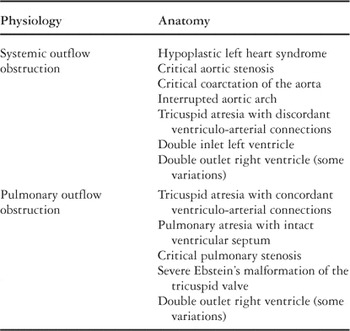
Table 1.

Arterial oxygen saturation (SaO2), mean arterial blood pressure (MAP), and mixed venous oxygen saturation (SmvO2) over time in a patient following Norwood palliation. The mixed venous saturation falls precipitously while pressures and systemic saturation do not change significantly. This can be explained by a rise in systemic vascular resistance, associated with an increase in pulmonary blood flow and a decrease in systemic blood flow. (Used with permission from Tweddell et al.64)

Pulmonary venous oxyhemoglobin saturation (SpvO2) plotted as a function of fractional inspired oxygen (FiO2), demonstrates that pulmonary venous desaturation occurs frequently following the Norwood operation. Pulmonary venous saturations of less than (points below the dashed line) most often occur at fractional inspirations of less than 0.21, but can also occur at values as high as 0.40. (Used with permission from Taeed et al.62)

The proportional error in the ratio of pulmonary to systemic blood flows as a function of pulmonary venous oxyhemoglobin saturation (SpvO2). Arterial oxyhemoglobin saturation (SaO2) is assumed to be 77%, and mixed venous oxyhemoglobin saturation is assumed to be 45%. The relationship is plotted for both a 5% and 10% error in pulmonary venous saturations. (Used with permission from Barnea et al.6)

Systemic oxygen availability (mlO2/min) as a function of the ratio of pulmonary to systemic blood flows (Qp/Qs)at different levels of cardiac output. Pulmonary venous oxyhemoglobin saturation (SpvO2) is assumed to be 96%, and oxygen consumption (VO2) is assumed to be 18 mlO2/min. (Used with permission from Barnea et al.5)
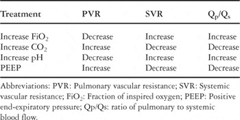
Table 2.
 You have
Access
- 23
- Cited by