Introduction
The Pediatric-Buccal-Epigenetic (PedBE) clock is a novel epigenetic tool that shows potential for evaluating the molecular impact of environmental and contextual factors on child development, and in turn, life course health and disease. The PedBE clock is quantified by two indicators: 1) epigenetic, or biological, age and 2) epigenetic age acceleration, indexed by the difference between one’s epigenetic and chronological age. As such, positive epigenetic age acceleration indicates faster biological aging relative to one’s chronological age and negative epigenetic age acceleration relays the converse. Prior to the inception of the PedBE clock, correlations between chronologic age and epigenetic age in pediatric samples conferred a high degree of variability and inaccuracy, as existing epigenetic clock construction such as the Horvath or Hannum clocks relied on adult-based biomarkers (Wang & Zhou, Reference Wang and Zhou2021). Specifically, these clocks did not account for the accelerated rate of change in epigenetic age during childhood, as compared to adulthood (Alisch et al., Reference Alisch, Barwick and Chopra2012). As a result, new clocks were constructed to generate a more precise estimate of epigenetic age in pediatric populations. For example, Knight’s clock estimates gestational age at birth via cord blood (Knight et al., Reference Knight, Craig and Theda2016). McEwen and colleagues developed the PedBE clock, based on buccal swab samples that are easier to collect in children of any age (McEwen et al., Reference McGill, Pokhvisneva and Clappison2020). Training datasets ranged in age from 2 months of age through 20 years of age. Questions remain about pediatric epigenetic clock utility when generated from methylation data from different tissue and cell types. Moreover, its validity in racially and ethnically diverse samples, and specific to the PedBE clock, in the neonatal period, remains unknown.
Birth outcomes, such as length of gestation and infant birthweight, predict short- and long-term health and development outcomes (Dhamrait et al., Reference Dhamrait, Christian, O’Donnell and Pereira2021; Gleason et al., Reference Gleason, Gilman and Sundaram2021; Hochstedler et al., Reference Hochstedler, Bell and Park2021). In a sample of uncomplicated, singleton pregnancies of infants born at term, evidence suggests that longer length of gestation at birth is associated with faster PedBE age acceleration at 3 and 9 months of age (McEwen et al., Reference McEwen, O’Donnell and McGill2020). In the same sample, PedBE age acceleration was unrelated to birthweight (adjusted for gestational age). However, in a separate study evaluating the impact of prenatal anxiety on PedBE age acceleration from 3 months to 4 years of age, gestational age at birth was positively associated with PedBE age acceleration, though birthweight was not included in this model (McGill et al., Reference McGill, Pokhvisneva and Clappison2022). The salutary effect of accelerated epigenetic age is provocative as it may suggest individual differences in rate of maturation in infancy but needs to be replicated.
The current study examined the concurrent association between PedBE age estimated from umbilical vein endothelial cells at the time of birth and length of gestation, extending the existing research in three crucial ways: 1) examining this association in a sample of endothelial cells, 2) earlier in the neonatal period than prior studies, and 3) assessing this relationship in a racially and ethnically diverse sample. Umbilical vein endothelial cells offer the advantage of a single cell type in contrast to mixed leukocytes found within cord blood. Single-cell approaches have recently emerged as advantageous for better understanding underlying aging in estimating biological clocks (He et al., Reference He, Memczak, Qu, Belmonte and Liu2020). We hypothesized that PedBE age would be positively associated with length of gestation based on prior studies. In addition, we examined whether infant characteristics (e.g., sex, birthweight), maternal sociodemographic and health characteristics (e.g., race, ethnicity, body mass index), and obstetric complications (e.g., pre-eclampsia) were associated with PedBE age acceleration. Given that we are applying the PedBE clock to a new tissue source there were no a priori hypotheses regarding these associations.
Method
Participants and procedure
Pregnant women were enrolled in two cohorts – the Stress and Health in Pregnancy (SHIP) cohort in North Carolina and the Prospective Research on Early Determinants of Illness and Children’s Health Trajectories (PREDICT) cohort in Florida. Both cohorts enrolled from university-affiliated obstetric clinics; a total of 577 were enrolled. Women were eligible if the mother was>18 years old, spoke and read English (SHIP), or English or Spanish (PREDICT), and planned to deliver at the study-affiliated hospital. Women were ineligible if the fetus had a known congenital anomaly or chromosomal abnormality, or if the mother had HIV, Hepatitis C, or Hepatitis B. Average gestational age at enrollment was 20.8 weeks (SD = 6.9) for SHIP and 24.6 weeks (SD = 6.3) for PREDICT. After enrollment, women completed questionnaires about demographics and health behaviors. After delivery, a cord blood sample from the umbilical cord vein was collected to yield human umbilical vein endothelial cells [HUVECs]. HUVECs were isolated following Davis et al. (Reference Davis, Crampton and Hughes2007)with minor modifications (please see Supplemental text for additional detail). Maternal obstetric and infant delivery records were abstracted following delivery. This study was approved by the two sites’ respective institutional review boards. Mothers provided written informed consent for themselves and their children.
DNA methylation measurement and preprocessing
DNA methylation data were collected for 459 samples, including 48 technical replicates, in two batches. Methylation levels were measured using the Illumina MethylationEPIC array (Pidsley et al., Reference Pidsley, Zotenko and Peters2016). Raw data were preprocessed and quantile normalized using the R package minfi (Aryee et al., Reference Aryee, Jaffe and Corrada-Bravo2014; Fortin et al., Reference Fortin, Triche and Hansen2017). We filtered samples according to the following criteria: samples with more than 1% (8,658) low-quality probes (i.e., probes with a detection p-value larger than 0.01); samples with mismatched sex between our record and prediction from methylation data; and samples with DNA contamination (i.e., log2 odds ratio of contamination larger than -2), resulting in the removal of 71 samples. After including the replicate sample with fewer low-quality probes for each pair 333 samples remained in the final analytic sample. At the probe level, the following probes were removed: probes with a detection p-value larger than 0.01 in more than 5% of total samples; probes with SNPs at CpG sites; probes on sex chromosomes; and cross-reactive probes (Chen et al., Reference Chen, Lemire and Choufani2013). In total, 64,254 out of 865,859 probes were removed, leaving 801,605 probes used in subsequent analyses. Lastly, functional normalization with control probes was adopted for batch effect correction of the data (please see supplementary text for additional details on DNAm extraction).
Epigenetic age and age acceleration
Infant’s epigenetic age was estimated using the PedBE clock described by McEwen and colleagues (McEwen et al., Reference McEwen, O’Donnell and McGill2020). Briefly, this clock consists of 94 CpG sites, only one of which overlaps with the Horvath epigenetic clock (Horvath, Reference Horvath2013). It is intended to estimate an epigenetic age in buccal cells for individuals aged 0-20 years. R code to generate the PedBE clock is available online: https://github.com/kobor-lab/Public-Scripts/. Epigenetic age acceleration was derived by regressing PedBE age estimates on gestational age at birth and extracting the resulting residuals; negative values indicate decelerated epigenetic aging and positive values indicate accelerated epigenetic aging.
Phenotypic measures
Sociodemographic factors
During the first study visit, mothers reported on their race (Black/African American [including those who described themselves as Black/African American and one or more additional racial group], or White), ethnicity (Hispanic/Latina or not Hispanic/Latina), and tobacco use during pregnancy (yes/no). Data on maternal pre-pregnancy body mass index (BMI) and infant sex were obtained from obstetric records.
Obstetric factors
Data on the presence or absence of pre-eclampsia, gestational diabetes, and gestational hypertension were obtained from obstetric records. A composite, binary variable was created combining these complications.
Statistical analyses
Although a total of 459 samples were processed for DNA methylation analysis, 333 samples remained after filtering on various quality control metrics (e.g., low-quality probes). Thus, our analytic sample was based on a sample size of 333. All analyses were conducted using R statistical software (R Core Team & Team, 2022). Prior to analyses, data were inspected for normality, missingness, and outliers. Results from Little’s test of missing completely at random (MCAR) suggest that data were MCAR (p = 0.43). Approximately 10% of data were missing in the present sample, with 5.6% of participants (n = 19) missing data on tobacco use during pregnancy, 3.6% on maternal education, and <1% of participants missing data on other study variables. Given the minimal amount of missing data and that the MCAR assumption was met, listwise deletion was used to handle missing data. We then examined the zero-order correlation between gestational age at birth and PedBE Age, as well as between birth weight (adjusted for gestational age) and PedBE Age acceleration. Next, informed by the presence of univariate associations, we used multivariate linear regression to examine the association of birthweight adjusted for gestational age and PedBE age acceleration, controlling for sociodemographic and obstetric factors. Finally, as a sensitivity analysis, we excluded preterm infants (i.e., <37 weeks’ gestation; n = 26). Significance was determined by a p-value ≤ 0.05.
Results
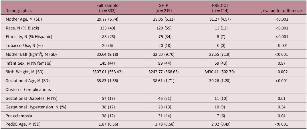
Sociodemographic and obstetric data for study participants are presented in Table 1. A total of 333 mother-infant dyads were included in the present study. Mothers were, on average, approximately 29 years old at the time of the child’s birth and the majority identified as non-Hispanic White. Approximately half of infants were male and infants were, on average, delivered at term and of normal birth weight. Eight percent of infants were born preterm and 7% at low birth weight.
Demographic and study characteristics

Note. Mother age reflects age at delivery. Race was coded as Black or White. Tobacco use reflects self-reported use during pregnancy. BMI = body mass index. Birth weight is measured in grams, gestational age is measured in weeks, and PedBE age is measured in years.
PedBE age
PedBE age and age acceleration were derived for all infants. Average PedBE age at birth was 1.87 years (SD = 0.57 years). PedBE age demonstrated a modest, positive association with gestational age at birth (r = 0.22, p < .001) and birth weight (r = 0.20, p < .001), but was not associated with any other sociodemographic or obstetric factors. Similarly, PedBE age acceleration demonstrated a modest, positive association with infant birth weight (r = 0.15, p = 0.01) and birth weight adjusted for gestational age (r = 0.18, p = .001; Fig 1). Correlations were similar when excluding preterm infants (data not shown).
Scatter plots of PedBE age and PedBE age acceleration with gestational age and birth weight.

Multivariate analyses
The omnibus multivariate model examining the prediction of PedBE age acceleration by birth weight adjusted for gestational age controlling for sociodemographic and obstetric factors, as well as birth cohort, was significant [F(9, 295) = 3.18, R 2 = 0.09, p = 0.001] suggesting significant variance in PedBE age acceleration was explained by this set of predictors. Specifically, infants with higher birth weight (adjusted for gestational age) had greater PedBE age acceleration (b = 0.0002, se = 0.0007, p = 0.002), though this association was small. In addition, findings suggested that infants in the SHIP cohort demonstrated PedBE age deceleration relative to infants in the PREDICT cohort (b = −0.26, se = 0.09, p = 0.003). At a trend level, having a mother with at least a high school education was associated with PedBE age deceleration (b = −0.19, se = 0.10, p = 0.07). There was no evidence of an association between PedBE age acceleration and infant sex (p = 0.93) or maternal race (p = 0.39), ethnicity (p = 0.51), BMI (p = 0.34), smoking status during pregnancy (p = 0.11), and presence of one or more obstetric complications (p = 0.52). Similar to bivariate associations, multivariate findings remained consistent in a sensitivity analysis when excluding preterm infants born before 37 weeks’ gestation.
Discussion
The PedBE clock was designed to estimate pediatric epigenetic age from buccal epithelial cells (McEwen et al., Reference McEwen, O’Donnell and McGill2020). Application of the PedBE clock in other single tissue specimens is limited to date; the current study examined PedBE age and age acceleration indexed from human umbilical vein endothelial cells. Consistent with prior work, greater gestational age at birth was associated with greater PedBE age. Similarly, higher birth weight was also associated with greater PedBE age. Gestational age and birth weight have been linked to neonatal morbidity and mortality and the timing of achievement of developmental milestones (Flensborg-Madsen et al., Reference Flensborg-Madsen, Grønkjær and Mortensen2019) thus PedBE age may yield similar utility as an indicator of neuromaturation at birth. Of note, we conducted a sensitivity analysis to ensure that the inclusion of preterm infants in our sample was not driving detection of these associations; associations remained unchanged with the exclusion of infants born prior to 37 weeks’ gestation. However robust, the majority of variance in PedBE age was independent of gestational age or size at birth. In addition to the modest nature of the association, we observed increased variability in PedBE age relative to gestational age. Therefore, it is plausible PedBE age may confer greater specificity in prediction of neonatal health and development.
Given that PedBE age acceleration is derived by extracted residuals from a linear model of PedBE age regressed on chronological age, we were unable to examine associations of gestational age at birth and PedBE age acceleration measured concurrently. However, findings suggest increased PedBE age acceleration was associated with higher birthweight, adjusted for gestational age at birth. Among expectant women, it has been shown that greater epigenetic age acceleration measured mid-gestation is associated with shorter gestational length and lower birthweight, independent of gestational age (Ross et al., Reference Ross, Carroll, Horvath, Hobel, Coussons-Read and Dunkel Schetter2020). However, our findings suggest an opposing directionality of association among infants, among whom more optimal birth outcomes are associated with greater epigenetic age acceleration, reinforcing the stance suggested by McEwen and colleagues that age acceleration in pediatric samples may reflect positive outcomes (McEwen et al., Reference McEwen, O’Donnell and McGill2020). Further, this aligns with Knight’s pediatric clock using cord blood samples which showed increased DNAm gestational age relative to clinical gestational age was associated with infant birthweight, independent of gestational age (Knight et al., Reference Knight, Craig and Theda2016).
Contrary to expectations, obstetric complications did not predict PedBE age acceleration in the current study. There is a robust and consistent literature linking exposure to gestational diabetes mellitus in utero to accelerated epigenetic age, and suggestive of a mechanism for intergenerational transmission of metabolic disease, in children and adolescents (Dłuski et al., Reference Dłuski, Wolińska and Skrzypczak2021; Franzago et al., Reference Franzago, Fraticelli, Stuppia and Vitacolonna2019; Shiau et al., Reference Shiau, Wang and Liu2020). Similar, yet more variable evidence is found among studies of hypertensive disorders of pregnancy including gestational hypertension and pre-eclampsia, with altered DNA methylation observed in cord blood (Kazmi et al., Reference Kazmi, Sharp and Reese2019). Some studies have found no association between epigenetic age and pre-eclampsia status (Heinsberg et al., Reference Heinsberg, Ray and Conley2021). The lack of association in the current study aligns with this and may reflect sample composition as this is not a clinical cohort of high-risk pregnancies.
In addition, we also did find support of demographic predictors of PedBE age acceleration at birth. This may be due in part to the observed impact of cohort in our study, where infants in the SHIP cohort experienced epigenetic age deceleration relative to infants in the PREDICT cohort. On average, the SHIP cohort was comprised of younger, Black or Hispanic expectant women, who were more likely to smoke cigarettes and presented with a higher pre-pregnancy BMI relative to women in the PREDICT cohort. Relatedly, infants in the SHIP cohort were born at earlier gestational age and lower birth weight compared to infants in the PREDICT cohort. There are also considerations outside of cohort effects. For example, prior research consistently shows underreporting of cigarette smoking in pregnancy (Dietz et al., Reference Dietz, Homa and England2011). Taken together, this highlights how sensitive clock constructions could be to cohort demographics and the bounds of generalizability. To yield the greatest precision in measurement, clock construction in diverse populations is paramount. Furthermore, there remains more to learn about functionality of the PedBE clock CpG sites. The original publication detailing construction of the PedBE clock notes the selected CpG sites were not enriched among particular genomic features (McEwen et al., Reference McEwen, O’Donnell and McGill2020). Yet, more recent evidence has shown a highly significant enrichment for dexamethasone-responsive PedBE CpGs relative to those found genome wide, suggestive of high susceptibility of the PedBE clock for stress signaling (Dammering et al., Reference Dammering, Martins and Dittrich2021). Thus, the PedBE clock CpGs may impart information about glucocorticoid regulation and biological aging perhaps via glucocorticoid receptor-induced genomic processes such as excision repair.
Our study presents several strengths and limiting factors to consider. Our focus on a single cell type to estimate epigenetic age confers greater precision relative to traditional Hannum and Horvath pan-tissue clocks utilized in the study of adult health and aging due to less cellular heterogeneity and the ability to quantify the rapid epigenetic changes in children, particularly in the first year. Yet, the PedBE clock has previously relied on buccal epithelial cells. Similar to buccal cells, umbilical vein endothelial cells measured in the current study offer the advantage of non-invasive collection and a single tissue. However, measurement is limited to birth. In addition, our analytic sample size does not permit the investigation of specific conditions of pregnancy (e.g., gestational diabetes, gestational hypertension, pre-eclampsia) with shared underlying physiology due to low observed frequencies. Relatedly, our nonsignificant associations between select confounders such as race or education and epigenetic age or age acceleration may be reflective of our ability to find a statistical effect rather than a true lack of association. It is important to replicate these findings in a larger sample.
In conclusion, the PedBE clock may have application to umbilical vein endothelial cells in addition to buccal epithelial cells to measure epigenetic aging in infants and children earlier in infancy than previously measured offering an anchoring sampling point at birth whereby to examine change over time. Future studies examining developmental drivers of PedBE age and age acceleration will expand our understanding of whether accelerated epigenetic age in early life is a reflection of adaptive maturation, or alternatively, foreshadows negative health sequelae.
Supplementary material
The supplementary material for this article can be found at https://doi.org/10.1017/S2040174424000333.
Acknowledgements
We would like to thank the families participating in the PREDICT and SHIP cohorts.
Funding Statement
Funding for this study was provided by the Johns Hopkins All Children’s Foundation to Sara Johnson and by NIH R01MD011746 to Cathrine Hoyo and Joan Kaufman.
Competing interests
The authors have no financial or non-financial conflicts of interest to disclose.