Introduction
The frontal sinus is described as the most difficult sinus to access surgically, given its challenging and variable anatomy, as well as its proximity to the cribriform plate, orbit and anterior ethmoidal artery. This complex space may be pneumatised by various surrounding cells. Although it is observed as a normal variant in much of the population, these cells may have the potential to cause frontal sinusitis, possibly due to obstruction of the frontal sinus outflow tract.
Various classifications have been given for frontal recess cells. In 1994, Bent, Kuhn and colleagues identified four frontal cell variations and classified them as type I–IV cells.Reference Bent, Cuilty-Siller and Kuhn1 In 1996, Kuhn described the modified Kuhn classification of fronto-ethmoidal cells.Reference Kuhn2 In 2004, the Kuhn–Citardi–Lee classification was described by Lee et al.Reference Lee, Kuhn and Citardi3 The recent International Frontal Sinus Anatomy Classification was proposed in 2016.Reference Wormald, Hoseman, Callejas, Weber, Kennedy and Citardi4 This latter consensus document was developed to provide a better description of frontal cells in relation to the frontal sinus drainage pathway, and to improve the surgeon's ability to: understand various possible variations, plan surgery, and communicate these complexities when teaching or reporting outcomes.
Many previous studies have investigated the clinical significance of fronto-ethmoidal cells described using older classification systems. Various studies have shown that the resultant narrowing of the frontal recess by Kuhn cells is associated with frontal sinusitis.Reference Kubota, Takeno and Hirakawa5–Reference Meyer, Kocak, Smith and Smith7 In addition, the presence of a fronto-ethmoidal cell (types 3–4), supra bullar cell and frontal bullar cell has been reported to significantly influence frontal sinusitis development.Reference Lien, Weng, Chang, Lin and Wang6,Reference Meyer, Kocak, Smith and Smith7
This study aimed to determine the prevalence of frontal cells according to the International Frontal Sinus Anatomy Classification, and to examine the association between cells classified via this system and frontal sinusitis by determining the frequency of different cell types in those with and without frontal sinusitis. We also studied the association between the different cell classifications and gender. In addition, the relationships between International Frontal Sinus Anatomy Classification cells and frontal sinus ostium and frontal recess diameters were explored.
Materials and methods
Ninety adults of either sex, with a clinical suspicion of rhinosinusitis, who had undergone a computed tomography (CT) scan of the paranasal sinuses, were included in the study. Ethical clearance was obtained from the institutional ethics committee (approval code: F No 17/IEC/MAMC/2017-Radio D-10).
With patients in the supine position, all patients underwent high-resolution CT of the paranasal sinuses and nasal cavity using a 128-slice single-source CT scanner (Somatom® Definition Adaptive Scanning (AS+) equipment; Siemens Healthineers, Erlangen, Germany). The following scanning parameters were used: 120 kVp, 80 mAs, 128 × 0.6 mm detector collimation, and matrix of 512 × 512. Thin axial sections of 0.6 mm thickness were obtained for each patient. Thin multiplanar images along coronal and sagittal planes were reconstructed from these axial images.
All patients in the study were aged over 18 years, as the size of the frontal sinus becomes stable after 18 years of age. Exclusion criteria included patients who had a previous history of sinus surgery, a maxillofacial fracture, sinonasal malignancy, fungal sinusitis or congenital anomalies, as these disrupt the frontal recess anatomy, hindering cell identification.
A total of 180 (left and right) sides of 90 patients were evaluated. Each scan was evaluated by two radiologists, one with 25 years of experience and another with 15 years of experience. In cases of discrepancy, a consensus was reached.
Fronto-ethmoidal cells were categorised according to the International Frontal Sinus Anatomy Classification, and the prevalence of each cell type was evaluated (Table 1).Reference Wormald, Hoseman, Callejas, Weber, Kennedy and Citardi4 The frontal ostium diameter was measured in the sagittal plane between the frontal beak and the anterior skull base (Figure 1a). The frontal recess diameter was measured in the sagittal plane from the agger nasi cell and nasofrontal beak anteriorly, to the ethmoid bulla, bulla lamella, supra bullar frontal cells and supra bullar cells posteriorly (Figure 1b).Reference Lai, Yang, Lee, Lin, Chu and Wang8 The right and left sides of each scan were evaluated for evidence of frontal sinusitis. Frontal sinusitis was evaluated on CT; it was defined as more than 3 mm of mucosal thickening involving the frontal sinus or the dependent portions of the frontal sinus.
(a) Non-contrast parasagittal computed tomography (CT) images of the paranasal sinus, showing the frontal sinus ostium (yellow double-headed arrow) located at the level of frontal beak (blue solid line), measured from frontal beak anteriorly to the skull base posteriorly (green solid line). (b) Non-contrast parasagittal CT images of the paranasal sinus, showing the frontal recess (yellow double-headed arrow), measured from posterior wall of the agger nasi anteriorly (blue solid line) to the ethmoidal bulla posteriorly (orange solid line).

International Frontal Sinus Anatomy Classification cell types

Statistical analysis
Data were analysed and statistically evaluated using SPSS software, version 17 (SPSS, Chicago, Illinois, USA). Quantitative data were expressed in terms of means and standard deviations, and differences between two comparable groups were tested using the student's t-test. Qualitative data were expressed in percentages. For quantitative data, analysis of variance was used to compare more than two groups, followed by a post-hoc test. For qualitative variables, the chi-square test was used. A p-value of less than 0.05 was considered statistically significant. Odds ratio and 95 per cent confidences were used to quantify the risk factors. A univariate analysis was conducted and the factors that were found to be significant with a p-value of 0.1 or less were entered in multivariate analysis.
Results
Demographics
The study group consisted of 90 patients (45 males (50 per cent) and 45 females (50 per cent)), with a male to female ratio of 1:1. The majority of patients (20 per cent) were in the age group of 26–30 years. As patients aged less than 18 years were excluded from the study, the youngest patient was 18 years old and the oldest was 62 years old. The mean age of the patients was 38 years.
Cell prevalence
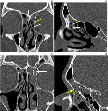
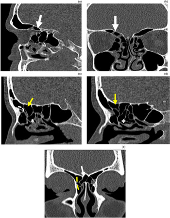
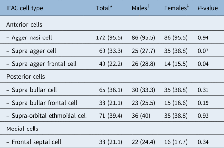
The prevalence of frontal cells (Table 2) was described, according to the new International Frontal Sinus Anatomy Classification system, in terms of anterior (Figure 2), posterior and medial cells (Figure 3). The agger nasi cell was the most commonly occurring cell, seen in 95.5 per cent of the patients. The prevalence of frontal septal cells and supra bullar frontal cells was lowest, with each cell type seen in 21.1 per cent of patients. The prevalence of supra agger cells, supra agger frontal cells, supra bullar cells and supra-orbital ethmoid cells was 33.3 per cent, 22.2 per cent, 36.1 per cent and 39.4 per cent, respectively.
Anterior International Frontal Sinus Anatomy Classification cells. Non-contrast coronal (a) and sagittal (b) computed tomography (CT) images of the paranasal sinus, showing a left agger nasi cell (white arrow) and a left supra agger cell (yellow arrow) in a patient with bilateral frontal sinusitis. Non-contrast coronal (c) and sagittal (d) CT images of the paranasal sinus, showing a left supra agger frontal cell (white arrow) in a patient with bilateral frontal sinusitis, which is seen to pneumatise into the frontal sinus through the frontal ostium and to sit above the frontal beak (yellow arrow). The cell pushes the drainage pathway of the frontal sinus posteriorly and medially (white dotted line). R = right; L = left; P = posterior

Posterior and medial International Frontal Sinus Anatomy Classification cells. Non-contrast sagittal (a) and coronal (b) computed tomography (CT) images of the paranasal sinus, showing a supra-orbital ethmoid cell (white arrow) in a patient with bilateral frontal sinusitis, which pneumatises over the orbit. Non-contrast sagittal CT images of the paranasal sinus of a different patient, showing a: (c) supra bullar cell (yellow arrow), which lies above the ethmoid bulla (asterisk) pushing the frontal drainage pathway anteriorly (white dotted line); and (d) a supra bullar frontal cell (yellow arrow), which lies above the ethmoid bulla (asterisk) pneumatising through the frontal ostium. (e) Non-contrast sagittal CT image, showing a right frontal septal cell (white arrow) attached to the inter-frontal sinus septum.

Prevalence of IFAC cell types

Data represent numbers (and percentages) of International Frontal Sinus Anatomy Classification cells, unless indicated otherwise. *n = 180; †n = 90; ‡n = 90. IFAC = International Frontal Sinus Anatomy Classification
Gender-based cell prevalence
The prevalence of the different International Frontal Sinus Anatomy Classification cells in males and females was also studied (Table 2). The occurrence of different cells varied in males and females. In our study, agger nasi cells had the highest prevalence, in both males and females, of 95.5 per cent. The prevalence rates of supra agger cells, supra agger frontal cells, supra bullar cells, supra bullar frontal cells, supra-orbital ethmoidal cells and frontal septal cells in males were 27.7 per cent, 28.8 per cent, 33.3 per cent, 25.5 per cent, 40 per cent and 24.4 per cent, respectively. In females, the prevalence rates were 38.8 per cent, 15.5 per cent, 38.8 per cent, 16.6 per cent, 38.8 per cent and 17.7 per cent, respectively. The supra agger frontal cells were found to be significantly more prevalent in males (28.8 per cent) than in females (15.5 per cent) (p < 0.04). There were no statistically significant differences between males and females in terms of the occurrence of the remaining International Frontal Sinus Anatomy Classification cells.
Associations with frontal ostium and recess diameters
The mean (± standard deviation) diameter of the frontal sinus ostium was 5.46 ± 2.11 mm and the mean diameter of the frontal recess was 1.94 ± 1.08 mm. The patients with supra bullar frontal cells had a significantly (p < 0.05) shorter anteroposterior diameter of the frontal ostium (4.81 ± 2.12 mm) compared to those without supra bullar frontal cells (5.60 ± 2.18 mm) (Table 3). However, there were no statistically significant differences in the anteroposterior diameters of the frontal ostium and frontal recess regarding the presence or absence of any other International Frontal Sinus Anatomy Classification cells.
Effect of presence of IFAC cells on mean anteroposterior diameters of frontal ostium and frontal recess*

*n = 180. IFAC = International Frontal Sinus Anatomy Classification; SD = standard deviation
Association with frontal sinusitis
In our study, frontal sinusitis was seen in 33.9 per cent of cases (61 out of 180) and absent in 66.1 per cent of cases (119 out of 180). The incidence of supra bullar cells and supra bullar frontal cells was greater in patients with frontal sinusitis (44.3 per cent and 26.2 per cent respectively) than in those without (31.9 per cent and 18.5 per cent respectively) (Table 4). However, there were no significant differences in the incidence rates of any of the International Frontal Sinus Anatomy Classification cells in patients with frontal sinusitis compared to those without frontal sinusitis (all p > 0.05).
Association of various IFAC cells with frontal sinusitis

*n = 119; †n = 61. IFAC = International Frontal Sinus Anatomy Classification; CI = confidence interval
Discussion
The frontal sinus is described as the most difficult sinus to access surgically because of its challenging and variable anatomy, as well as its proximity to the cribriform plate, orbit and anterior ethmoidal artery. The frontal sinus drains into the frontal recess through the frontal ostium. The frontal recess is a complex space, with the approximate shape of an inverted funnel or cone whose apex is at the frontal ostium. This complex space may be pneumatised by various surrounding cells. Although it is observed as a normal variant in much of the population, these cells may have the potential to cause frontal sinusitis, possibly due to obstruction of the frontal sinus outflow tract.
During functional endoscopic sinus surgery (FESS), complete removal of these frontal cells is necessary to ensure adequate opening of the frontal sinus, and to enable proper physiological drainage and ventilation. Incomplete removal of the cells in the frontal recess is one of the most common causes of FESS failure.
The pathophysiology of frontal sinusitis is associated with disturbance of its drainage and ventilation of the sinus via the ostium. Numerous studies have documented the prevalence of frontal cells, using the various frontal sinus classification systems, and examined their association with frontal sinusitis development. However, only one study in the literature investigated the association of International Frontal Sinus Anatomy Classification cells with frontal sinusitis.Reference Sommer, Hoffmann, Harter, Döscher, Kleiner and Lindemann9
Cell prevalence findings
The agger nasi cell, which is the anterior most ethmoid cell, was documented in 95.5 per cent of the frontal sinuses in our Indian population. This is comparable to published findings, which range from 89 per cent to 94 per cent.Reference Lee, Kuhn and Citardi3,Reference Sommer, Hoffmann, Harter, Döscher, Kleiner and Lindemann9 In a study by Choby et al.Reference Choby, Thamboo, Won, Kim, Shih and Hwang10 on a North American population and a study by Tran et al.Reference Tran, Ngo and Psaltis11 on a Vietnamese population, where the fronto-ethmoidal cells were categorised according to the International Frontal Sinus Anatomy Classification, the prevalence rates of agger nasi cells were 96.5 per cent and 95.7 per cent respectively. The consistent presence of agger nasi cell is the primary reason why this cell is used as a reference cell for all the anteriorly based cells in the International Frontal Sinus Anatomy Classification system.
In our study, the prevalence rates of supra agger cells and supra agger frontal cells were 33.3 per cent and 22.2 per cent respectively, which are closer to the rates reported for the North American population, of 30 per cent and 20 per cent respectively.Reference Choby, Thamboo, Won, Kim, Shih and Hwang10 The prevalence rates for supra agger cells and supra agger frontal cells were 16.3 per cent and 13 per cent respectively for the Vietnamese population, which are lower than the rates in our study.Reference Tran, Ngo and Psaltis11 Direct comparison of these cells with earlier studies using an older classification system was difficult, because of their different anatomical description. Tran et al. assumed that type 1 and type 2 cells (categorised using the Bent and Kuhn classification) likely correspond to supra agger cells, with type 3 and type 4 (Bent and Kuhn classification) likely representing supra agger frontal cells.Reference Bent, Cuilty-Siller and Kuhn1,Reference Tran, Ngo and Psaltis11 The quoted prevalence rates for these cells were highly variable, ranging from 16.3 per cent to 56 per cent for T1 and T2 cells, and from 5.6 per cent to 13 per cent for T3 and T4 cells, respectively.
The prevalence of supra bullar cells in our study was 36.1 per cent. This prevalence rate is lower than that reported in the study by Choby et al.Reference Choby, Thamboo, Won, Kim, Shih and Hwang10 (another International Frontal Sinus Anatomy Classification based study) of a North American population, which reported the prevalence rate of 72 per cent.Reference Choby, Thamboo, Won, Kim, Shih and Hwang10 According to Tran et al., supra bullar cells were noted in 46.2 per cent of patients, which is also higher than in our study.Reference Tran, Ngo and Psaltis11 The prevalence of supra bullar frontal cells in our study was 21.1 per cent. This prevalence rate was higher than those in the North American and Vietnamese groups, which were 5.5 per cent and 4.3 per cent respectively.Reference Choby, Thamboo, Won, Kim, Shih and Hwang10,Reference Tran, Ngo and Psaltis11
The prevalence of supra-orbital ethmoidal cells varied significantly across studies, when examining the previously published literature. In our study, supra-orbital ethmoidal cells were seen in 39.4 per cent of frontal sinuses, which is a higher rate than that reported for the North American population (28.5 per cent). In the study by Tran et al., the prevalence of supra-orbital ethmoidal cells was 17.3 per cent, which was lower than in our population.Reference Tran, Ngo and Psaltis11 The varying prevalence rates of supra-orbital ethmoidal cells may be a result of its similar radiological appearance to supra bullar cells.
The differences in these posterior cell prevalence rates could also be because of differences in ethnicity. Furthermore, 33 per cent of the patients in our study had frontal sinusitis, which could result in a higher prevalence of posterior cells.
In our study, frontal septal cells were seen in 21.1 per cent of the patients; this rate is closer to the 30 per cent reported for the North American population.Reference Choby, Thamboo, Won, Kim, Shih and Hwang10
Table 5 shows a comparison of the various studies.
Comparison of IFAC cell prevalence among various ethnic groups

‘Sides’ refers to patients’ left and right sides, subjected to computed tomography scanning. Data represent percentages (and numbers) of International Frontal Sinus Anatomy Classification cells. IFAC = International Frontal Sinus Anatomy Classification
Gender-based cell prevalence findings
To best of our knowledge, no previous study has investigated the association between different International Frontal Sinus Anatomy Classification cells and gender. Gender differences may be important predictors of frontal sinus disease in males versus females. In our study, males had a higher proportion of frontal sinusitis (24.5 per cent) than females (13.3 per cent). Males also had a significantly higher number of supra agger frontal cells (28.8 per cent) than females (15.5 per cent) (p < 0.05), which may explain the increased incidence of frontal sinusitis in males. There were no statistically significant differences between males and females in terms of the occurrence of the remaining International Frontal Sinus Anatomy Classification cells. In a study by House et al., males had a significantly higher number of type 4 cells (categorised based on the Bent and Kuhn classificationReference Bent, Cuilty-Siller and Kuhn1), with a higher incidence of frontal sinusitis in males.Reference House, Stringer and Seals12
Frontal ostium and recess diameter findings
We studied the associations between anterior, posterior and medial International Frontal Sinus Anatomy Classification cells and the mean diameters of the frontal ostium and frontal recess. In our study, the mean anteroposterior diameter of the frontal ostium in patients with supra bullar frontal cells was 4.81 ± 2.12 mm, whereas the diameter in patients without supra bullar frontal cells was 5.60 ± 2.18 mm; this difference was statistically significant (p < 0.05). This result is similar to that of Lien et al., who found that the presence of supra bullar frontal cells was associated with a significantly narrower frontal ostium diameter.Reference Lien, Weng, Chang, Lin and Wang6 Thus, the existence of supra bullar frontal cells might lead to narrowing of the frontal sinus drainage pathway and produce significant obstruction. However, there were no statistically significant associations between the anteroposterior diameters of the frontal ostium and frontal recess and the presence or absence of other International Frontal Sinus Anatomy Classification cells.
Frontal sinusitis findings
We studied the association between International Frontal Sinus Anatomy Classification cells and frontal sinusitis. In our study, frontal sinusitis was present in 33.9 per cent of cases and absent in 66.1 per cent of cases. The odds ratio of having supra bullar cells was 1.69 times higher for those with frontal sinusitis than for those without. However, this finding did not reach statistical significance in our study. The incidence rates of supra bullar cells and supra bullar frontal cells were greater in patients with frontal sinusitis (44.3 per cent and 26.2 per cent respectively) than in those without (31.9 per cent and 18.5 per cent respectively). However, there was no significant association between these cells and the presence of frontal sinusitis. On univariate analysis, the prevalence rates of agger nasi cells, supra agger cells, supra agger frontal cells, supra-orbital ethmoidal cells and frontal septal cells also failed to show any association with the presence of frontal sinusitis. Sommer et al. demonstrated no increased occurrence of frontal sinus opacification associated with the presence of International Frontal Sinus Anatomy Classification cells (p > 0.05).Reference Sommer, Hoffmann, Harter, Döscher, Kleiner and Lindemann9
Meyer et al. categorised the cells according to the Bent and Kuhn classification and found a higher incidence of frontal sinusitis in patients with frontal cell types 3 and 4.Reference Meyer, Kocak, Smith and Smith7 DelGaudio et al. also classified the cells according to the Bent and Kuhn classificationReference Bent, Cuilty-Siller and Kuhn1 and reported no significant difference in the frequency of frontal sinusitis for patients with or without agger nasi cells or frontal cells.Reference DelGaudio, Hudgins, Venkatraman and Beningfield13 A study by Langille et al. demonstrated a significant association between frontal sinus mucosal thickening and the presence of frontal cells type 2 and type 3.Reference Langille, Walters, Dziegielewski, Kotylak and Wright14 Eweiss and Khalil demonstrated no significant difference between frontal sinus mucosal disease regarding the presence or absence of agger nasi and frontal cells.Reference Eweiss and Khalil15
Hashimoto et al. classified the cells according to the modified Kuhn classification and found that the presence of frontal recess cells did not influence frontal sinusitis development.Reference Hashimoto, Tsuzuki, Okazaki and Sakagami16
The frontal cells in the following studies were classified according to the Kuhn–Citardi–Lee classification and varying results were reported.Reference Lee, Kuhn and Citardi3 Lien et al. reported a significant association between the presence of supra bullar cells, frontal bullar cells and supra-orbital ethmoidal cells and the development of frontal sinusitis, owing to significant shortening of the anteroposterior diameters of the frontal ostium and frontal recess.Reference Lien, Weng, Chang, Lin and Wang6 According to Kubota et al., the presence of frontal bullar cells was significantly associated with frontal sinusitis development.Reference Kubota, Takeno and Hirakawa5 Lai et al. reported a higher incidence of supra bullar cells and supra bullar frontal cells in patients with frontal sinusitis, but there was no significant association between the presence of various fronto-ethmoidal cells and frontal sinusitis development.Reference Lai, Yang, Lee, Lin, Chu and Wang8 These discrepancies can be explained as due to differences in ethnicity between the various studies, different classifications of fronto-ethmoidal cells and our small sample size.
• The agger nasi cell was the most commonly occurring cell, seen in 95.5 per cent of patients
• Occurrence of International Frontal Sinus Anatomy Classification cells varied in males and females
• There was no significant difference in occurrence of any International Frontal Sinus Anatomy Classification cells in patients with frontal sinusitis compared to those without
• High-resolution computed tomography (CT) is the reference standard for evaluation prior to functional endoscopic sinus surgery
• High-resolution CT enables delineation of anatomical details such as frontal recess cell, which is critical for pre-operative evaluation and treatment of frontal sinus pathology
There were a few limitations of our study. The sample size was small because of study time restraints, which may have masked or enhanced the differences observed. In order to obtain more conclusive results, a larger population needs to be studied. Our study included patients clinically suspected of having chronic rhinosinusitis who were referred to our department for CT scanning of the paranasal sinuses; thus, our findings may not be directly applicable to patient populations without chronic frontal sinusitis. Racial differences in frontal anatomy are possible, which makes it difficult to extrapolate the findings to populations of different racial origins.
Conclusion
This study documents the prevalence of International Frontal Sinus Anatomy Classification cells in patients undergoing CT of the paranasal sinuses for clinically suspected chronic rhinosinusitis. The presence of any of the International Frontal Sinus Anatomy Classification cells was not significantly associated with frontal sinusitis.
Competing interests
None declared