Introduction
The rise in incidence of gastroesophageal reflux disease has been accompanied by an increase in the presentation of benign and malignant oesophageal strictures. The pathophysiology of these strictures is thought to be attributed to Barrett's metaplasia due to acid reflux.Reference Holtmann1 Initially, treatment of such strictures included endoscopic insertion of a plastic prosthesis to relieve dysphagia. However, these prostheses had high complication rates, mainly due to oesophageal perforation, with procedure-related mortality rates of between 2 and 16 per cent.Reference Tan, Mason and Adam2 Newer, expandable, metallic stents have wider internal diameters, easier deployment mechanisms and markedly safer efficacy.Reference Siersema, Hop, Dees, Tilanus and van Blankenstein3 There is a wide variety of metallic stents available, including such features as anti-reflux valves, internal plastic coatings and retrievable threads.
We hereby present the case of an upper airway complication of an expandable, covered, metallic oesophageal anti-reflux stent. The Hanaro anti-reflux stent (MI Tech Co. Ltd., Kyunggi-do, Korea), designed by Shim et al.,Reference Shim, Jung and Cheon4 has an S-type valve with long leaflets inside its body designed to reduce acid reflux, and is suitable for the treatment of tumours at the gastroesophageal junction. Our patient presented four months after insertion of this stent with multiple broken fragments, one of which had migrated to the nasopharynx, giving symptoms of chronic sinusitis and deafness in both ears caused by middle-ear effusion.
Case report
A 58-year-old woman presented complaining of the gradual onset of dysphagia symptoms. She had a past medical history of cleft palate repair 30 years ago, rheumatoid arthritis, a 10-year history of gastroesophageal reflux disease, and hiatus hernia.
In September 2005, gastroesophagoscopy had demonstrated Barrett's oesophagus and a stricture at 35 cms. At that time, radiological investigations (i.e. computed tomography and endo-oesophageal ultrasound scanning) and repeated biopsies had confirmed that the stricture was benign.
The patient had experienced gradual worsening of her dysphagia symptoms and also weight loss, despite multiple endoscopic dilatations.
In August 2006, an anti-reflux Hanaro-Shim (covered) oesophageal stent had been placed at the gastric cardia (Figure 1). This had resulted in a marked reduction in dysphagia and a general improvement in the patient's nutritional state.
Barium swallow following stent insertion.

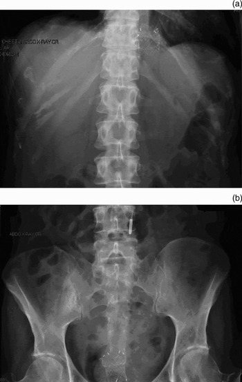
Four months after stent insertion, the patient presented with a four-day history of coughing green sputum, nausea and vomiting, and was diagnosed with pneumonia. Abdominal radiography demonstrated that the stent was broken, with a fragment lying at the gastroesophageal junction and another migrated to the rectum (Figure 2), which was eventually excreted rectally. The patient spent three weeks in the intensive care unit with septic pneumonia and multiple organ failure, as well as profuse oral secretions, sore throat, and persistent nausea and vomiting. Blood cultures isolated Serracia marcesens and Candida albicans. The patient recovered with antibiotics and was discharged.
Abdominal radiograph showing broken stent fragments (a) at the gastroesophageal junction and (b) in the rectum.

Two months after discharge (April 2007), the patient was readmitted with recurrent pneumonia, sinus symptoms (facial pain, green nasal discharge and hyposmia) and bilateral, progressive deafness. Blood cultures isolated Streptococcus oralis. She recovered with antibiotics and was referred for an ENT out-patient review.
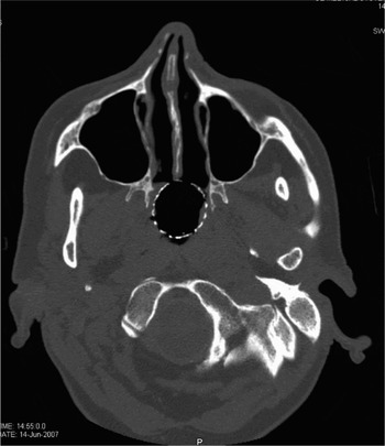
On ENT review in June 2007, examination of the patient's ears revealed retracted tympanic membranes bilaterally with fluid in the middle ears. Flexible naso-endoscopy revealed thick, mucous discharge in the nasal cavities and a wire mesh embedded in the postnasal space. Computed tomography scanning of the sinuses illustrated the exact location of the wire mesh (Figure 3).
Axial computed tomography scan showing stent fragment in the postnasal space.

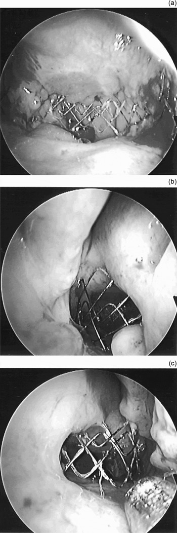
In August 2007, the patient was taken to theatre. Bilateral myringotomy produced fluid. Grommets were inserted on both sides, and the wire mesh was removed piecemeal from the postnasal space (Figure 4). The patient was discharged the following day.
Intra-operative photographs showing stent fragments found in the postnasal space. (a) Postnasal space via left nostril; (b) naso-endoscopy right nostril; (c) naso-endoscopy left nostril.

On one-month post-operative out-patients review, there was marked improvement in the patient's sinus symptoms and an improvement in her hearing.
Discussion
To the best of our knowledge, and after a thorough literature search, the upper airway complication presented has not previously been documented in the literature. The Hanaro stent used in this case for symptom relief of a benign oesophageal stricture produced marked initial improvement in symptoms. However, four months after insertion the stent fractured, further complicated by fragment migration with one fragment lodging proximally in the nasopharynx. Local reaction to this foreign body caused the patient's symptoms and signs of persistent nausea and vomiting, sore throat, increased oral and nasal secretions, and chronic sinusitis. Eustachian tube dysfunction gave rise to bilateral otitis media with effusion, causing hearing loss in both ears.
Other serious complications of metallic stents have been documented in the literature.Reference Lee5, Reference Cwikeil, Tanberg, Cwikeil and Lillo-Gil6 Early complications include chest pain after stent insertion, usually resolving after the first week. However, approximately 10–20 per cent of patients have a major complication such as bleeding, perforation, aspiration, fever or fistula.Reference Cwikeil, Tanberg, Cwikeil and Lillo-Gil6–Reference Acunas, Rozanes, Akpinar, Tunaci, Tunaci and Acunas8 Stent migration is common when stents are placed at the cardia, and more so when covered stents are used.Reference Cwikeil, Tanberg, Cwikeil and Lillo-Gil6, Reference Song, Do, Han, Sung, Choi and Sohn7 Late complications include haemorrhage, oesophageal ulceration, perforation or fistula, stent torsion, stent migration, and stent fracture. Lower airway complications, such as airway compression and tracheoesophageal fistula, are commoner with stents placed in the proximal third of the oesophagus.Reference Wang, Sze, Wang, Wang, Gao and Dake9, Reference Kawasaki, Sano and Matsumoto10
• This paper describes a rare upper airway complication of an oesophageal stent failure
• A portion of fragmented oesophageal stent was located in the nasopharynx, where it had caused sinusitis and otitis media with effusion
• This case highlights the importance of examining the upper airways in such cases, and also the need for further, long term studies of complications of metallic, expandable stents
Metallic anti-reflux stents have been proven to significantly reduce acid oesophageal exposure and dysphagia symptoms, when compared with open stents.Reference Shim, Jung and Cheon4 However, Schoppmeyer et al. argue against their use for tumours of the gastroesophageal junction, due to the high rates of migration seen in their study of Gianturco-Z anti-reflux stents (MI Tech Co. Ltd., Kyunggi-do, Korea).Reference Schoppmeyer, Golsong, Schiefke, Mössner and Caca11 As there is a lack of data regarding long term complication rates and efficacy, we may in future see more upper airway complications of fractured or migrated oesophageal stents. We therefore put forward a case for routine inspection of the upper airways following stent fracture (especially if some fragments cannot be traced) when there are localising symptoms such as sore throat, persistent nausea and vomiting, nasal discharge, and sinusitis.