Introduction
Being born small for gestational age significantly increases the risk of developing insulin resistance and type 2 diabetes later in life.Reference Barker, Winter, Osmond, Margetts and Simmonds 1 – Reference Hales, Barker and Clark 4 Skeletal muscle insulin resistance has been associated with impaired skeletal muscle mitochondrial biogenesis (synthesis of new mitochondria) and metabolism.Reference Kelley, He, Menshikova and Ritov 5 – Reference Patti 7 Mitochondria are the primary controllers of cellular energy metabolism and impairments in skeletal muscle mitochondrial content and function have been shown in adult rats born small.Reference Wadley, Siebel and Cooney 8 , Reference Selak, Storey, Peterside and Simmons 9 Our laboratory has shown that foetal growth restriction reduces gene and protein markers of skeletal muscle mitochondrial biogenesis [e.g. peroxisome proliferator-activated receptor (PPAR)-γ coactivator-1α (PGC-1α), mitochondrial transcription factor A (Tfam), cytochrome c oxidase subunits 3 and 4 (COX III and IV)] in 6-month-old adult rats, with males more affected than females.Reference Wadley, Siebel and Cooney 8 This potential impairment in skeletal muscle metabolism may provide an important mechanistic link to skeletal muscle insulin resistanceReference Kelley, He, Menshikova and Ritov 5 – Reference Patti 7 associated with being born small.
The developmental time course for the reduction in markers of skeletal muscle mitochondrial biogenesis following uteroplacental insufficiency is unclear. One study reported that the master regulator of mitochondrial biogenesis, PGC-1α, mRNA was downregulated in the slow twitch soleus muscle but upregulated in the fast twitch extensor digitorum longus muscle of small birth weight offspring at 21 days of age, with males more affected than females.Reference Lane, Maclennan and Daood 10 It is important to consider that the control group used in these studies had their litter size reduced at birth,Reference Lane, Maclennan and Daood 10 , Reference Lane, Chandorkar, Flozak and Simmons 11 previously shown to alter postnatal growth and impair skeletal muscle markers of mitochondrial biogenesis at 6 months of ageReference Wadley, Siebel and Cooney 8 and therefore impacts on the interpretation of these findings. Consequently, the impact of foetal growth restriction on markers of skeletal muscle mitochondrial biogenesis and the developmental time course in early life remains unclear.
Low birth weight in humans has also been associated with reduced muscle mass and strength that persists from childhoodReference Hediger, Overpeck and Kuczmarski 12 – Reference Sayer, Syddall and Dennison 15 through to adult life.Reference Sayer, Syddall and Dennison 16 – Reference Yliharsila, Kajantie and Osmond 18 Evidence suggests that the structural and functional development of muscles and muscle fibres is altered in babies born smallReference Yliharsila, Kajantie and Osmond 18 – Reference Alves, Damasco and Pai 23 and this is likely to play an important role in the programming of adult disease. Animal studies show that foetal undernutrition is associated with reduced postnatal muscle weight and myofibre density,Reference Ward and Stickland 20 , Reference Dwyer, Madgwick, Ward and Stickland 21 , Reference Costello, Rowlerson and Astaman 24 along with impaired morphological and contractile characteristicsReference Wilson, Ross and Harris 22 , Reference Alves, Damasco and Pai 23 that may persist into adulthood.Reference Prakash, Fournier and Sieck 25 In the rat, skeletal muscle develops during mid-to-late gestation and continues to grow some months postnatally.Reference Harris, Duxson, Fitzsimons and Rieger 26 – Reference Zhang and McLennan 28 During gestation, skeletal muscle development coincides with the expression of myogenic regulatory factors (MRFs), which are temporally expressed throughout perinatal development and control an array of regulatory and structural genes.Reference Braun and Arnold 29 – Reference Kassar-Duchossoy, Gayraud-Morel and Gomes 32 Myogenic differentiation 1 (MyoD), myogenin and MRF4 are involved in skeletal muscle proliferation and differentiation with expression detected from mid-to-late gestation.Reference Thompson, Condon and Astrow 27 , Reference Cossu, Kelly and Tajbakhsh 33 – Reference Rawls, Morris and Rudnicki 35 In the rat, myogenesis continues for up to 2 weeks postnatally after which skeletal muscle hypertrophy persists. Insulin-like growth factor I (IGF-I) plays a well-defined role in skeletal muscle hypertrophy,Reference Baker, Liu, Robertson and Efstratiadis 36 – Reference Powell-Braxton, Hollingshead and Warburton 38 but evidence also implicates IGF-I in skeletal muscle proliferation and differentiation.Reference Coolican, Samuel, Ewton, McWade and Florini 39 Owing to the temporal pattern of MRF expression, the timing of an insult may be an important determinant of the muscle's phenotypic profile. An insult, during gestation or after birth, may alter the expression of these myogenic regulatory and growth factors and may impact on later skeletal muscle mass and function.
Our laboratory has shown that uteroplacental insufficiency in pregnant rats not only impairs the growth of the foetus but also impairs maternal mammary development resulting in poor lactation,Reference Wlodek, Lorenc and O'Dowd 40 further compromising the growth of the offspring after birth.Reference O'Dowd, Kent, Moseley and Wlodek 41 In low birth weight humans, improved growth between birth and 2 years has been shown to reduce the risk of developing adult metabolic disease.Reference Hales, Barker and Clark 4 , Reference Eriksson 42 – Reference Forsén, Eriksson and Tuomilehto 44 Indeed, Siebel et al. have shown that improved postnatal nutrition by cross-fostering growth-restricted pups onto a control mother with normal lactation, improved early postnatal growth and ameliorated the impaired glucose toleranceReference Siebel, Mibus and De Blasio 45 and pancreatic β-cell massReference Siebel, Gallo, Guan, Owens and Wlodek 46 observed at 6 months of age. Furthermore, at 7 days of age, restricted pups fostered onto a control mother showed upregulation of key pancreatic genes important for β-cell growth and maintenance.Reference Siebel, Gallo, Guan, Owens and Wlodek 46 These results highlight the potential for early life interventions that may lessen the adverse consequences of being born small.
Therefore, Study 1 determined the impact of uteroplacental insufficiency on gene expression of skeletal muscle markers of mitochondrial biogenesis and myogenic regulatory and growth factors in late gestation and during postnatal life. Study 2 determined whether improved lactation after birth by cross-fostering could ameliorate any adverse consequences of uteroplacental insufficiency on skeletal muscle gene expression in early life. Finally, due to multiple and significant gender differences in metabolic disease outcomes in response to impaired foetal growth reported previously by ourselvesReference Wadley, Siebel and Cooney 8 , Reference Siebel, Mibus and De Blasio 45 and others,Reference Owens, Thavaneswaran and De Blasio 47 – Reference Owens, Gatford and De Blasio 49 we determined the impact of uteroplacental insufficiency and cross-fostering in males and females separately. On the basis of our and others findings in adulthood, we hypothesized that key genes involved in skeletal muscle development and mitochondrial biogenesis would be altered following intrauterine growth restriction in rats, with different developmental profiles between males and females.
Methods
Animals
All procedures were approved by The University of Melbourne Animal Experimentation Sub-Committee. In both studies, Wistar Kyoto rats (aged 11 weeks) were obtained from the Australian Resource Centre (Murdoch, WA, Australia) and house with a 12-h light dark cycle with access to water and normal chow ad libitum. On day 18 of gestation (term 22 days), pregnant rats were anesthetized by intraperitoneal injection of ketamine (225 mg/kg) and Ilium Xylazil-20 (30 mg/kg). Bilateral uterine vessel ligation of both the artery and vein supplying each uterine horn was performed to induce late gestation uteroplacental insufficiency (Restricted) or sham surgery was performed to generate the Control group.Reference Wlodek, Westcott and O'Dowd 50 , Reference Wlodek, Mibus and Tan 51
Study 1: developmental timeline study
At day 20 of gestation (E20), mothers that underwent either bilateral uterine vessel ligation or sham surgery were anesthetized by intraperitoneal injection of ketamine (225 mg/kg) and Ilium Xylazil-20 (30 mg/kg) and the uterus exposed. In Restricted litters at E20, the ligation integrity was confirmed. Foetuses at E20 or pups at postnatal day 1 (PN1) and 7 (PN7) were weighed, killed by decapitation and hindlimb skeletal muscle rapidly excised, pooled within litters and separated by sex (n = 8–10 litters/group). Skeletal muscle was not weighed at ages E20, PN1 or PN7 due to potential inaccuracies in dissection of the muscle in the small animals. Because of the increased body mass at postnatal day 35 (PN35) individual male and female offspring were anesthetized by intraperitoneal injection of ketamine (225 mg/kg) and Ilium Xylazil-20 (30 mg/kg) and the gastrocnemius muscle rapidly excised and weighed (n = 8–10 offspring/group). The gastrocnemius muscle was examined at PN35 as it is the largest muscle of the lower hindlimb and is also a mixed fibre muscle making it an appropriate hindlimb muscle for comparing to mixed hindlimb muscle collected at earlier ages. All samples collected were immediately frozen in liquid nitrogen and stored at −80°C.
Study 2: cross-fostering study
In a separate cohort of offspring exposed to uteroplacental insufficiency or sham surgery, Restricted or Control pups were cross-fostered 1 day after birth onto either a mother who had undergone bilateral uterine vessel ligation or a sham-operated Control mother to yield four treatment groups. Control pups fostered onto a Control mother (Cont-on-Cont), Control pups on Restricted mother (Cont-on-Rest), Restricted pups on Control mother (Rest-on-Cont) and Restricted pups on Restricted mother (Rest-on-Rest).Reference Siebel, Gallo, Guan, Owens and Wlodek 46 To determine the early effects of foetal growth restriction and cross-fostering before the majority of proliferation and differentiation was complete, hindlimb skeletal muscle was collected at PN7 as described above (n = 7–10 litters/group).
Analysis for studies 1 and 2: real-time polymerase chain reaction (PCR) analysis
For the Developmental Timeline Study, total RNA was extracted from skeletal muscle using the Tri-Reagent (Ambion Inc., Austin, TX, USA) method due to the small amounts of pooled tissue obtained from pups at E20 and PN1. RNA was extracted from skeletal muscle of the Cross-foster Study using the Invitrogen micro–to-midi RNA extraction kits (Invitrogen, Carlsbad, CA, USA). RNA yield was measured using the NanoDrop™ 1000 Spectrophotometer (NanoDrop Technologies, Wilmington, DE, USA) at 260 nm absorbance. Integrity was confirmed from the 260/280 nm ratio and random samples were also selected for gel electrophoresis with high levels of 18S and 28S detected.
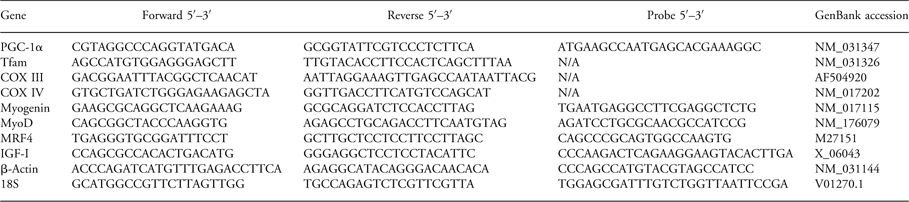
First-strand cDNA was synthesized from 1 μg of RNA using superscript III first-strand system (Invitrogen). Primer and Taqman® probe sequences for PGC-1α, Tfam, COX III, COX IV, myogenin, MyoD, MRF4, IGF-I, β-actin and 18S are presented in Table 1. Depending on probe availability, real-time PCR using either SYBR greenReference Wadley, Siebel and Cooney 8 or TaqMan® Reference Vodstrcil, Wlodek and Parry 52 chemistry was performed, as described previously, using the sequence detector software (Rotor-gene v6, Corbett Research, Sydney, NSW, Australia). Samples from each age within an experimental group were amplified on the same run to eliminate inter-assay variability, whereas a positive control sample was used as a reference to make inter-assay comparisons within an age group. In cases when SYBR green chemistry (Tfam, COX III and COX IV) was utilized, samples were subjected to a heat dissociation protocol following the final amplification cycle to ensure that only a single product was detected. Relative gene expression was quantified using the comparative C t (ΔΔC t) method.Reference Wadley, Siebel and Cooney 8 , Reference Siebel, Gallo, Guan, Owens and Wlodek 46 18S was used as the endogenous control for the Developmental Timeline Study (Study 1) as it was stably expressed across age groups, whereas β-actin was used for the Cross-foster Study (Study 2) due to stable expression between treatment groups at that age.
Primer and probe sequences used for genes quantified using real-time PCR relative to 18S or β-actin and GenBank accession numbers

PGC-1α, peroxisome proliferator-activated receptor (PPAR)-γ coactivator-1α; Tfam, mitochondrial transcription factor A; COX III and IV, cytochrome c oxidase subunits 3 and 4; MyoD, myogenic differentiation 1; MRF4, myogenic regulatory factor 4; IGF-1, insulin-like growth factor 1.
Studies 1 and 2: statistical analysis
At each age, body weight and dimensions were analysed by a two-way ANOVA for sex and treatment. For the Developmental Timeline Study, gene expression data were analysed separately for sex using a two-way ANOVA for age and treatment. If a statistically significant interaction was observed, data were then split and analysed by one-way ANOVA with Duncan's post hoc test where appropriate. For the Cross-foster Study, gene expression data were analysed separately for sex using a one-way ANOVA for treatment. All data are presented as the mean ± s.e.m. The level of significance was set at P < 0.05.
Results
Study 1: developmental timeline study
Body weight, dimensions and litter size
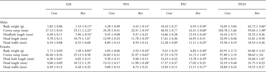
Litter size was reduced by uteroplacental insufficiency during gestation at E20 (8.1 ± 0.7 v. 10.5 ± 0.9; P = 0.05) and after birth at PN1 (5.2 ± 0.6 v. 8.2 ± 0.5; P < 0.05). Restricted offspring were lighter (P < 0.05) with shorter crown-rump length (P < 0.05) than Control at all ages in males and at E20, PN1 and PN7 in females (Table 2). Hindlimb length was shorter in Restricted males at E20 and in both sexes at PN7 (Table 2). Head length was shorter in Restricted at PN1 and head width was shorted in Restricted at PN7 compared with Controls in both sexes (Table 2). Foetal growth restriction reduced PN35 absolute gastrocnemius weight in males (0.26 ± 0.01 g v. 0.20 ± 0.01 g; P < 0.05), but not in females (0.20 ± 0.02 g v. 0.21 ± 0.02 g). Relative (to body weight) gastrocnemius weight was unaffected by foetal growth restriction in both males (0.34 ± 0.02 g v. 0.32 ± 0.01 g) and females (0.31 ± 0.02 g v. 0.32 ± 0.03 g).
Effect of uteroplacental insufficiency on body weight and dimensions in males and females at E20, PN1, PN7 and PN35

E20, day 20 of gestation; PN1, postnatal day 1; PN7; postnatal day 7; PN35, postnatal day 35; Cont, Control; Rest, Restricted.
*Denotes significantly (P < 0.05) different from Cont within that age.
Body weight, crown-rump length, hindlimb length, head length and head width measured for male and female offspring. Data are expressed as mean ± s.e.m. (n = 8–10/group).
Skeletal muscle gene expression
Skeletal muscle mitochondrial biogenesis markers
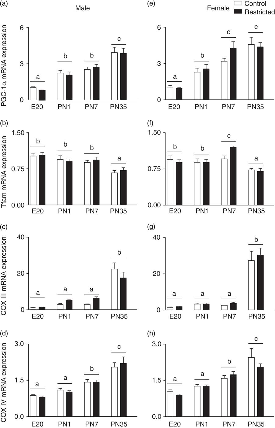
Uteroplacental insufficiency had no impact on skeletal muscle gene expression of mitochondrial biogenesis markers regardless of sex (Fig. 1). In both males and females, PGC-1α mRNA was lowest at E20 (P < 0.05) and showed increasing (P < 0.05) expression such that by PN35 was more than four-fold higher (P < 0.05) than E20 (Fig. 1a and 1e). In females, Tfam mRNA was similar between E20 and PN1 and peaked (P < 0.05) at PN7 (Fig. 1f). In males, Tfam mRNA was similar at E20, PN1 and PN7 and decreased (P < 0.05) only at PN35 (Fig. 1b). COX III mRNA levels were similar at E20, PN1 and PN7 and increased (P < 0.05) substantially by PN35 in both males and females (Fig. 1c and 1g). Finally, COX IV mRNA in males and females was similar at E20 and PN1 and progressively increased such that it was highest (P < 0.05) at PN35 (Fig. 1d and 1h).
Effect of uteroplacental insufficiency on skeletal muscle markers of mitochondrial biogenesis. Hindlimb skeletal muscle peroxisome proliferator-activated receptor (PPAR)-γ coactivator-1α (PGC-1α), mitochondrial transcription factor A (Tfam), cytochrome c oxidase subunits 3 and 4 (COX III and IV), mRNA relative to 18S in male (a–d, respectively) and female (e–h, respectively) offspring at day 20 of gestation (E20), postnatal days 1 (PN1), 7 (PN7) and 35 (PN35). Values are mean ± s.e.m. (n = 8–10/group). Different lowercase letters above the bars indicate significant differences (P < 0.05) between ages (‘a’ is different from ‘b’).

Myogenic regulatory and growth factors
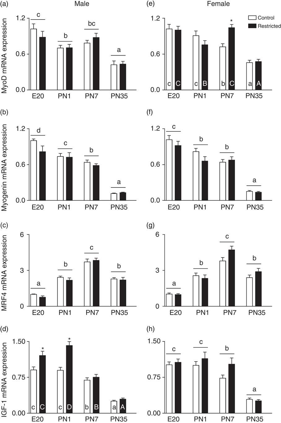
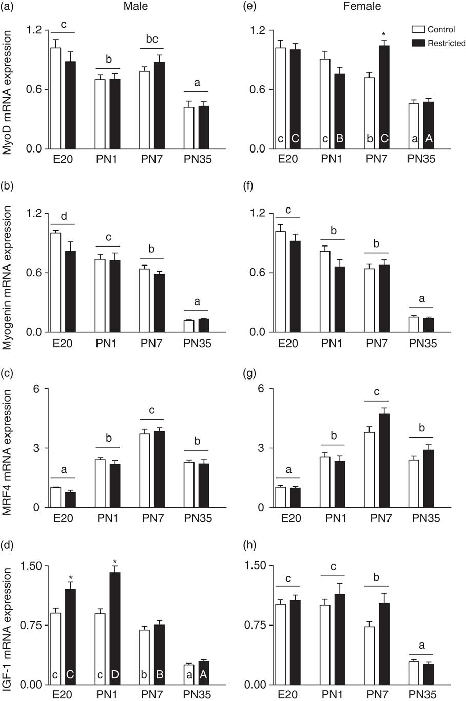
In both males and females, MyoD and myogenin mRNA progressively decreased such that it was highest at E20 (P < 0.05) and lowest (P < 0.05) at PN35 (Fig. 2a, 2b, 2e and 2f, respectively). Uteroplacental insufficiency had no impact on MyoD and myogenin mRNA in either sex (Fig. 2a, 2e, 2b and 2f, respectively) except in females at PN7, MyoD mRNA was ∼40% higher (Fig. 2e; P < 0.05). MRF4 mRNA was lowest (P < 0.05) at E20 and progressively increased (P < 0.05) to peaked (P < 0.05) at PN7 in both sexes (Fig. 2c and 2g). In males and females, IGF-I mRNA was highest (P < 0.05) at E20 and PN1 and progressively decreased such that expression was lowest (P < 0.05) at PN35 (Fig. 2d and 2h). Uteroplacental insufficiency increased IGF-I mRNA by approximately 50% (P < 0.05) at E20 and PN1 in male offspring only (Fig. 2d).
Effect of uteroplacental insufficiency on skeletal muscle myogenic regulatory and growth factors. Hindlimb skeletal muscle myogenic differentiation 1 (MyoD), myogenin, myogenic regulatory factor 4 (MRF4) and insulin-like growth factor 1 (IGF-I) mRNA relative to 18S in male (a–d, respectively) and female (e–h, respectively) offspring at day 20 of gestation (E20), postnatal days 1 (PN1), 7 (PN7) and 35 (PN35). Values are mean ± s.e.m. (n = 8–10/group). Different lowercase letters above the bars indicate significant differences (P < 0.05) between ages (‘b’ is different from ‘c’ but not different from ‘bc’). Different lowercase letters within the bars indicate significant differences (P < 0.05) between ages within the Control group (‘a’ is different from ‘b’). Different uppercase letters within the bars indicate significant differences (P < 0.05) between ages within the Restricted group (‘A’ is different from ‘B’). *Denotes significant (P < 0.05) differences between Control and Restricted offspring at that age.
Study 2: cross-foster study
Weights and dimensions
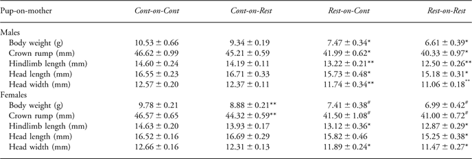
The effect of uteroplacental insufficiency and cross-fostering on body weight and dimensions in the male cohort has previously been published.Reference Siebel, Gallo, Guan, Owens and Wlodek 46 Briefly, uteroplacental insufficiency reduced litter size compared with sham-operated Controls (5.6 ± 0.5 v. 8.2 ± 0.7; P < 0.05). Both male (3.42 ± 0.05 g v. 4.12 ± 0.06 g) and female (3.42 ± 0.07 g v. 4.04 ± 0.07 g) Restricted offspring were smaller (P < 0.05) than Control at PN1 and remained smaller (P < 0.05) at PN7 regardless of cross-fostering (Table 3). At PN7, female restricted pups fostered onto a control (Rest-on-Cont) mother showed intermediate head length. Male Rest-on-Cont showed improved growth displaying increased (P < 0.05) hindlimb length and head width compared with Rest-on-Rest and intermediate head length between Cont-on-Cont and Rest-on-Rest (Table 3).
Effect of uteroplacental insufficiency and cross-fostering on body weight and dimensions in male and female rats at PN7

PN7; postnatal day 7; Cont, Control; Rest, Restricted.
*Denotes significantly (P < 0.05) different form Cont-on-Cont.
Denotes significantly (P < 0.05) different from Cont-on-Cont and Cont-on-Rest.
**Denotes significantly (P < 0.05) different form all other groups.
Body weight, crown-rump length, hindlimb length, head length and head width measured for male and female offspring. Data are expressed as mean ± s.e.m. (n = 7–10/group).
Skeletal muscle gene expression
Skeletal muscle mitochondrial biogenesis markers
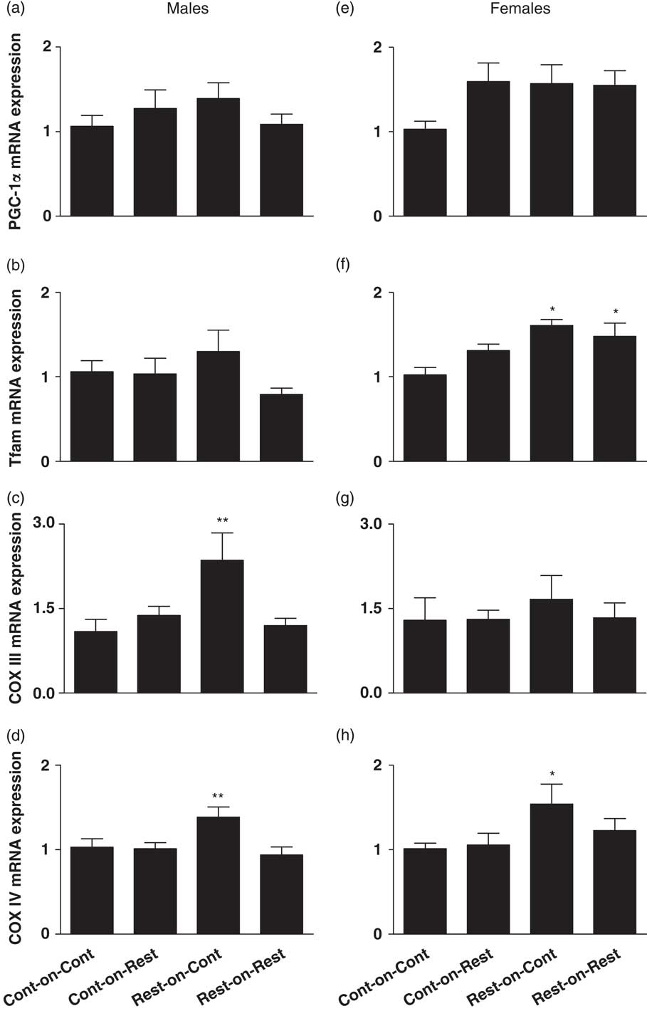
PGC-1α mRNA was similar for all groups regardless of the postnatal environment (Fig. 3a and 3d). Female Rest-on-Rest pups showed higher (P < 0.05) Tfam mRNA compared with Cont-on-Cont offspring (Fig. 3e) and there was no impact of cross-fostering. Interestingly, even though COX III and IV mRNA levels were similar between Cont-on-Cont and Rest-on-Rest offspring (Fig. 3), when restricted pups were fostered onto a control (Rest-on-Cont) mother with normal lactation, COX III increased (P < 0.05) by up to 50% in males (Fig. 3c), whereas COX IV mRNA increased (P < 0.05) by up to 50% above Cont-on-Cont levels in both sexes (Fig. 3d and 3h).
Effect of uteroplacental insufficiency and cross-fostering on skeletal muscle markers of mitochondrial biogenesis. Hindlimb skeletal muscle peroxisome proliferator-activated receptor (PPAR)-γ coactivator-1α (PGC-1α), mitochondrial transcription factor A (Tfam), cytochrome c oxidase subunits 3 and 4 (COX III and IV) mRNA relative to β-actin in male (a–d, respectively) and female (e–h, respectively) offspring at postnatal day 7. Control and Restricted offspring were fostered onto a different Control or Restricted mother to yield four experimental groups (pup-on-mother): Cont-on-Cont, Cont-on-Rest, Rest-on-Cont and Rest-on-Rest. Values are mean ± s.e.m. (n = 7–10/group). *Denotes significantly (P < 0.05) different from Cont-on-Cont. **Denotes significantly (P < 0.05) different from all other groups.

Myogenic regulatory and growth factors
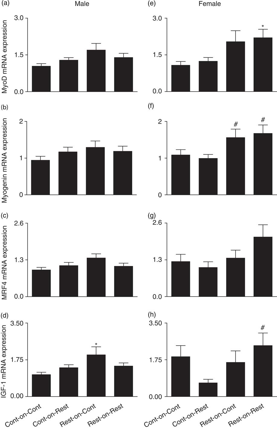
In general, there were few effects of foetal growth restriction or cross-fostering on MyoD, myogenin and MRF4 mRNA in males and females (Fig. 4a–4c and 4e–4g, respectively). In females, myogenin was higher (P < 0.05) in Rest-on-Rest and Rest-on-Cont compared with Cont-on-Rest that were subjected to a suboptimal postnatal nutritional environment (Fig. 4f). Consistent with data from Study 1, female Rest-on-Rest MyoD expression was higher (P < 0.05) compared with Cont-on-Cont offspring (Fig. 4e). There was no impact of foetal growth restriction on IGF-I (Fig. 4d and 4h); however, IGF-I was altered in a sex-specific manner upon exposure to a mismatched pre- and postnatal environment. In females, fostering control pups onto a restricted mother (Cont-on-Rest) with impaired lactation lowered (P < 0.05) IGF-I mRNA compared with Rest-on-Rest females (Fig. 4h). Whereas in males, restricted pups fostered onto a control mother (Rest-on-Cont) with normal lactation increased (P < 0.05) IGF-I mRNA compared with Cont-on-Cont males (Fig. 4d).
Effect of uteroplacental insufficiency and cross-fostering on skeletal muscle myogenic regulatory and growth factors. Hindlimb skeletal muscle myogenic differentiation 1 (MyoD), myogenin, myogenic regulatory factor 4 (MRF4) and insulin-like growth factor 1 (IGF-I) mRNA relative to β-actin in male (a–d, respectively) and female (e–h, respectively) offspring at postnatal day 7. Control and Restricted offspring were fostered onto a different Control or Restricted mother to yield four experimental groups (pup-on-mother): Cont-on-Cont, Cont-on-Rest, Rest-on-Cont and Rest-on-Rest. Values are mean ± s.e.m. (n = 7–10/group). *Denotes significantly (P < 0.05) different from Cont-on-Cont. ‘’ denotes significantly (P < 0.05) different from Cont-on-Rest.

Discussion
These studies have shown that skeletal muscle gene expression of mitochondrial biogenesis markers and myogenic regulatory and growth factors display age-dependent expression patterns throughout development. Surprisingly, there was a remarkably small effect of growth restriction and cross-fostering. However, foetal growth restriction did trigger a compensatory upregulation of specific myogenic regulatory and growth factor genes in both males and females. Improved postnatal lactation in restricted offspring (Rest-on-Cont) upregulated the mRNA expression of the mitochondrial proteins COX III and IV above levels observed in Cont-on-Cont pups. Furthermore, altered postnatal nutrition, particularly when it was mismatched with the prenatal environment, caused sex-specific alterations in IGF-I expression, which were in parallel with sex-specific changes in body weight and dimensions. These data suggest that skeletal muscle is susceptible to nutritional intervention during early postnatal life; however, the functional consequences later in life may not become apparent.
Skeletal muscle gene expression during pre- and postnatal life (Study 1)
A novel finding of this study was that gene expression of the master regulator of mitochondrial biogenesis, PGC-1α, and the mitochondrial electron transfer chain proteins, COX III and IV, increased in skeletal muscle from late gestation and throughout early postnatal life indicative of increasing mitochondrial biogenesis after birth and into the juvenile period. These data are consistent with previous studies that have shown in both rats and humans that skeletal muscle oxidative enzyme activity and mitochondrial DNA (mtDNA) content is up to 50% higher in adulthood compared with early postnatal life.Reference Colling-Saltin 53 – Reference Glatz and Veerkamp 55 Interestingly, the gene expression profile of Tfam, the transcription factor responsible for mtDNA replication, is not reflective of the PGC-1α and COX IV expression profiles. It is possible that Tfam's additional role in maintenance of mtDNA,Reference Kang, Kim and Hamasaki 56 and not just mtDNA replication, may account for its unique expression profile during skeletal muscle development.
Consistent with the literature,Reference Thompson, Condon and Astrow 27 , Reference Braun and Arnold 29 – Reference Coolican, Samuel, Ewton, McWade and Florini 39 the timing of myogenic regulatory and growth factor expression seems to reflect their proposed individual roles in skeletal muscle development. Indeed, the high levels of MyoD and myogenin during late gestation and early postnatal life suggest roles in proliferation and differentiation,Reference Hasty, Bradley and Morris 31 , Reference Nabeshima, Hanaoka and Hayasaka 57 – Reference Rudnicki, Schnegelsberg and Stead 59 whereas the expression of MRF4, which peaks around PN7, is consistent with a role in primary and secondary fibre formation.Reference Thompson, Condon and Astrow 27 , Reference Braun and Arnold 29 Finally, the higher expression of IGF-I from gestation to at least PN7 may reflect its role in early skeletal muscle development and growth,Reference Coolican, Samuel, Ewton, McWade and Florini 39 which is then downregulated by the early juvenile period when skeletal muscle differentiation is complete.
It is important to consider that although in the human the majority of skeletal muscle growth and differentiation occurs in the last 3 months of gestation, compared with the newborn rat, human skeletal muscle development is more advanced at birth. However, skeletal muscle differentiation in the human does continue after birth with up to 20% of fibres undifferentiated in the newborn, which drops to adult levels by 1 year of age.Reference Colling-Saltin 53
The impact of uteroplacental insufficiency (Study 1)
Following uteroplacental insufficiency and growth restriction, skeletal muscle markers of mitochondrial biogenesis were largely intact during late gestation and postnatal life. It was particularly surprising to find that foetal growth restriction had no impact on PGC-1α gene expression as uteroplacental insufficiency causes foetal hypoxia and in skeletal muscle the hypoxia inducible factor 1 activity and expression is tightly coupled to PGC-1α.Reference O'Hagan, Cocchiglia and Zhdanov 60 It therefore appears that the deficit in skeletal muscle markers of mitochondrial biogenesis previously reported in small birth weight males at 6 months of ageReference Wadley, Siebel and Cooney 8 likely develop after weaning, during juvenile or early adult life. Consequently, the impact of foetal growth restriction on skeletal muscle mitochondrial biogenesis and its time course in the perinatal period remains unclear.
Specific myogenic regulatory and growth factors showed a compensatory upregulation following uteroplacental insufficiency in an age- and sex-specific manner. Female Restricted offspring showed a significant increase in MyoD gene expression at PN7. MyoD's proposed role in skeletal muscle specification and proliferation suggest that the elevated mRNA levels observed in Restricted females at PN7 may represent a delayed myogenic process. Indeed, previous studies found that offspring of malnourished mothers had delayed differentiation of muscle fibres at day 7 and to a lesser extent at day 14 after birth;Reference Alves, Damasco and Pai 23 however, this was only investigated in male offspring. The current study reports that male Restricted offspring showed an upregulation of IGF-I gene expression at E20 and PN1. Other studies have found that following foetal growth restriction in sheep, skeletal muscle IGF-I levels were either unchangedReference Brameld, Mostyn and Dandrea 61 or reduced.Reference Kind, Owens and Robinson 62 Importantly, these studies imposed foetal growth restriction on pregnant ewes for 60Reference Brameld, Mostyn and Dandrea 61 and 121Reference Kind, Owens and Robinson 62 days of gestation, whereas we report changes in IGF-I as little as 2 days following the induction of growth restriction. Indeed, more recently Costello et al.Reference Costello, Rowlerson and Astaman 24 reported that following late gestation undernutrition in sheep, reduced myofibre density was associated with a compensatory upregulation of the IGF-I receptor at 127 days gestation (term 147 days). The upregulation of IGF-I mRNA reported in the current study may be a mechanism to drive skeletal muscle growth and hypertrophy to compensate for potential reductions in fibre number as a consequence of the suboptimal uterine environment.
The effects of cross-fostering and altered postnatal lactation (Study 2)
Cross-fostering growth-restricted pups onto a mother with normal lactation and consequently improved postnatal nutrition has been shown to increase milk intakeReference O'Dowd, Siebel and Wlodek 63 and, in the current study, was associated with improved postnatal growth in males only, indicated by improved head width and hindlimb length.Reference Siebel, Gallo, Guan, Owens and Wlodek 46 It is possible that males are more responsive to intervention due to their apparently more severe growth restriction, observed at all ages investigated, and increased adult disease susceptibility.Reference Wadley, Siebel and Cooney 8 , Reference Siebel, Mibus and De Blasio 45 , Reference Owens, Thavaneswaran and De Blasio 47 – Reference Owens, Gatford and De Blasio 49 , Reference Siebel, Westcott and Mibus 64
Despite foetal growth restriction (Rest-on-Rest) having little impact on markers of mitochondrial biogenesis, cross-fostering growth-restricted pups onto a mother with normal lactation (Rest-on-Cont) was associated with an upregulation of skeletal muscle COX III mRNA in males only and COX IV mRNA in males and females above that of Cont-on-Cont levels. COX III is a mitochondrial-encoded protein, whereas COX IV is a nuclear-encoded protein, both involved in the electron transfer chain on the inner mitochondrial membrane and, therefore, the upregulation of COX III and IV mRNA may contribute to increased electron transfer chain components to improve mitochondrial respiration. This upregulation may be a compensatory response following uteroplacental insufficiency in the face of improved substrate availability due to improved postnatal nutrition. How this will impact on the adult skeletal muscle metabolic profile is unknown, but as cross-fostering has been associated with improved glucose tolerance in adulthood of small birth weight rats,Reference Siebel, Mibus and De Blasio 45 the gene changes in skeletal muscle may also contribute to improved adult health.
Finally, female control offspring fostered onto a restricted mother (Cont-on-Rest) with impaired lactation displayed significantly reduced skeletal muscle IGF-I mRNA compared with Rest-on-Rest offspring. This observation was in parallel with slowed growth in Cont-on-Rest females displaying lower body weight and crown-rump length compared with Cont-on-Cont. In contrast, male restricted offspring fostered onto a control mother (Rest-on-Cont) and therefore subjected to improved nutrition after birth, displayed significantly elevated IGF-I mRNA levels compared with Cont-on-Cont offspring, along with improved growth indicated by larger hindlimb length and head width compared with Rest-on-Rest males. Although foetal growth restriction alone had no impact in IGF-I mRNA, it appears that expression is altered in both males and females when the pre- and postnatal nutritional environments were mismatched and was associated with specific changes in body weight and dimensions. Furthermore, it is possible that the IGF-I gene may be subject to a predictive adaptive response to nutritional status during gestation, which is altered in the face of mismatched postnatal nutrition. Along with the COX III and IV data, we suggest that skeletal muscle may be susceptible to specific interventions in early life. Whether these relatively minor gene changes following cross-fostering will contribute to later disease prevention, observed at 6 months of age,Reference Siebel, Mibus and De Blasio 45 , Reference Siebel, Gallo, Guan, Owens and Wlodek 46 , Reference Wlodek, Mibus and Tan 51 is unclear; but perhaps an intervention that occurs at an age when deficits in skeletal muscle mitochondrial biogenesis markers are present would provide greater benefits to improved skeletal muscle mitochondrial biogenesis and insulin sensitivity.
These unique studies have shown novel developmental patterns of key genes involved in skeletal muscle mitochondrial biogenesis and development in early life. It is important to acknowledge that mRNA data are limited in its interpretation without accompanying protein, histological or functional analysis. Because of limitations in the amount of muscle tissue available, this analysis was not possible in the current study. Future studies should attempt to confirm the current gene data with protein analysis as well as histological analysis to determine whether the increased MyoD and IGF-I following uteroplacental insufficiency in our rat model is associated with impaired muscle fibre development, distribution and number.
Summary
The current studies have shown that skeletal muscle markers of mitochondrial biogenesis remain intact in early life following foetal growth restriction despite our previous data showing marked impairments at 6 months of age in males.Reference Wadley, Siebel and Cooney 8 It is therefore likely that the deficits in mitochondrial biogenesis develop later in life. Indeed, the upregulation of MyoD and IGF-I following foetal growth restriction may be indicative of delayed myogenesis in a sex-specific manner. Improved postnatal nutrition by cross-fostering improved growth in males early in life and was associated with upregulation of the electron transfer chain proteins, COX III and IV in small birth weight males and females. Whether these subtle changes will have consequences for later disease outcomes in adulthood are unknown.
Acknowledgements
The authors thank Andrew Jefferies and Kerryn Westcott for assistance with animal surgery and handling and Dr Andrew Siebel for assistance with cross-fostering and real-time PCR. This research was supported by a grant from the Diabetes Australia Research Trust and the National Health and Medical Research Council (no. 454570). RCL was supported by Sheppard M Lowe Scholarship (Faculty of Medicine, Dentistry and Health Science, The University of Melbourne, 2007) and co-funded NHMRC/NHF Biomedical Postgraduate Scholarship (2008–2010).