INTRODUCTION
Trichinella spiralis is an intracellular parasitic nematode of mammalian striated muscles. It is responsible for trichinellosis, a zoonosis caused by the consumption of raw or undercooked meat from infected animals (e.g. pork, game animals). Human outbreaks have been regularly reported during the last century (Ancelle et al. Reference Ancelle, De Bruyne, Poisson and Dupouy-Camet2005; Khumjui et al. Reference Khumjui, Choomkasien, Dekumyoy, Kusolsuk, Kongkaew, Chalamaat and Jones2008) and it has been estimated that more than 11 million people could be infected worldwide (Dupouy-Camet, Reference Dupouy-Camet2000). Trichinellosis is regarded as an emerging or re-emerging disease in some parts of the world (Eastern Europe, Asia, etc.). Trichinella infections are mainly due to dietary or culinary habits, pork being the major source of contamination for humans (Devine, Reference Devine2003; Blaga et al. Reference Blaga, Durand, Antoniu, Gherman, Cretu, Cozma and Boireau2007).
The T. spiralis life cycle is completed within a single host and begins within the stomach with the release of the infective larval stage of the parasite (L1) from the muscle fibres of contaminated meat. The parasites then move to the small intestine, undergo 4 moults (L1 to L4), and develop into their adult form within 30–36 h of ingestion (Kozek, Reference Kozek1971a,Reference Kozekb). At day 4, female worms start shedding newborn larvae (Liu et al. Reference Liu, Zhu, Xu, Lu and Boireau2001) which migrate to muscular tissue via the vascular systems. There, larvae can persist for years and remain infective (Wakelin and Goyal, Reference Wakelin and Goyal1996).
The host's immune response strongly influences the duration of the parasites’ intestinal phase and the degree of establishment in the muscles, thereby determining the final outcome of infection. It has been demonstrated that T. spiralis stimulates the host's immune system (Bell, Reference Bell1998) and expresses many immunodominant antigens. In addition, several studies have shown that Trichinella antigens are stage-specific and can induce a specific host immune response (James and Denham, Reference James and Denham1975; James et al. Reference James, Moloney and Denham1977; Denkers et al. Reference Denkers, Wassom, Krco and Hayes1990; Boireau et al. Reference Boireau, Vayssier, Fabien, Perret, Calamel and Soule1997, Reference Boireau, Mingyuan, Baoquan, Le Guerhier, Le Rhun, Hernandez-Bello, Bahuon and Vallee2006; Liu et al. Reference Liu, Wang, Fu, Li, Wu, Le Rhun, Chen and Boireau2007). This is in agreement with qualitative and quantitative changes of proteins observed on the cuticle surface (Philipp et al. Reference Philipp, Parkhouse and Ogilvie1980, Reference Philipp, Taylor, Parkhouse and Ogilvie1981; Boireau et al. Reference Boireau, Vayssier, Fabien, Perret, Calamel and Soule1997) and in excretion-secretion (ES) products (Takahashi, Reference Takahashi1997). Muscle larvae (ML), adult worms (Ad) and newborn larvae (NBL) are considered to be the 3 major Trichinella antigenic stages (Boireau et al. Reference Boireau, Vayssier, Fabien, Perret, Calamel and Soule1997; Wu et al. Reference Wu, Fu, Wang, Yu, Yu, Deng, Liu, Boireau, Wang and Liu2009). Therefore, only a small number of studies have focused on the antigenicity of Trichinella intestinal larval stages (Bell et al. Reference Bell, McGregor and Despommier1979). Indeed, it has been demonstrated that pre-adults (intestinal larval stages) and adults are specifically expelled in less than 24 h from rats immunized against Trichinella (Bell and McGregor, Reference Bell and McGregor1979). These stages might then correspond to targets of the host immune system and could express specific antigenic products.
The aim of the present study was to identify very early antigens from Trichinella intestinal stages. Thus, 3 original cDNA libraries of pre-adult and young adult T. spiralis worms were constructed and immuno-screened with serum from pigs experimentally infected with T. spiralis. This paper presents for the first time the different antigenic proteins identified at the very early invasive stages of Trichinella.
MATERIALS AND METHODS
Parasites
Trichinella spiralis (ISS 004) was maintained by serial passage in OF1 female mice (Charles River Laboratories, l'Arbresle, France). Muscle larvae (ML) were recovered from infected mice at 35 days p.i. by artificial digestion according to a standard protocol (Gamble et al. Reference Gamble, Bessonov, Cuperlovic, Gajadhar, Van Knapen, Nockler, Schenone and Zhu2000). After recovery, the worms were washed 3 times with water and then used to inoculate 90 OF1 female mice, aged 8–10 weeks, orally, with 1500 ML per mouse in a volume of 20 μl.
Three groups of 30 mice were euthanized at 14 h, 20 h and 48 h p.i. The small intestine was collected, cut along its entire length, and washed in pre-warmed PBS (phosphate-buffered saline) supplemented with antibiotics (200 UI/ml penicillin and 200 μg/ml streptomycin). Then, the small intestine was cut into pieces and incubated in PBS at 37°C for 1 h 30 min on a 300 μm sieve. The released parasites were separated from intestinal debris by filtration through a 200 μm sieve and differential sedimentation for 30 min. After several washes in PBS, the worms were centrifuged at 600 g for 10 min and stored at −80°C until use.
Preparation of antisera
Three 10-week-old SPF pigs (German Yorkshire) were experimentally infected per os with 20 000 T. spiralis ML. Blood samples were taken twice: prior to infection (negative control) and at 30 days p.i. Serum was extracted by centrifugation at 2000 g for 30 min and diluted at 1:100 prior to examination by use of an anti-Trichinella-IgG ELISA based on ES antigens from T. spiralis ML, according to the protocol of Nockler et al. (Reference Nockler, Voigt, Protz, Miko and Ziedler1995).
RNA isolation and preparation of T. spiralis cDNA libraries
Total RNA (Tot.RNA) was extracted from T. spiralis worms using an acid guanidinium thiocyanate-phenol-chloroform mixture method (Chomczynski and Sacchi, Reference Chomczynski and Sacchi1987, Reference Chomczynski and Sacchi2006). Then, mRNA was purified using the Dynabeads mRNA Purification Kit (Dynal, Compiègne, France) and RNA quality was monitored by formaldehyde gel electrophoresis (Sambrook and Russell, Reference Sambrook and Russell2001). The cDNA libraries were prepared using the cDNA Synthesis Kit and the ZAP-cDNA Gigapack III Gold Cloning Kit (Stratagene, Amsterdam Zuidoost, Holland). Briefly, mRNA was primed in the first-strand synthesis with a hybrid oligo(dT) linker–primer and reverse-transcribed using AccuScript reverse transcriptase (Stratagene). After second-strand synthesis, cDNA was size-fractionated with Chromaspin-400 columns (Ozyme, St Quentin-en-Yvelines, France) and unidirectionally ligated into a λ ZAP II vector (Stratagene). The titre of the packaged lambda library was determined and the percentage of non-recombinant clones evaluated by blue-white colour selection. The cDNA library was amplified in the Escherichia coli XL1-Blue MRF´ strain and amplified libraries were stored at −80°C until use.
Immuno-screening of cDNA libraries
First, pseudo-screenings were performed in order to remove cross-reactive antibodies from pre-immune and immune pig serum according to the method described by Sambrook and Russell (Reference Sambrook and Russell2001). Then 100 000 plaques per cDNA library were immuno-screened according to conventional methods (Sambrook and Russell, Reference Sambrook and Russell2001) with serum from pigs experimentally infected with 20 000 T. spiralis ML and collected at 30 days p.i. Positive clones, appearing as spots on membranes, were picked and kept for future analysis. Isolation of selected positive clones was tested by PCR-amplification of the cloned cDNA fragment with T3 and T7 primers (T3 primer 5′-ATTAACCCTCACTAAAG-3′; T7 primer 5′-ATTACGACTCACTATAG-3′). Then, pBluescript SK(-) plasmids containing cDNA insert were excised from the lambda ZAP phage vector and propagated in an E. coli SOLR strain before being stored at −80°C until use.
Immuno-screenings included parallel runs with the corresponding pre-immune serum, in order to exclude non-specific antibody binding.
Sequence analysis
T3/T7 amplified PCR products were sequenced (Qiagen, Hilden, Germany). Multiple alignments were performed with Multalin software (Corpet, Reference Corpet1988). After removal of flanking vector regions, sequences were analysed using the BLAST tool for homology searches. SignalP 3.0 and SMART were used for signal peptide and protein family domain predictions. Expasy (http://www.expasy.org/) was used for the analysis of protein sequences and structures.
RESULTS
Preparation of T. spiralis cDNA libraries and validation
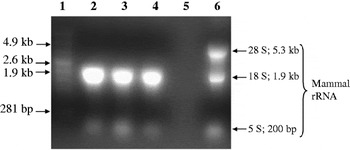
From the 45 000 T. spiralis ML given to each group of 30 mice, between 30% and 80% of parasites were recovered at 14 h, 20 h and 48 h p.i. Trichinella's characteristic profile (Zarlenga and Dame, Reference Zarlenga and Dame1992) was observed for the 3 periods of infection (Fig. 1, Lanes 2, 3 and 4) and compared to the Tot.RNA extracted from murine intestinal epithelial cells (Fig. 1, Lane 6). Thus, it confirmed the absence of any visible contamination by RNA from the host and allowed purification of mRNA. The 3 cDNA libraries which were established contained approximately 1×10⁶ independent clones (Table 1). After amplification, the libraries were tested to analyse the average size of inserts, which ranged from 500 to 1500 bp (data not shown).
Electrophoresis of Tot.RNA extracted from Trichinella spiralis worms 14 h, 20 h and 48 h p.i. Molecular weight markers (Lane 1). Tot.RNA extracted from worms 14 h p.i (Lane 2), 20 h p.i (Lane 3), and 48 h p.i (Lane 4) Negative control (Lane 5). Tot.RNA extracted from murine intestinal epithelial cells (Lane 6) (provided by Dr Appleton).

Main characteristics of the 14 h, 20 h and 48 h p.i. Trichinella spiralis cDNA libraries ligated into the λ ZAP II vector

Analysis of positive clones
The lowest number of positive clones (n=29) was obtained with the 14 h p.i. cDNA library compared to the 20 h and 48 h p.i. cDNA libraries (n=167 and n=91, respectively). After alignment analysis, redundant sequences were removed. As a result, 8, 12 and 7 different sequences were obtained from the 14 h, 20 h and 48 h p.i. cDNA libraries, respectively.
Analysis of positive clones from the 14 h p.i. T. spiralis cDNA library
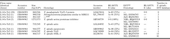
Clone L14 h-Ts1 represents 70% of isolated clones (n=20) (Table 2) and showed 73% identity with an antigenic T. pseudospiralis ES Tp22-3 protein (Nagano et al. Reference Nagano, Wu, Nakada, Boonmars and Takahashi2002). The full ORF was sequenced and the deduced amino acid (AA) sequence was 2 residues longer compared to the T. pseudospiralis protein. The first 28 residues were predicted to be a signal peptide. One clone, L14 h-Ts2, was similar to the meiotic recombination 11 homologue A (MRE11) complex and contained a metallophosphoesterase motif (E-value=1·90e-14). Clone L14 h-Ts3 showed no significant matches with known sequences and primary structure analysis revealed that it was composed by more than 70% of hydrophilic AA. Clone L14 h-Ts4 was (100% identity) a T. spiralis serine proteinase inhibitor (Nagano et al. Reference Nagano, Wu, Nakada, Matsuo and Takahashi2001b). SMART analysis revealed the presence of a putative serpin domain from position 14 to 373. Clone L14 h-Ts5 shared identity with a T. spiralis structural protein known as actin. Clone L14 h-Ts8 showed identity (92%) with a T. spiralis SML-3 protein that could be involved in nurse cell formation (Guiliano et al. Reference Guiliano, Oksov, Lustigman, Gounaris and Selkirk2009). The deduced protein sequence contained a hydrophobic N-terminal signal sequence of 20 AA.
Analysis of clones isolated from 14 h p.i. cDNA library by immuno-screening with Trichinella spiralis-infected pig serum

Analysis of positive clones from the 20 h p.i. T. spiralis cDNA library
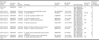
The sequence with the highest redundancy was the clone L20 h-Ts1 (n=111) containing the complete ORF (Table 3). It encodes a short polypeptide of 67 residues with a signal peptide prediction of 20 AA and a predicted hydrophilic region of 35 AA. In addition, this polypeptide shared no identity with known proteins but appeared frequently in T. spiralis expressed sequence tags (ESTs) from 3-day-old adults (Mitreva et al. Reference Mitreva, Appleton, Mccarter and Jasmer2005). Clone L20 h-Ts3 showed identity (91%) with a T. spiralis SML-3 protein (Guiliano et al. Reference Guiliano, Oksov, Lustigman, Gounaris and Selkirk2009). One clone, L20 h-Ts4, encoded a protein of 172 AA sharing 88% identity with a T. spiralis hypothetical ORF 17.20 and a T. pseudospiralis 21 kDa ES protein (Nagano et al. Reference Nagano, Wu, Nakada, Matsuo and Takahashi2001a). Primary structure prediction revealed an N-terminal signal peptide. Clone L20 h-Ts6 aligned with a previously identified antigenic sequence: a putative T. spiralis serine protease (Wu et al. Reference Wu, Fu, Wang, Yu, Yu, Deng, Liu, Boireau, Wang and Liu2009). Clone L20 h-Ts7 shared high identity (99%) with a T. spiralis tropomyosin (Nakada et al. Reference Nakada, Nagano, Wu and Takahashi2003). Sequence homology searches indicated that clone L20 h-Ts9 was similar to a putative T. spiralis porin precursor (Wu et al. Reference Wu, Fu, Wang, Yu, Yu, Deng, Liu, Boireau, Wang and Liu2009). Clone L20 h-Ts10 encoded a T. spiralis multi-cystatin-like domain protein precursor (Robinson et al. Reference Robinson, Massie and Connolly2007) and showed high identity (99%) with a hypothetical protein (Wu et al. Reference Wu, Fu, Wang, Yu, Yu, Deng, Liu, Boireau, Wang and Liu2009). Moreover, it appeared frequently in T. spiralis ESTs from 3-day-old adults and ML (Mitreva et al. Reference Mitreva, Appleton, Mccarter and Jasmer2005). Clone L20 h-Ts11 was similar in sequence (58%) to a RH56418p mRNA from Tribolium castaneum. Clone L20 h-Ts12 shared identity (67%) with a T. spiralis heat shock protein (HSP) 70 (Wang et al. Reference Wang, Zhu, Yang, Yang, Gu, Wei, Hao, Boireau and Cui2009a) and a T. nativa HSP70 (97%).
Analysis of clones isolated from 20 h p.i. cDNA library by immuno-screening with Trichinella spiralis-infected pig serum

Analysis of positive clones from the 48 h p.i. T. spiralis cDNA library
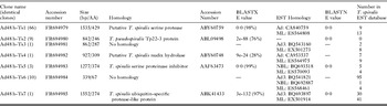
Among the 7 different sequences, clone Ad48 h-Ts1 represents 70% of the 91 positive clones isolated from the 48 h p.i. cDNA library (Table 4). It showed high identity (98%) with a putative T. spiralis serine protease family also isolated from the 3-day-old adult T. spiralis cDNA library (Wu et al. Reference Wu, Fu, Wang, Yu, Yu, Deng, Liu, Boireau, Wang and Liu2009). SMART analysis revealed a trypsin-like serine protease domain (E-value: 2·79e-64) and a signal peptide of 18 AA was predicted. Clone Ad48 h-Ts3 did not share identity with any known protein but was found in T. spiralis ESTs from 3-day-old adults and ML (Mitreva et al. Reference Mitreva, Appleton, Mccarter and Jasmer2005). Most of the predicted AA sequence was composed of hydrophilic residues. Clone Ad48 h-Ts5 aligned with a T. spiralis serine proteinase inhibitor (Nagano et al. Reference Nagano, Wu, Nakada, Matsuo and Takahashi2001b). Sequence homology searches indicated that clone Ad48 h-Ts7 had 97% identity with a T. spiralis ubiquitin-specific protease-like protein (Liu et al. Reference Liu, Wang, Fu, Li, Wu, Le Rhun, Chen and Boireau2007). Clone Ad48 h-Ts6 did not share identity with any known proteins but was also identified in the 20 h p.i T. spiralis cDNA library. Clone Ad48 h-Ts2 showed identity with a T. pseudospiralis ES Tp22-3 protein (Nagano et al. Reference Nagano, Wu, Nakada, Boonmars and Takahashi2002) which had also been identified in the 14 h p.i. T. spiralis cDNA library.
Analysis of clones isolated from 48 h p.i. cDNA library by immuno-screening with Trichinella spiralis-infected pig serum

DISCUSSION
Following Trichinella infection, the host elicits a strong, fast and specific immune response that leads to the expulsion of intestinal worms within weeks of the primary infection (Boireau et al. Reference Boireau, Vallee, Le Guerhier, Blaga, Baoquan, Hernandez-Bello, Ortega Pierres and Mingyuan2004; Mitreva and Jasmer, Reference Mitreva and Jasmer2006). The complex interactions between the parasite and the host's immune system are not yet fully understood and very few Trichinella antigens have been fully characterized so far (Nagano et al. Reference Nagano, Wu and Takahashi2009). Recently, immuno-screening of cDNA libraries of ML, NBL and adults permitted the identification of Trichinella proteins bearing antigenic functions (Nagano et al. Reference Nagano, Wu, Nakada, Matsuo and Takahashi2001b; Zarlenga et al. Reference Zarlenga, Boyd, Lichtenfels, Hill and Ray Gamble2002; Nakada et al. Reference Nakada, Nagano, Wu and Takahashi2003; Boireau et al. Reference Boireau, Mingyuan, Baoquan, Le Guerhier, Le Rhun, Hernandez-Bello, Bahuon and Vallee2006; Trap et al. Reference Trap, Fu, Le Guerhier, Liu, Le Rhun, Romand, Perret, Blaga and Boireau2006).
This study focuses on pre-adult/young adult T. spiralis worms and screened genes according to the antigenicity of expressed proteins. As reported by Khan (Reference Khan1966), observations on the moulting patterns in T. spiralis in mice indicate that 14 h p.i. worms are L2 larvae and 20 h p.i. worms are L3 larvae (pre-adults), with 48 h p.i. worms being young adults. It is difficult to assert that the entire populations studied at 14 h and 20 h p.i. actually correspond to the stages quoted previously, in particular because of the use of a different murine model, massive infestations for cDNA library construction as well as the poor availability of statistical evidence. However, at 48 h p.i. copulatory papillae of males were clearly visible (data not shown) signifying the sexual maturation of larvae into adult worms. Thus, it seemed more sensible to name those ‘stages’ according to the duration of infection. Numerous nucleotide sequences were identified by immuno-screening, showing that despite the short life of the enteric stages of Trichinella, an immune response was well induced with specific antigens, some of which have never been identified up to now. These results are in agreement with the studies of Bell and McGregor (Reference Bell and McGregor1979) which showed that pre-adults and adults are potentially targeted by the host's immune system since they are specifically expelled from immunized rats. In addition, the identification of different genes at 14 h, 20 h and 48 h p.i. is consistent with the use of 3 different developmental stages of Trichinella. The high discordance is surprising though, as it could be expected that most hit targets would be expressed at the 3 time-points chosen. The parasite should hypothetically make use of very similar gene products into the intestine, having a more or less close location with identical periparasitic physiology. The method used to identify antigenic targets could explain a part of these results. Antigens were selected randomly among the high number of clones, and not every clone was picked for analysis. Thus, antigens expressed at a low rate have less chance to be identified by immuno-screening as they are present in fewer clones. Besides, antigens selected with this method are continuous and only reflect a percentage of available epitopes as previously demonstrated (Boireau et al. Reference Boireau, Vayssier, Fabien, Perret, Calamel and Soule1997). If the same number of positive clones had been sequenced for each library, perhaps more antigens would have been in common. Moreover, several studies showed that Trichinella antigens from ML, 3-day-old adults and NBL are stage-specific (Boireau et al. Reference Boireau, Mingyuan, Baoquan, Le Guerhier, Le Rhun, Hernandez-Bello, Bahuon and Vallee2006; Wu et al. Reference Wu, Fu, Wang, Yu, Yu, Deng, Liu, Boireau, Wang and Liu2009) allowing the parasite to evade the host immune response (Bruschi, Reference Bruschi2002). Liu et al. (Reference Liu, Wang, Fu, Li, Wu, Le Rhun, Chen and Boireau2007) also identified many different antigenic proteins from 3- and 5-day-old T. spiralis adults although they are both present in the intestine. This observation shows that even within a single stage, Trichinella could express different protein profiles probably in order to adapt to the local environment (Bruschi, Reference Bruschi2002). Therefore, the discordance between the targets found in this study could reveal stage-specificity of the antigens and major physiological differences for Trichinella intestinal stages associated with qualitative changes in gene products as it was observed for 3- and 5-day-old T. spiralis adults (Liu et al. Reference Liu, Wang, Fu, Li, Wu, Le Rhun, Chen and Boireau2007).
Among these genes, the sequence with the highest redundancy was a small polypeptide of 67 AA identified from the 14 h and 20 h p.i. cDNA libraries, which is the shortest antigenic peptide of the Trichinella genus identified so far (Wu et al. Reference Wu, Nagano, Boonmars and Takahashi2007; Yang et al. Reference Yang, Jian and Qin2010). It is the first time this new gene has been described in T. spiralis. The deduced AA sequence does not exhibit any tag sequence allowing a particular function to be identified. However, this sequence was strongly represented in the ESTs of 3-day-old adult T. spiralis (Mitreva et al. Reference Mitreva, Appleton, Mccarter and Jasmer2005) which could strengthen the hypothesis that this polypeptide is important for worm persistence and survival in the intestine. A large portion of the antigenic proteins isolated from 48 h p.i. worms are serine proteases which are among the most abundant group of proteins within the ES products of T. spiralis (Cwiklinski et al. Reference Cwiklinski, Meskill, Robinson, Pozio, Appleton and Connolly2009). Serine proteases have been detected at different stages (Criado-Fornelio et al. Reference Criado-Fornelio, De Armas-Serra, Gimenez-Pardo, Casado-Escribano, Jimenez-Gonzalez and Rodriguez-Caabeiro1992; Moczon and Wranicz, Reference Moczon and Wranicz1999; Trap et al. Reference Trap, Fu, Le Guerhier, Liu, Le Rhun, Romand, Perret, Blaga and Boireau2006; Boireau et al. Reference Boireau, Mingyuan, Baoquan, Le Guerhier, Le Rhun, Hernandez-Bello, Bahuon and Vallee2006; Liu et al. Reference Liu, Wang, Fu, Li, Wu, Le Rhun, Chen and Boireau2007) and might be involved in a wide variety of essential biological processes including host tissue and cell invasion (Dzik, Reference Dzik2006), blood coagulation and fibrinolysis (Ros-Moreno et al. Reference Ros-Moreno, Vazquez-Lopez, Gimenez-Pardo, De Armas-Serra and Rodriguez-Caabeiro2000), moulting and/or digestive functions (Trap et al. Reference Trap, Fu, Le Guerhier, Liu, Le Rhun, Romand, Perret, Blaga and Boireau2006). Interestingly, serine proteases were poorly detected at 14 h and 20 h p.i. A serine protease inhibitor was isolated from the 14 h and 48 h p.i. cDNA libraries. Serpins and their homologues are a group of structurally related proteins performing diverse functions such as blood coagulation, angiogenesis, and modulation of inflammation (Zang and Maizels, Reference Zang and Maizels2001). These functions could explain how the parasite is able to evade the host's immune response and survive in the intestine. An HSP70 was identified in the 20 h p.i. cDNA library. HSPs are produced as an adaptive response of the parasite to the hostile environment encountered in the host intestine (Vayssier et al. Reference Vayssier, Le Guerhier, Fabien, Philippe, Vallet, Ortega-Pierres, Soule, Perret, Liu, Vega-Lopez and Boireau1999; Wang et al. Reference Wang, Chuang, Yeh, Wang, Liu, Chang and Hung2009b). The HSP70’s precise mechanism of action is still unknown but it acts as a chaperone protein (Kampinga and Craig, Reference Kampinga and Craig2010). Thus, the HSP70 could promote the stability of transcriptional factors, which in turn directly regulate gene expression and trigger off rapid changes (moulting) from ML to adult stage in the host intestine. The HSP70 also acts as a modulator of the environmental stress (Wieten et al. Reference Wieten, Broere, Van Der Zee, Koerkamp, Wagenaar and Van Eden2007) being directly or indirectly involved in the development of several organisms (Boscheinen et al. Reference Boscheinen, Lyck, Queitsch, Treuter, Zimarino and Scharf1997). Therefore the HSP70 expression could be critical for the parasite during the intestinal phase. HSPs are antigenic during infections caused by a number of pathogens (Kanamura et al. Reference Kanamura, Hancock, Rodrigues and Damian2002; Mohamed et al. Reference Mohamed, Aosai, Chen, Mun, Norose, Belal, Piao and Yano2003) including Trichinella (Vayssier et al. Reference Vayssier, Le Guerhier, Fabien, Philippe, Vallet, Ortega-Pierres, Soule, Perret, Liu, Vega-Lopez and Boireau1999). A putative ES product showing identity with the T. pseudospiralis Tp22-3 protein was identified. ES products are exposed to the host's immune system and have been shown to be antigenic in Trichinella species (Gamble et al. Reference Gamble, Rapic, Marinculic and Murrell1988). A sequence encoding a porin-like protein was identified in the 20 h p.i. cDNA library. A porin-like protein in ES products of Trichuris trichiura has been confirmed to be highly immunogenic (Barker and Bundy, Reference Barker and Bundy1999).
In conclusion, pre-adult and young adult T. spiralis worms express specific antigenic proteins, some of which were identified in the present study. In addition, the results obtained illustrate the abundance of serine proteases during the intestinal phase of Trichinella infection and presume the essential role of proteins such as HSPs and serine protease inhibitors. Thus, sequences presented herein could constitute attractive targets of research to improve diagnostic tools or develop vaccines.
ACKNOWLEDGEMENTS
We would like to thank W. Xiuping and D. HongKuan for their advice and technical assistance. We would also like to thank J. Appleton for providing the murine intestinal epithelial cells.
FINANCIAL SUPPORT
This work was supported by a TRICHIMED grant (MPY13/04-27) under the MedVetNet project (FOOD-CT-2007 506122).