Aberrant right subclavian artery is the most common aortic arch anomaly,Reference Molz and Burri 1 , Reference Freed and Low 2 which concerns around 1% of population.Reference Molz and Burri 1 – Reference Haesemeyer and Gavant 7 The prevalence of aberrant right subclavian artery depends on the evaluated population characteristics, inclusion criteria, and the method used for its detection.Reference Polguj, Chrzanowski and Kasprzak 8 The invention of CT enabled non-invasive detection of aberrant right subclavian artery with a diagnostic sensitivity of 100%,Reference Yang, Mo and Jin 9 , Reference Chen, Qu and Peng 10 which is superior compared with ultrasound and X-ray sensitivities of 92 and 20%, respectively.Reference Yang, Mo and Jin 9 – Reference Branscom and Austin 11 The development of multi-slice CT angiography allowed for the three-dimensional visualisation of an aberrant right subclavian artery and surrounding structures with high spatial and temporal resolution.Reference Haesemeyer and Gavant 7 The majority of people with an aberrant right subclavian artery remain asymptomatic; however, if the clinical symptoms occur, the most common symptom caused by an aberrant artery is dysphagia.Reference Freed and Low 2 , Reference Natsis, Tsitouridis and Didagelos 4 , Reference Van Dyke and White 6 , Reference Polguj, Chrzanowski and Kasprzak 8 , Reference Myers, Fasel and Kalangos 12 It was first reported by Bayford in 1787,Reference Bayford 13 who described a case of a 62-year-old woman who died after suffering from severe dysphagia. The subsequently performed autopsy revealed oesophageal compression secondary to an aberrant right subclavian artery, which clinically presented with dysphagia named by the author as “dysphagia lusoria”.
Apart from clear oesophageal compression, other explanations for the onset of dysphagia lusoria were proposed. They are all potentially evaluable in CTReference Pelberg and Mazur 14 and include the presence of atherosclerosis causing increased stiffness of the aberrant right subclavian artery wall, increased lumen area of the aberrant right subclavian artery, and elongation of aorta.Reference Polguj, Chrzanowski and Kasprzak 8 , Reference Klinkhamer 15 , Reference Bennett, Cock and Heddle 22 In the published CT-based analyses of aberrant right subclavian artery,Reference Haesemeyer and Gavant 7 , Reference Yang, Mo and Jin 9 , Reference Chen, Qu and Peng 10 the authors concentrated mostly on its incidence and coexistence of other aortic anomalies than on detailed morphometric evaluation of aberrant right subclavian artery anatomy. Haesemeyer et alReference Haesemeyer and Gavant 7 performed CT-based study of aberrant right subclavian artery occurrence and its clinical consequences in population with suspected acute aortic tear. However, this study was performed using old-generation helical scanner; moreover, the authors were not able to precisely evaluate the incidence of clinical symptoms, including dysphagia, and concentrated mostly on aberrant right subclavian artery occurrence and its impact on repair procedure. Qualitative and quantitative analysis of morphometric details of aberrant right subclavian artery anatomy using spiral multi-slice CT and occurrence of dysphagia is a new perspective, which is missing in the existing published literature. Therefore, the aim of the study was to perform CT angiography-based evaluation of the aberrant right subclavian artery prevalence, anatomy, and its influence on occurrence of clinical symptoms.
Materials and methods
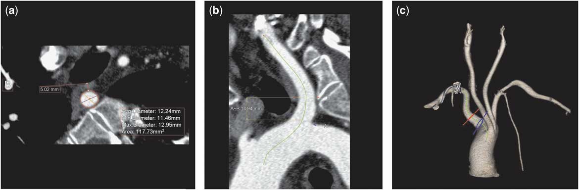
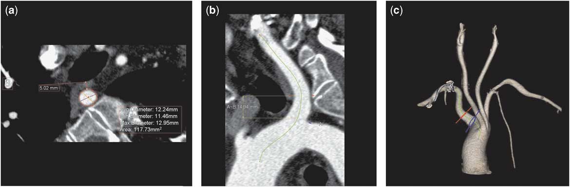
A total of 6833 consecutive patients, who underwent CT angiography of the carotid, vertebral, subclavian arteries (4811 patients), and thoracic aorta (2022 patients) were retrospectively screened for the presence of aberrant right subclavian artery. The CT examinations were performed in the tertiary care hospital within 10 consecutive years. A total of 923 patients were evaluated using 64-slice CT; Somatom Sensation 64 Cardiac, Siemens, Erlangen, Germany, and 5910 patients using dual-source CT; Somatom Definition, Siemens, Erlangen, Germany. The contrast-enhanced acquisitions were performed during inspiratory breath-hold with the collimation of 0.75 mm. The field of view extended from the level below the aortic arch to the base of the cranium for carotid, vertebral, and subclavian arteries angiography and from the level above the aortic arch to the level below the diaphragm for the thoracic aorta. The bolus tracking method was used with the region of interest in the aortic arch and the scan initiated when the density reached 180 Hounsfield Units. An iodinated contrast agent was injected with an injection rate of 5 ml/second for carotid, vertebral, and subclavian arteries and 3.5 ml/second for aorta.Reference Pelberg and Mazur 14 Images were reconstructed with an image matrix of 512 × 512 pixels. The post-processing and study evaluation was performed using a dedicated workstation Aquarius; TeraRecon, San Mateo, United States of America. Patients were assessed for the presence of an aberrant right subclavian artery using axial, curved multi-planar and volume-rendered technique reconstructions. The exclusion criteria to include the patients in the analysis were known gastroesophageal disease, as well as prior aortic, oesophageal, and tracheal interventions including surgical and endoscopic therapy. The CT evaluation concerned qualitative and quantitative analysis of aberrant right subclavian artery. Qualitative evaluation included aberrant right subclavian artery course, comprising retroesophageal, between trachea and oesophagus, pre-tracheal; potential oesophageal and tracheal compression; concomitant anatomical variants and presence of atherosclerotic lesions in aberrant right subclavian artery. Quantitative analysis included the distance between the outer outline of aberrant right subclavian artery and the trachea, as well as the lumen area of aberrant right subclavian artery at its origin and at the level of oesophagus (Fig 1). Available medical records of patients with an aberrant right subclavian artery were analysed for the prevalence and characteristics of clinical symptoms. Subsequently, the patients were contacted by an observer blinded to CT analysis results and asked for the occurrence of clinical symptoms at the time of the CT examination and during follow-up. Informed consent was obtained from all individual participants included in the study. Local bioethics committee approved the protocol and classified it as a primarily retrospective study. The clinical analysis included demographic data such as age, gender; indication to perform CT; presence of clinical symptoms related to aberrant right subclavian artery,Reference Polguj, Chrzanowski and Kasprzak 8 including dysphagia, dyspnoea, cough, retrosternal pain, back pain, numbness of the upper right limb, stomach ache and weight loss, and potential vascular intervention on aberrant right subclavian artery in the follow-up.Reference Kopp, Wizgall and Kreuzer 16 Next, an analysis of the relationship between the data derived from CT and clinical symptoms was performed. Variables were presented as median, including Q1 and Q3, and compared using the Mann–Whitney test. In the qualitative analysis of anatomic features, χ2-test with Yates modification was used. The value p<0.05 was considered as statistically significant. A statistical analysis was performed using the software Statistica 10; StatSoft Inc.
Multi-planar (a,b) and volume-rendered technique reconstructions (c) of aberrant right subclavian artery. Quantitative evaluation (a) of distance between the outer outline of aberrant right subclavian artery and trachea and lumen area of aberrant right subclavian artery at the level of oesophagus.
Results
Aberrant right subclavian artery was found in 32 (0.47%) patients consisting of 13 males and 19 females with mean age of 60.8±13.4 years. Of these, 22 (69%) patients underwent CT angiography of carotid, vertebral, and subclavian arteries owing to suspected arterial stenosis. In the remainder 10 (31%) patients, aortic CT angiography was performed owing to suspected aortic aneurysm. The course of aberrant right subclavian artery was retroesophageal in all patients with compression of oesophagus in 16 (50%) patients and trachea in four (12%) cases. The aberrant right subclavian artery in all the described cases was the ultimate branch of the aortic arch. Concomitant vascular abnormalities were truncus bicaroticus, Kommerell’s diverticulum, aberrant right subclavian artery aneurysm, and aortic origin of right vertebral artery, which occurred in nine cases (28%), nine cases (28%), four cases (12%), and one case (3%), respectively. Atherosclerotic lesions were present in 17 (53%) cases and total occlusion of the aberrant right subclavian artery was observed in two (6%) cases.
In the follow-up [1990±903; 1894 days (mean ± SD; median)], 30 (94%) patients were contacted and interviewed. The prevalence of clinical symptoms in the evaluated group at the time of CT examination were: dysphagia in nine cases (30%), dyspnoea in six cases (20%), numbness of the upper right limb in six cases (20%), cough in five cases (17%), and retrosternal pain in five cases (17%); and in the follow-up dysphagia was found in nine cases (30%), dyspnoea in seven cases (23%), numbness of the upper right limb in five cases (20%), cough in five cases (17%), and retrosternal pain in six cases (20%). At the time of CT, 12 (40%) patients were asymptomatic and none of the patients presented with back pain, stomach ache, or weight loss; four (44%) patients with Kommerell’s diverticulum revealed dysphagia, (p = 0.15). The mean age of patients with and without dysphagia were 63.3±14.7 and 59.8±12.6 years, respectively. Atherosclerotic lesions were present in nine (30%) patients without dysphagia and six (20%) patients with dysphagia (p = 0.07). In the follow-up none of the patients revealed aortic dissection and none underwent surgery owing to the presence of aberrant right subclavian artery.
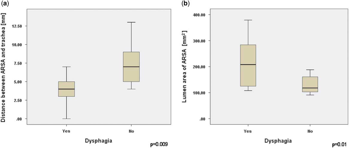
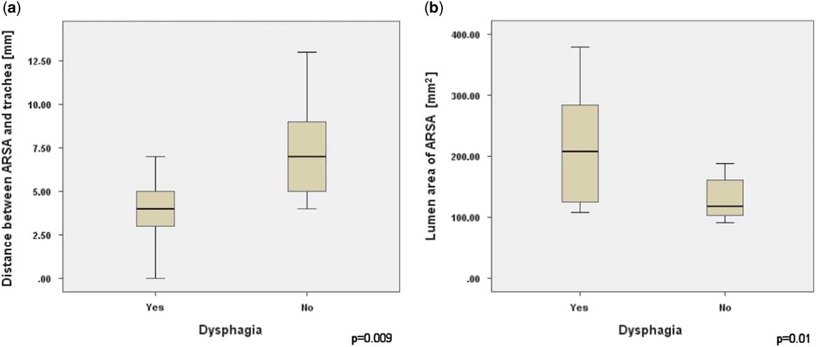
In the follow-up group oesophageal compression was observed in 14 cases (47%) and tracheal compression in three cases (10%). The median distance between aberrant right subclavian artery and trachea and median lumen area of aberrant right subclavian artery at the level of oesophagus revealed 5.5 mmReference Natsis, Tsitouridis and Didagelos 4 – Reference Polguj, Chrzanowski and Kasprzak 8 and 128 (107–185) mm2, respectively. All patients with dysphagia revealed oesophageal compression, and in those patients the median distance between aberrant right subclavian artery and trachea was lower at 4 (3.5–5.5) mm than in individuals without dysphagia at 7 (4.5–9) mm (p = 0.009) (Table 1, Fig 2). The median lumen area of aberrant right subclavian artery at the level of oesophagus was higher in patients with dysphagia [208 (120.5–313.5) mm2] than individuals without dysphagia [108 (101.5–162.5) mm2] (p = 0.01) (Table 1, Fig 2). The median lumen area of the aberrant right subclavian artery at its origin tended to be higher in patients with dysphagia [268 (146.5–360) mm2] than individuals without dysphagia [180 (110.5–225) mm2] (p = 0.08).
The value of distance between aberrant right subclavian artery and trachea and lumen area of aberrant right subclavian artery at the level of oesophagus in patients with and without dysphagia.

ARSA = aberrant right subclavian artery.
Box-and-whisker plot. The value of distance between aberrant right subclavian artery and trachea ( a ) and lumen area of aberrant right subclavian artery ( b ) at the level of oesophagus in patients with and without dysphagia.
Discussion
The incidence of aberrant right subclavian artery in the evaluated adult group is similar to previously published analyses.Reference Molz and Burri 1 – Reference Van Dyke and White 6 This observation strongly supports almost equal incidence of aberrant right subclavian artery in the screened population (0.47%) in this study compared to the previously published CT-based analysis by Haesemeyer et alReference Haesemeyer and Gavant 7 that revealed occurrence of aberrant right subclavian artery in 0.4% of the studied group. The other similarities between the two analyses concerned are the number of evaluated patients of 6833 patients in this study versus 7174 in the above cited article, and screening of patients without suspicion of aberrant right subclavian artery. However, few discrepancies between the studies should also be elucidated. First, we performed examination in patients with various indications, while Haesemeyer et alReference Haesemeyer and Gavant 7 included subjects with suspected acute traumatic aortic tear. Second, our examinations were performed a decade later on a more modern scanner with higher spatial resolution that enabled for a more detailed analysis of aberrant right subclavian artery anatomy. Interestingly, this research confirms previously observed female predominance of aberrant right subclavian artery.Reference Molz and Burri 1 , Reference Polguj, Chrzanowski and Kasprzak 8 The study results showed 59% of women in this study group of patients with aberrant right subclavian artery compared to 55 and 58% of female predominance in analyses by Polguj et alReference Polguj, Chrzanowski and Kasprzak 8 and Molz et al,Reference Molz and Burri 1 respectively.
This study results remain consistent with the analysis by Klinkhamer et al,Reference Klinkhamer 15 where this study analysis showed 28% with regard to the concomitant presence of an aberrant right subclavian artery and truncus bicaroticus, whereas 29% in the above cited study. However, other authors revealed lower coincidence of those vascular anomalies with prevalence of 19% by Polguj et alReference Polguj, Chrzanowski and Kasprzak 8 and 7% by Hartyanszky et al.Reference Hartyánszky, Lozsadi and Marcsek 17 The occurrence of Kommerell’s diverticulumReference Kommerell 18 was different in this study and Polguj et al,Reference Polguj, Chrzanowski and Kasprzak 8 with prevalence of 27 and 14%, respectively. Epstein et alReference Epstein and DeBord 19 revealed different results with 60% of coexistence of aberrant right subclavian artery and Kommerell’s diverticulum. Kieffer et alReference Kieffer, Bahnini and Koskas 20 described presence of aberrant right subclavian artery aneurysm of 30%, which was less common than in this study of 12%. All those differences may result from different definitions of listed anatomical variations, which were applied in the above cited publications.
The retro-oesophageal course of aberrant right subclavian artery, which is known to be the most common,Reference Natsis, Tsitouridis and Didagelos 4 , Reference Klinkhamer 15 , Reference Barry 21 was present in all individuals of this study. This may be related to the high average age in this study group, which was over 60 years while individuals with early respiratory symptoms related to the other course – between trachea and oesophagus and pre-tracheal – are more common in childrenReference Bennett, Cock and Heddle 22 , Reference Natsis, Didagelos and Manoli 23 and might have already been diagnosed due to severity of symptoms. This theory supports the fact that in spite of the occurrence of clinical symptoms, none of the patients in this study group underwent surgery related to aberrant right subclavian artery in the follow-up. This may be attributed to the fact that symptoms might not have been severe enough to warrant the need for the surgical treatmentReference Myers, Fasel and Kalangos 12 , Reference Kopp, Wizgall and Kreuzer 16 as no substantial change in the prevalence of clinical symptoms related to aberrant right subclavian artery was observed in the follow-up. To that authors’ best knowledge, we are the first to perform clinical follow-up analysis in adult patients with aberrant right subclavian artery, in addition to our observation that exceeded 5 years.
Contrary to previous analyses,Reference Freed and Low 2 , Reference Natsis, Tsitouridis and Didagelos 4 , Reference Van Dyke and White 6 , Reference Polguj, Chrzanowski and Kasprzak 8 the majority of patients (60%) in this study group were symptomatic; but in accordance with the above-mentioned analyses, the most common symptom was dysphagia, which concerned nine patients at the time of examination and in the follow-up. The higher prevalence of symptomatic individuals in this study may again result from age over 60 years and occurrence of retrosternal pain in five cases and dyspnoea in six cases, which are not only related to aberrant right subclavian artery but also to cardiac and pulmonary diseases. In children aberrant right subclavian artery manifests the above-mentioned respiratory symptoms owing to decreased rigidity of trachea, whereas in adults trachea becomes more resistant to compression.Reference Donadel, Lontra and Cavazzola 24 On the contrary, rigidity of aberrant right subclavian artery and oesophagus also increase with age, and according to some theories it is responsible for increased prevalence of dysphagia lusoria in older population.Reference Polguj, Chrzanowski and Kasprzak 8 , Reference Bennett, Cock and Heddle 22 Several other explanations for late onset of dysphagia lusoria in patients with aberrant right subclavian artery were proposed, which include the presence of atherosclerosis causing increased stiffness of the aberrant right subclavian artery wall, increased lumen area of the aberrant right subclavian artery with age, and elongation of aorta.Reference Polguj, Chrzanowski and Kasprzak 8 , Reference Klinkhamer 15 , Reference Bennett, Cock and Heddle 22
In the observation of this study, there is no significant difference between the age of patients with and without dysphagia of 63 versus 60 years, respectively. Moreover, we found no significant relation between the occurrence of dysphagia and the presence of atherosclerosis in aberrant right subclavian artery, which was proposed by some researchers.Reference Polguj, Chrzanowski and Kasprzak 8 , Reference Klinkhamer 15 , Reference Bennett, Cock and Heddle 22 As not all patients with oesophageal compression revealed dysphagia, we subsequently performed a comparative quantitative analysis of the value of the lumen area of the aberrant right subclavian artery at its origin and at the level of oesophagus and the distance between the aberrant right subclavian artery and trachea in patients with and without dysphagia. To the best of authors’ knowledge, this analysis was performed for the first time and in patients with dysphagia the authors found significantly a higher aberrant right subclavian artery lumen area at the level of oesophagus and a lower distance between aberrant right subclavian artery and trachea than in non-dysphagia patients. Those measurements strongly support the theory of an increased lumen area of an aberrant right subclavian arteryReference Polguj, Chrzanowski and Kasprzak 8 , Reference Klinkhamer 15 at the level of oesophagus leading to dysphagia lusoria. No correlation was found between the occurrence of Kommerell’s diverticulum and dysphagia. It was also found that not all patients with oesophageal compression on CT present with dysphagia, but dysphagia was present in those patients with severe oesophageal compression by aberrant right subclavian artery and its proximity to posterior wall of trachea. To the best of authors’ knowledge this is the first study to perform qualitative and quantitative morphometric analysis of aberrant right subclavian artery anatomy in reference to dysphagia.
The small population included in this study limits statistical significance of the analysis. However, in the literature it was found that there is only one study with an equal aberrant right subclavian artery patients sample of 32 patientsReference Freed and Low 2 and no single analysis exceeding such number of population. The CT-based article by Haesemeyer et alReference Haesemeyer and Gavant 7 concerned a population of only one patient less than this analysis, but in contrary to this study they did not perform clinical symptoms evaluation. Another limitation is failure to contact two (6%) patients in the follow-up, which resulted in lack of collection of clinical symptoms in those individuals. Lastly, the evaluation of the oesophagus was diminished because of a lack of endoscopic examination, as well as a non-dynamic radiological examination without oral contrast administration.
Conclusions
Aberrant right subclavian artery is a rare-occurring anomaly in CT angiography and its incidence is comparable to previously published analyses based on other diagnostic methods. In the evaluated adult population with aberrant right subclavian artery, dysphagia is the most common clinical syndrome at the time of CT examination and in the follow-up. Dysphagia occurs in patients with decreased distance between aberrant right subclavian artery and trachea and increased lumen area of aberrant right subclavian artery at the level of compressed oesophagus.
Acknowledgements
None.
Financial Support
This research received no specific grant from any funding agency, or from commercial or not-for-profit sectors.
Conflicts of Interest
None.
Ethical Standards
The authors assert that all procedures contributing to this work comply with the ethical standards of the relevant national guidelines on human experimentation (Poland) and with the Helsinki Declaration of 1975, as revised in 2008, and has been approved by the institutional committees (Kraków).