Whereas rheumatic mitral valve disease still accounts for the majority of mitral valve stenosis and obstruction of the left ventricular outflow tract in general,Reference Deshpande, Vaideeswar, Amonkar and Vasandani 1 discrete membranous supravalvular mitral stenosis is a very rare congenital cardiac defect. It is mostly associated with other congenital heart defects such as in Shone’s Complex, coarctation of the aorta, ventricular septum defect or left ventricular outflow tract obstructions.Reference Carpentier, Branchini and Cour 2 It was first described by FischerReference Fischer 3 in 1902 and has been reported only about 100 times till the 1990s.Reference Mychaskiw, Sachdev, Braden and Heath 4 The first surgical correction was performed in 1962 where the membranous supravalvular ring, which was adherent to the mitral valve annulus, could be removed completely.Reference Glaser, Yakirevich and Vidne 5 , Reference Lynch, Ryan and Williams 6
Owing to a high variability and difficulties in pre-operative diagnosis, only few data are available on the actual incidence of supravalvar mitral rings.Reference Vaideswaar, Baldi and Warghade 7 A supravalvular mitral membrane could be predicted only in ~45% of the patients by routine pre-operative echocardiography. Retrospective detailed frame-by-frame analysis revealed a possibility of such a membrane in 91% of the analysed cases. However, owing to a significant effort of time, secondary post-hoc knowledge of the disease, and specific echocardiographic knowledge, it seems to be clinically impractical to reveal as many of those entities pre-operatively as described retrospectively.Reference Mychaskiw, Sachdev, Braden and Heath 4 , Reference Sullivan, Robinson, de Leval and Graham 8
This malformation may occur approximately in 17–31% of congenital anomalies of the mitral valve, and is reported to be 71% in patients with a parachute mitral valve.Reference Watraida, Shiraishi and Katsuyama 9 , Reference Banerjee, Kohl and Silverman 10 Hoashi et al found a supravalvular ring in four out of 20 patients with congenital mitral valve stenosis, whereas Vaideeswar et al even reported 37.5% – 24 out of 64 congenital left ventricular inflow tract obstructions from 1633 autopsied examples.Reference Hoashi, Bove, Devaney, Hirsch and Ohye 11
Clinically, it has to be differentiated between a complete circumferential ring and an incomplete supravalvular mitral ring. Owing to the fact that it is not easily seen on routine echocardiography, in many patients the discrete supravalvular mitral membrane adherent to the mitral valve is only detected during surgical investigation and repair for other congenital heart disease.Reference Shone, Sellers, Anderson, Adams, Lillehei and Edwards 12 , Reference Brickner, Hillis and Lange 13 The membrane is of a thin fibrous structure only; often, it is adherent to a leaflet of the mitral valve on the atrial site and frequently only one edge of it is visible.
The main characteristic in echocardiography is the Doppler flow acceleration beginning just before the mitral valve annulus. The visualisation of the membrane can be enhanced by trans-oesophageal echocardiography in the four-chamber view. In many cases, it appears like a valvular stenosis, but the differentiation between the two is of major surgical importanceReference Pettersen and Berger 14 because the supravalvular mitral ring has a good prognosis if treated in time. Valvular mitral stenosis, that is, in the form of a parachute valve, mitral arcade, hammock valve or double orifice mitral valve, needs a technically difficult reconstruction or even replacement of the valve, whereas a supramitral ring and a discrete membrane can be removed.
In addition, the supravalvular mitral ring has to be differentiated from a cor triatriatum sinistrum, where the membrane is fibromuscular and located further above the mitral valve and has usually only one perforation.Reference Mychaskiw, Sachdev, Braden and Heath 4 It separates the atrium in a proximal and a distal chamber.
We report about the typical echocardiographic findings in three young patients with the rare form of a discrete membranous supravalvular stenosis caused by a membrane adherent to the mitral valve. We believe that this echocardiographic sign is an excellent marker to differentiate this anatomy and provide a pre-operative diagnosis. On the basis of the typical colour Doppler appearance, we propose to name it Polar Light Sign (see Fig 1).
Typical Polar Lights (http://honasurd.livejournal.com/24283.html).

Case presentations
The first patient was a 10-month-old girl with Shone’s Complex. She presented with multiple ventricular septum defects, a moderate hypoplasia of the aortic arch, an atrial septum defect and a minimal mitral stenosis, retrospectively caused by a supravalvular membrane. She developed clinical symptoms of congestive heart failure at the age of 5 months and was treated accordingly. Catheter examination revealed pulmonary hypertension with a mean pressure of 55 mmHg and an increased resistance of 7.9 iWE due to the mitral valve stenosis.
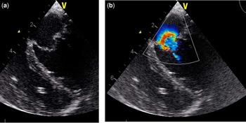
Echocardiographic findings showed a small left ventricle with a mitral valve diameter of 8–9 mm and a small left ventricular outflow tract and aortic valve of 6–7 mm diameter. The mean Doppler gradient across the mitral valve was not significantly elevated and there was no regurgitation. In the supravalvular region, no clear membranous structure could be detected in two-dimensional echo. Colour Doppler flow, however, showed a colour flow inversion beginning just above the mitral valve annulus as a surrogate of a supravalvular mitral ring resembling a “polar light” (Fig 2a and b).
( a ) Appearance of the mitral valve in diastole. No clear membrane is visible. ( b ) In colour Doppler flow, there is a clear flow acceleration above the mitral valve annulus.

During corrective surgery, the proposed supravalvular membrane was confirmed and removed, and the ventricular septum defects were closed. The post-operative course was uneventful and she was discharged 15 days later in good clinical condition. Echo investigation revealed a mean pressure gradient of 7 mmHg across the mitral valve after exertion. Unfortunately, the membrane reappeared about 1 year later and again the same echocardiographic findings were visible. The patient had to be operated again because of severe pulmonary hypertension with a transmitral gradient of 25 mmHg. A thickened supravalvular fibrous membrane attached to the posterior mitral valve leaflet was diagnosed and removed. Again the post-operative course was uneventful and control echocardiography 4 months later showed an excellent result (Fig 2).
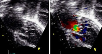
The second patient was a 2-year-old boy with a variant of Shone’s complex including coarctation of the aorta, bicuspid aortic valve and mitral stenosis leading to pulmonary hypertension with consecutive right heart failure. The coarctation had been repaired at the age of 1 month without any problems. The mitral stenosis was initially not significant; however, it increased in haemodynamic severity and thus clinical signs of heart failure with failure to thrive, sweating and fatigue became obvious. Finally, he presented with syncope and cyanosis after excitement and was transferred for surgical repair. Echocardiography showed a dysplastic mitral valve with a barely opening posterior leaflet and little regurgitation; the mean Doppler gradient was 12–13 mmHg, the left atrium was enlarged and there was severe pulmonary hypertension (right ventricular systolic pressure of 100 mmHg). Again, echocardiography revealed a flow acceleration above the mitral valve opening and without a clearly visible membrane, indicating a supravalvular mitral stenosis (i.e. “Polar Light Sign”, see Fig 3). This finding was confirmed by surgery; the supramitral membrane was identified and removed without any complications. Post-operative echocardiography showed no residual stenosis, the mean Doppler gradient was 4 mmHg, and maximum flow velocity was 1.3 m/second with trivial regurgitation.
( a ) Appearance of the mitral valve in diastole. No clear membrane is visible. ( b ) In colour Doppler flow, there is clear flow acceleration above the mitral valve annulus.

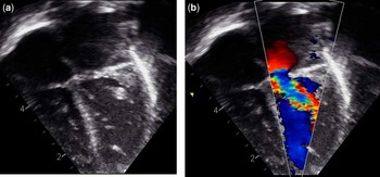
The third patient was a 2-month-old female baby with a double-outlet right ventricle and a malalignment ventricular septum defect. Pre-operatively, she was diagnosed with a hypoplastic mitral valve, significant stenosis and left atrial enlargement. Echocardiography revealed a discrete supravalvular membrane again with flow acceleration above the mitral valve (see Fig 3). She was operated upon at the age of 5 months with full repair of the double-outlet right ventricle, intra-operative confirmation and removing of the membrane. At control investigation 9 months after surgery, she was doing well and echocardiography shows no significant mitral stenosis with a flow velocity of 1.9 m/s. The size of the left atrium is nearly back to normal and there is only a minimal regurgitation of the mitral valve (Fig 4).
( a ) Appearance of the mitral valve in diastole, atypical view to present the supravalvular membrane. ( b ) In colour Doppler flow, there is clear flow acceleration above the mitral valve annulus.

Discussion
Congenital mitral valve stenosis is a very rare condition often resulting in significant secondary pulmonary hypertension. In many cases, other congenital cardiac defects are combined such as ventricular septum defects and various forms of left ventricular outflow tract obstructions and malformations of the mitral valve. This combination is often named as Shone’s Complex or its variants. Whereas typical supravalvular mitral stenosis caused by cor triatriatum or a clearly visible supravalvular ring is easily detectable by echocardiography, a discrete supravalvular membrane adjacent to the mitral valve leaflets resembling valvular mitral stenosis is difficult to differentiate by routine echocardiography.Reference Alvares, Sa Melo and Antunes 15 – Reference La Corte, Harada and Williams 17 , Reference Mychaskiw, Sachdev, Braden and Heath 4 , Reference Watraida, Shiraishi and Katsuyama 9
These various forms are important to differentiate from the anatomical variation we describe here in our patients, that is, the discrete form of supravalvular membranous stenosis adherent to the mitral valve. This entity is very rare in childhood, and only a few cases have been described in the literature. Sullivan et al described 14 cases in 1986, out of which 12 received a successful removal of the membrane and two underwent valve replacement. The three patients we report here also received a surgical removal of the supramitral membrane without complications.
The typical echocardiographic findings are best visualised in the apical four-chamber view. In those cases where the stenosis is caused by a clearly visible supravalvular ring or a cor triatriatum sinistrum, this anatomical structure is easily detectable by the distance from the mitral valve annulus and flow acceleration by Doppler also starts clearly above the mitral valve annulus. In rheumatic mitral valve stenosis, as well as in other forms of congenital valve dysplasia – that is, parachute mitral valve, hammock valve – flow acceleration usually is visible below the mitral valve annulus, that is, at the level of the valvular apparatus.Reference La Corte, Harada and Williams 17 , Reference Ma, Huang, Liang, Liu and Jia 18
In the cases described here, the main characteristic in echocardiography is the Doppler flow acceleration beginning just at the level of the mitral valve annulus without a clearly visible obstruction in two-dimensional echocardiography. This finding seems characteristic for this discrete form of supravalvular membranous stenosis caused by the adhesion of the membrane to the mitral valve.
This typical colour Doppler feature has already been described in the literature before by Banerjee in 1995,Reference Hoashi, Bove, Devaney, Hirsch and Ohye 11 as well as by other authors such as Lai et alReference Lai, Mertens, Cohen and Geva 19 Therefore, it needs to be pointed out that we do not describe a novelty in the echocardiographic diagnosis of the supravalvular mitral stenosis. We, however, focus our descriptions on a distinct figurative similarity to the typical and well-known polar lights with the flow acceleration starting just before the mitral annulus.Reference Eidem, Cetta and O’Leary 20 Even though it may not exclude a supramitral ring, it may help to detect this specific type of supramitral membranous stenosis more easily. Despite the typical echocardiographioc picture, there may still be the need for a more extensive search, that is, with trans-oesophageal echocardiography in some doubtful cases, where it is indeed challenging to distinguish a membrane closely adherent to the valve leaflets in case of intramitral ring.
In summary, the appearance of the colour flow similar to polar lights may help to recognise this aspect in supravalvular membranous mitral stenosis pre-operatively even by less-experienced paediatric cardiologists.
Conclusion
Mitral valve stenosis caused by a discrete supravalvular membrane is a rare congenital malformation haemodynamically leading to significant mitral valve stenosis. When the supravalvular mitral stenosis consists of a discrete supravalvular membrane adherent to the mitral valve, it is usually not clearly detectable by routine echocardiography. These cases, however, present a typical echocardiographic sign in colour Doppler generated by the pathognomonic supramitral flow acceleration. In our opinion, this colour phenomenon does resemble the visual impression of polar lights in the Northern hemisphere; owing to its typical appearance, we suggest that it may therefore be named “Polar Light sign”. This phenomenon may help to detect this anatomical entity by echocardiography in time and therefore improve the prognosis for repair.