INTRODUCTION
American cutaneous leishmaniasis (ACL) is an infectious disease characterized by the presence of skin or mucosal lesions (Reithinger et al. Reference Reithinger, Dujardin, Louzir, Pirmez, Alexander and Brooker2007). In Brazil, 260 573 cases were reported between 2000 and 2009. ACL is caused by protozoa of the genus Leishmania, obligate intracellular parasites affecting the macrophages of vertebrate hosts. So far, seven species of Leishmania have been recognized to cause ACL in the Brazilian Amazon. Among them, Leishmania (Leishmania) amazonensis and Leishmania (Viannia) braziliensis are the main pathogenic species for humans in Brazil and Leishmania (Viannia) shawi has been getting more attention due to the increased frequency of cases associated with this agent (Silveira et al. Reference Silveira, Ishikawa, De Souza and Lainson2002). However, despite the medical and epidemiological significance, little is known about its interaction with the host.
Previous studies demonstrated the presence of phagocytes infected with Leishmania (Leishmania) amazonensis in the brain parenchyma in experimental cutaneous leishmaniasis in murine models and anti-parasitic antibodies were detected in the cerebrospinal fluid of naturally infected humans and animals (Garcia-Alonso et al. Reference Garcia-Alonso, Nieto, Blanco, Requena, Alonso and Navarrete1996; Vinuelas et al. Reference Vinuelas, Garcia-Alonso, Ferrando, Navarrete, Molano, Miron, Carcelen, Alonso and Nieto2001; Abreu-Silva et al. Reference Abreu-Silva, Calabrese, Tedesco, Mortara and Goncalves da Costa2003). Microglia are phagocytes that represent the first line of defence against invading pathogens in the central nervous system (CNS) (Olson and Miller, Reference Olson and Miller2004; Zuiderwijk-Sick et al. Reference Zuiderwijk-Sick, van der Putten, Bsibsi, Deuzing, de Boer, Persoon-Deen, Kondova, Boven, van Noort, t Hart, Amor and Bajramovic2007). In response to microbial invasion or injury, microglia undergo morphological changes, enhance the expression of surface proteins [e.g. toll-like receptors (TLR), molecules of major histocompatibility complex class II and costimulatory molecules] and begin to produce a range of cytokines/chemokines, contributing to the defence of the CNS or leading to neuropathogenesis (Rock et al. Reference Rock, Gekker, Hu, Sheng, Cheeran, Lokensgard and Peterson2004). A single in vitro experimental study on interactions with L. (L) amazonensis suggested that microglia has high potential to eliminate this parasite (Baetas-da-Cruz et al. Reference Baetas-da-Cruz, Macedo-Silva, Santos-Silva, Henriques-Pons, Madeira, Corte-Real and Cavalcante2004); however, little is known about the factors that modulate their effectiveness.
In this paper we demonstrated that microglia have a greater efficiency in controlling infection by these parasites as compared with macrophages, and this higher efficiency is associated at least in part with ultrastructural and cytokine profile changes during interactions with L. (L.) amazonensis, L. (V.) braziliensis and L. (V.) shawi.
MATERIALS AND METHODS
Animals
The experimental procedures were in accordance with the National Institutes of Health Guide for the Care and Use of Laboratory Animals (8th edition, 2002) and approved by the Ethics Committee on Animal Research of the IEC, Case No. 014/2005/CEPAN/IEC/SVS/MS.
The inbred BALB/c (female, 4–8 weeks old) and the outbred Swiss albino mice strains, within 2 days of life, used in our study were obtained from the animal care facility at IEC. The mice were maintained in standard mouse cages and fed pelleted food and water ad libitum.
Biosafety procedures
All procedures involving the manipulation of parasites were conducted in compliance with biosafety standards, which include the use of personal protective equipment and collective protection equipment.
Parasites
Metacyclic promastigotes of L. (V.) shawi (MHOM/2001/BR/M19703), L. (V.) braziliensis (MHOM/BR/95/M15280), and Leishmania (L.) amazonensis (MHOM/BR/68/H21) were grown in Novy-McNeal-Nicolle medium and subsequently in RPMI 1640 medium, pH 7·2 supplemented with 10% foetal bovine serum (FBS), 0·2 m glutamine (Gibco BRL, Grand Island, NY, USA), 0·125 m sodium pyruvate (Sigma, St. Louis, MO, USA), 100 IU mL−1 penicillin, and 100 μg mL−1 streptomycin (Gibco) to obtain large quantities of parasites. The parasites were obtained from the cryobank of Programa de Leishmanioses at IEC. The samples of L. (V.) shawi, L. (V.) braziliensis, and L. (L.) amazonensis were identified and characterized by electrophoresis of isoenzymes using standard strains (Gardener et al. Reference Gardener, Chance and Peters1974; Cupolillo et al. Reference Cupolillo, Grimaldi and Momen1995). The infectivity of the parasites was maintained by regular passages in hamsters (Mesocricetus auratus).
Peritoneal macrophage cultures
Murine macrophages were obtained from adult BALB/c mice by harvesting the peritoneal cavity. The cell suspensions were plated in 24-well culture dishes and incubated in RPMI 1640 medium (Gibco) in a humidified 95% air/5% CO2 atmosphere at 37 °C for 1 h. Then, non-adherent cells were removed by washing and the adherent cells were cultured in complete medium.
Primary glial cell cultures
Mixed primary glial cultures were prepared as described previously (Moura Neto et al. Reference Moura Neto, Mallat, Jeantet and Prochiantz1983), with slight modifications. Briefly, the brains of anaesthetized 2-day-old mouse pups were removed and disrupted by mechanical trituration in sterile phosphate buffered saline supplemented with 0·6% D-glucose (Sigma). Then non-dissociated tissue was allowed to settle for 3–5 min, and the cell supernatant was centrifuged for 5 min at 350 g. The pellet was resuspended in Dulbecco's modified Eagle's medium/nutrient mixture F-12 (Gibco) supplemented with 10% FBS (Gibco, São Paulo, SP, Brazil), 2 mm glutamine (Sigma, St. Louis, MO, USA), 0·6% D-glucose (Gibco), 3 mm sodium bicarbonate (Gibco), 100 IU mL−1 penicillin, and 100 μg mL−1 streptomycin (Gibco), henceforth referred to as ‘glial medium’. The dissociated cells were transferred to 75-cm2 plastic culture flasks at a concentration of 2×10⁶ cells mL−1 in glial medium. Culture flasks were previously treated with a solution of 6·25 μg mL−1 cold poly-l-ornithine (Sigma) (0·16 mL cm−2 surface area) overnight. The cultures were incubated at 37 °C in a humidified 5% CO2 atmosphere and cells were observed daily with an Axiovert S100 (Zeiss) microscope. These cultures usually reached confluence within 10–14 days.
Microglial cell cultures
Microglial cultures were obtained following a protocol described elsewhere (Lima et al. Reference Lima, Gervais, Colin, Izembart, Neto and Mallat2001), with minor modifications. Briefly, isolated bright cells located over the culture monolayer of culture flasks were obtained after rinsing and manual shaking, followed by centrifugation of the supernatant at 350 g for 5 min. After re-suspension, microglial cells were plated and maintained in glial medium as described above.
Infection of macrophages and microglial cells
Normal cells from mice were plated on 12 mm2 glass cover slips (Thomas Scientific, State College, PA, USA) in 24-well tissue culture plates (TPP, Trasadingen, Switzerland). Non-adherent cells were removed after 1 h at 37 °C in 5% CO2 and macrophages and microglial cells were incubated overnight in RPMI 1640 medium supplemented with 10% FBS (v/v) (Gibco) under the same conditions described above. Promastigotes taken at stationary phase during in vitro growth were washed in phosphate buffered saline and added to the adhered macrophages and microglial cells at a parasite/cell ratio of 10 : 1. After 1 h at 37 °C in 5% CO2, free parasites were removed by extensive washing and incubated again under the same temperature and CO2 conditions. At 24, 48 and 72 h of interaction, the cells were fixed with methanol (Merck, Rio de Janeiro, RJ, Brazil) and culture supernatants were removed and stored at −70 °C until use. Experiments were performed three equal and independent times. To determine the infectivity as the cultures progressed, we estimated a percentage of infected cells and number of parasites per cell. On each cover slip 200 cells were counted, with two cover slips per experiment.
Giemsa staining
Fixed cells were stained by Giemsa (Merck, Darmstadt, Germany), dehydrated with ethanol–xylol (Merck, RJ) solutions, and mounted in Entellan (Merck, Darmstadt, Germany).
Scanning electron microscopy
After infection with leishmaniasis, macrophages and microglial cells were fixed for 1 h at room temperature with a solution containing 1% glutaraldehyde (Electron Microscopy Sciences – EMS, Fort Washington, PA), 4% freshly prepared paraformaldehyde (Sigma), and 1 mm CaCl2 (Sigma) in 0·1 m cacodylate buffer (Sigma), pH 7·2. The cells were post-fixed for 1 h in a solution containing 1% OsO4, 0·8% potassium ferrocyanide (Sigma), and 1 mm CaCl2 in 0·1 m cacodylate buffer, pH 7·2. Then, the cells were rinsed, dehydrated in acetone (Merck, RJ), dried by the critical point method with the Emitech K850 (Emitech, Kent, UK), mounted on stubs with double-sided carbon tape (EMS), metallized with a thin layer of about 20 nm of gold (Emitech) using an Emitech K550 (Emitech), and observed with the 1450VP LEO scanning electron microscope.
Cytokine detection
The production of interleukin 12 (IL-12 p40/p70), tumour necrosis factor alpha (TNF-α) and tumour growth factor beta 1 (TGF-β1) was quantified from culture supernatants of peritoneal macrophages and microglia, in interaction with promastigotes of L. (V.) shawi, L. (V.) braziliensis and L. (L.) amazonensis, after 48 h of culture using ELISA capture. ELISA Set was used for each specific cytokine (BD Biosciences, San Diego, CA, USA) according to the manufacturer's instructions. To measure concentration of total TGF we have used Human TGF-b1 ELISA Set (BD Biosciences, San Diego, CA, USA). The optical density was determined using an 800 El Bio-Tek Microplate Reader (Bio-Tek Instruments, Winooski, VT, USA). The data were analysed with KC Junior software (Bio-Tek Instruments). The concentrations of each cytokine were determined using a previously established standard curve. Experiments were performed three equal and independent times.
Statistical analysis
The data were evaluated by analysis of variance (ANOVA) and Bonferroni post-test using GraphPad Prism 5.0, with a significance level of P<0·05.
RESULTS
The interactions of parasites with macrophages and microglia were examined by light microscopy from specimens stained with Giemsa (Fig. 1). The kinetics of infection were monitored by the rate that phagocytes were infected with L. (V.) shawi, L. (V.) braziliensis and L. (L) amazonensis at 24, 48 and 72 h. The initial phase of interaction was also observed by phase contrast microscopy; some specimens were processed for scanning electron microscopy to observe the process of internalization of the parasite.
Photomicrographs from cell cultures. Microglia infected with promastigotes of L. (L.) amazonensis (A), L. (V.) braziliensis (B), and L. (V.) shawi (C) are shown after 24 h. Amastigotes (arrows) are observed in the interior of parasitophorous vacuoles.

The leishmania engulfing process by microglia presented distinct nuances from that of the peritoneal macrophage
As previously described in macrophages similar promastigote stages of adhesion and internalization and amastigote survival have been observed within the parasitophorous vacuoles (Fig. 1) (Zenian et al. Reference Zenian, Rowles and Gingell1979). Early after the addition of promastigotes to macrophage cultures, parasites were found adhered to host cells by both cell bodies and flagellum. Five minutes after this interaction, internalization of the parasites through the classical phagocytic pathway (‘zipper’ type) (Fig. 2) was observed. No morphological differences in the cell surface were detected between uninfected and infected cultures (Fig. 2).
Scanning electron micrographs of peritoneal macrophages in vitro after 5 min of infection to illustrate internalization of parasite through phagocytosis. Uninfected controls (A) and macrophages infected with L. (V.) shawi (B), L. (V.) braziliensis (C) and L. (L.) amazonensis (D) are shown. No differences were detected in ruffles (arrowheads) from uninfected vs infected cultures. M = Macrophage, P = Parasite.

In contrast to macrophages, microglia showed significant change in morphology at the initial stage of the interaction, with dark undulations at the cell edges observed by phase contrast microscopy (not illustrated) and confirmed by scanning microscopy images (see Fig. 3). Scanning electron microscopy images of control cultures showed elongated microglia with many small filopodia and lamellipodia giving a rough aspect to the cell surface (Fig. 3A). Immediately after inoculation, microglia began to exhibit a smoother appearance on the cell surface and large cytoplasmic projections similar to lamellipodia were observed (Fig. 3B). These structures probably correspond to the dark projections previously mentioned.
Scanning electron micrographs of uninfected (A) and infected (B) microglia 15 min after infection. The microglia were infected with promastigotes of L. (V.) shawi. Similar results were observed after infection with L. (L.) amazonensis and L. (V.) braziliensis (not illustrated). A ‘curly’ surface, presenting with many filopodia (arrowheads) and small lamellipodia are observed on the microglial surface in the uninfected culture (A). Note a reduction of this curly aspect after interaction with the parasite and larger lamellipodia in the infected culture after 15 min (arrows in B).

Figure 4A and B shows microglia engulfing isolated promastigotes (‘zipper’ type phagocytosis); whereas Fig. 5 illustrates groups of parasites being engulfed at the same time, surrounded by large lamellipodia.
Scanning electron micrographs showing different time points of the engulfing process. (A, B) The internalization of promastigotes of L. (V.) shawi (green) by microglia (magenta), illustrating a typical ‘zipper’ phagocytosis.

Scanning electron micrographs of microglia engulfing promastigotes. Promastigotes of L. (V.) shawi (A–D), L. (L.) amazonensis (E) and L. (V.) braziliensis (F) are engulfed 30 min after inoculation. Similar to those observed in macropinocytosis, larger lamellipodia (arrowheads) project from the cell surface to engulf the parasites (A, B, C, D, E, F). Parasites are indicated in green colour and microglia in magenta.

Figure 6 illustrates a group of L. (V.) shawi within a large parasitophorous vacuole, which is not common for members of the subgenus Viannia.
Photomicrography from cell culture. Microglia infected with amastigotes of L. (V.) shawi is shown after 72 h. Amastigotes (arrows) are observed in the interior of large parasitophorous vacuoles.

Microglia were more efficient than macrophages in the clearance of Leishmania
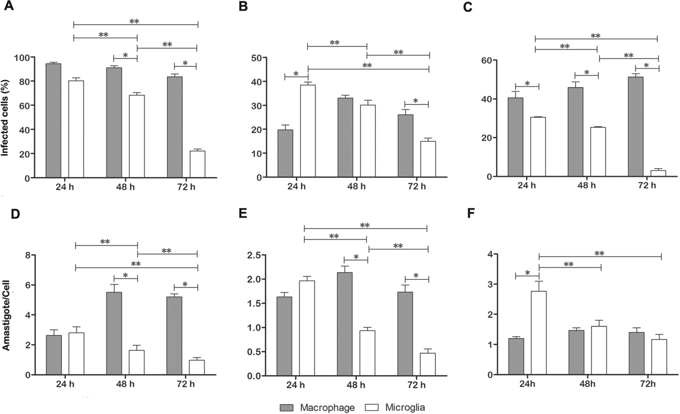
Kinetic studies were performed to compare the ability of each parasite to infect macrophages and microglia. The mean number of parasites per cell in L (L.) amazonensis was affected by both time post-infection (pi) and cell type (2-way ANOVA, P<0·0001). As the cultures progressed, the infectivity decreased significantly in the microglia but not in the macrophages (2-way ANOVA, P<0·0001). In macrophage cultures, the percentage of cells infected by L. (L.) amazonensis decreased from 94·3±2·25% at 24 h pi to 83·5±4% at 72 h pi; whereas the percentage of cells infected by L. (L) amazonensis in microglial cultures decreased from 80±4·3% (n = 6) at 24 h to 22±2·8% (n = 6) after 72 h (1-way ANOVA, P<0·05). The parasite burden per cell was then determined and there were significant differences between cell types (2-way ANOVA, P<0·0001). The mean values of amastigotes per macrophage increased from 2·6±0·65 (n = 3) at 24 h to 5·2±0·35 (n = 6) at 72 h (1-way ANOVA, P<0·05); whereas the amastigotes per cell decreased from 2·8±0·7 (n = 6) at 24 h to 0·9±0·32 (n = 6) at 72 h (1-way ANOVA, P<0·05) in microglial cultures.
In macrophage cultures, the percentage of cells infected by L. (V.) braziliensis increased from 19·7±3·5% at 24 h pi to 33±1·98% at 48 h pi, but no differences were detected after 72 h. On the other hand, a progressive significant decline in microglial infection was detected (38·43±2·1% (n = 6) at 24 h; 30·16±3·33% (n = 6) after 48 h, and 14·93±2·4% at 72 h; 1-way ANOVA, P<0·05). The parasite burden per cell was then determined and there were no significant differences between time windows after interaction with this species.
After interaction with L. (V.) shawi, the mean number of infected macrophages did not change significantly (40·53±5·8 after 24 h, 45·83±5·0 after 48 h, and 51·3±2·86 after 72 h; 1-way ANOVA, P>0·05); whereas the mean number of infected cells in microglia cultures significantly decreased (30·5±0·5, after 24 h, 25·3±0·6 after 48 h, and 3±1·7 after 72 h). In macrophage cultures, analysis of the number of parasites per cell in different time windows revealed no significant differences; whereas there was a significant decrease in the parasite burden in infected microglia in different time windows, except between 48 and 72 h.
Figure 7A–C illustrates the percentage of infected cells 24, 48 and 72 h after inoculation of macrophages or microglia cultures with L. (L.) amazonensis (A), L. (V.) braziliensis (B), and L. (V.) shawi (C). Figure 7D–F illustrates the number of parasites/cell after infection with L. (L.) amazonensis (D), L. (V.) braziliensis (E), and L. (V.) shawi (F). Microglia seem to be equally efficient in clearing all tested species, while macrophages are differentially sensitive to the species. For example, L. braziliensis infected fewer and L. (L.) amazonensis infected more macrophages. On the other hand, the lowest number of parasites per macrophage was observed with L. (V.) shawi infection, whereas the highest number occurred after L. (L.) amazonensis infection. As compared with macrophages, the clearance of parasites was much more efficient in microglia for all the Leishmania species tested, especially L. (V.) shawi.
The percentage of infected macrophages and microglia and mean number of amastigotes per cell (A–F). (*) indicates a significant difference (P<0·001), (**) indicates a significant difference (P<0·05). (A, D) = L. (L.) amazonensis; (B, E) = L. (V.) braziliensis; (C, F) = L. (V.) shawi. Note that microglia seem to reduce the number of parasites more rapidly than macrophages, independent of the Leishmania species. Errors bars indicate standard error (s.e.m.).

Leishmania was able to induce anti-inflammatory cytokine profile in macrophage but not in microglia
Supernatants from macrophage cultures infected with L. (V.) shawi and L. (V.) braziliensis promastigotes had significantly (1-way ANOVA, P<0·0001) increased levels of IL-12, TNF-α and TGF-β compared with respective controls, whereas there were no significant changes in TNF-α and IL-12 (P>0·05) after interaction with L. amazonensis, but a significant increase in TGF-β (1-way ANOVA, P<0·05) was observed (Fig. 8). In microglia, there was a significant increase in TNF-α and especially IL-12 (P<0·05) with all species of Leishmania tested. However, the levels of TGF-β in cell cultures infected with Leishmania were below the detection level of the ELISA immunoassay.
Graphic representations of TNF-α (A), IL-12 (B) and TGF-β (C) in culture supernatants. Macrophages and microglia were infected with L. (V.) shawi, L. (L.) amazonensis and L. (V.) braziliensis. The results represent the mean of three individual experiments. (*) = significant difference between cell types (P<0·001); (**) = significant difference between cell types (P<0·01); (***) = significant difference between cell types (P<0·05). Errors bars indicate standard error (s.e.m.).

DISCUSSION
The present study describes in vitro interactions of L. (V.) shawi, L. (V.) braziliensis and L. (L) amazonensis with mice peritoneal macrophages and microglia. The interactions were monitored by phase contrast and scanning electron microscopy, beginning at the initial contact of promastigotes with the host cells, until complete internalization of the parasites. We confirmed the notion that microglial cells show faster clearance of the parasites, compared with macrophages, in L. (L) amazonensis and expanded this finding to L. (V.) shawi and L. (V.) braziliensis. Although these phagocytes share the same cellular origin, some differences in morphology and cytokine production were revealed during the interactions. The novel findings of the present work are: (i) the demonstration of morphological differences in the phagocytosis; (ii) faster clearance of parasites by microglia; and (iii) higher levels of pro-inflammatory cytokines in microglia compared with macrophages.
Leishmania contact with microglia induces large ruffles on its cell surfaces
Unlike macrophages, the inoculation of promastigotes caused changes on the surface of microglia, which were independent of the Leishmania species. The rough aspect of the surface of microglia became smoother, with large cytoplasmic projections. These large cytoplasmic projections seem to participate in the process of internalization, allowing the involvement and engulfment of one or more parasites. The internalization of all species by microglia seemed to occur by both receptor-mediated phagocytosis and an association of this type of phagocytosis with large ruffles. Ruffles are sheet-like extensions of cell surfaces that form through localized assembly of actin filaments beneath the plasma membrane (Doherty and McMahon, Reference Doherty and McMahon2009). They are usually observed in macropinocytosis or circular dorsal ruffles, important fluid-phase endocytic pathways (Buccione et al. Reference Buccione, Orth and McNiven2004; Ladwein and Rottner, Reference Ladwein and Rottner2008; Lim and Gleeson, Reference Lim and Gleeson2011). Previous report (Wanderley et al. Reference Wanderley, Moreira, Benjamin, Bonomo and Barcinski2006) showed that, similar to what happens with apoptotic cells, amastigotes of L. (L.) amazonensis are internalized by macropinocytosis in macrophages. Furthermore, morphological studies demonstrated that after the interaction of the infective forms of T. cruzi with phagocytic or non-phagocytic cell types, plasma membrane can form protrusions, showing similarity with those observed during canonical phagocytosis or macropinocytic events (Barrias et al. Reference Barrias, Reignault, De Souza and Carvalho2012, Reference Barrias, de Carvalho and De Souza2013). The presence of these ruffles in microglia during the internalization of three Leishmania species and of large parasitophorous vacuoles containing L. (V.) shawi suggest a typical engulfment by macropinocytosis or a variation in the process of classical phagocytosis (canonical phagocytosis). However, to understand in detail the molecular mechanisms of the internalization process of promastigotes of Leishmania by microglia, further studies are necessary.
Leishmania live less time inside microglia compared with macrophages
Microglia are resident macrophages of the CNS with higher plasticity than the mature macrophage. Mature macrophages differ from microglia in the final stage of maturation and cannot proliferate or change their cellular phenotypes (Prinz et al. Reference Prinz, Priller, Sisodia and Ransohoff2011). Although the presence of amastigotes has been described in the liquor of an infant with clinical signs of kalazar and various case reports of visceral leishmaniasis with neurological signs (Prasad and Sen, Reference Prasad and Sen1996; Diniz et al. Reference Diniz, Duani, Freitas, Figueiredo and Xavier2010) (see Petersen and Greenlee, Reference Petersen and Greenlee2011), no clinical reports of cutaneous leishmaniasis were described. In addition, the presence of Leishmania in the human CNS has not been described, except in immunosuppressed patients (Walker et al. Reference Walker, Kublin and Zunt2006). In line with these reports, only a few others demonstrated CNS involvement in leishmaniasis in dogs (Garcia-Alonso et al. Reference Garcia-Alonso, Nieto, Blanco, Requena, Alonso and Navarrete1996; Nieto et al. Reference Nieto, Vinuelas, Blanco, Garcia-Alonso, Verdugo and Navarrete1996; Machado et al. Reference Machado, Melo, Moraes, Souza, Marcondes, Perri and Vasconcelos2010; Melo and Machado, Reference Melo and Machado2011) and the presence of amastigotes in the CNS of hamsters (Oliveira et al. Reference Oliveira, Oshiro, Pinto, Castro, Daniel, Oliveira, Junior, Guimaraes, Silva and Dorval2011) and mice (Abreu-Silva et al. Reference Abreu-Silva, Calabrese, Tedesco, Mortara and Goncalves da Costa2003). These occurrences were detected only at later stages of disease progression where infected subjects showed severe clinical signs or were immunosuppressed. In the present report, it was clear after 72 h that the number of parasites per cell was far less in microglial cultures than in macrophages. Indeed, the percentage values of infected cells in microglia cultures were 3, 15 and 22% compared with 51, 26 and 83% in macrophages after interaction with L. (V.) shawi, L. (V.) braziliensis and L (L.) amazonensis, respectively. In agreement with previous descriptions of L. (L) amazonensis (Baetas-da-Cruz et al. Reference Baetas-da-Cruz, Macedo-Silva, Santos-Silva, Henriques-Pons, Madeira, Corte-Real and Cavalcante2004), we confirmed that microglial cells are much more efficient than macrophages in clearing leishmania in vitro. Although Baetas-da-Cruz et al. (Reference Baetas-da-Cruz, Macedo-Silva, Santos-Silva, Henriques-Pons, Madeira, Corte-Real and Cavalcante2004) adopted pharmacological treatments to increase infectivity and used a single, different strain of leishmania, they also found microglia are more efficient in clearing parasites. Considering previously mentioned results and the present data, we suggest the absence of cerebral forms of human leishmaniasis may be related to the more efficient surveillance and clearance of the parasite by microglia.
Microglial cytokine profile may contribute to a higher clearance of Leishmania
To evaluate the possible cytokine profile of macrophages and microglia after infection by L. (V.) shawi, L. (V.) braziliensis and L. (L) amazonensis, we assessed the levels of TNF-α, IL-12 and TGF-β after 48 h of infection. Both macrophages and microglia produced TNF-α and IL-12; while microglia, unlike macrophages, did not produce TGF-β after infection with any of the species investigated. It is important to highlight that TNF-α and IL-12 levels in macrophages were dependent on the species used for infection. Compared with the control group, we did not find any changes in TNF-α and IL-12 levels after L (L.) amazonensis infection; while higher levels of these cytokines were systematically detected after L. (V.) braziliensis and L. (V.) shawi infections. On the other hand, production of IL-12 by microglia was significantly greater than by macrophages to all parasites tested.
It is relevant to mention that the control of Leishmania infection requires generation of a strong Th1 response (Alexander and Bryson, Reference Alexander and Bryson2005), which is driven by the presence of IL-12 at the initiation of the immune response (Scott et al. Reference Scott, Natovitz, Coffman, Pearce and Sher1988; Boom et al. Reference Boom, Liebster, Abbas and Titus1990; Heinzel et al. Reference Heinzel, Sadick, Mutha and Locksley1991; Ghalib et al. Reference Ghalib, Whittle, Kubin, Hashim, El-Hassan, Grabstein, Trinchieri and Reed1995; Chandra and Naik, Reference Chandra and Naik2008). Furthermore, the treatment of susceptible mice with recombinant IL-12 led to a curative response to Leishmania major infection via the up-regulation of IFN-γ production and inhibition of Th2 cytokines (Liew et al. Reference Liew, Millott, Parkinson, Palmer and Moncada1990; Heinzel et al. Reference Heinzel, Schoenhaut, Rerko, Rosser and Gately1993; Sypek et al. Reference Sypek, Chung, Mayor, Subramanyam, Goldman, Sieburth, Wolf and Schaub1993). The induction of IFN-γ production from Th1 and NK cells by IL-12 results in macrophage activation leading to nitric oxide (NO)-dependent killing of Leishmania parasites (Liew et al. Reference Liew, Millott, Parkinson, Palmer and Moncada1990; Wei et al. Reference Wei, Charles, Smith, Ure, Feng, Huang, Xu, Muller, Moncada and Liew1995; Stenger et al. Reference Stenger, Donhauser, Thüring, Röllinghoff and Bogdan1996). However, it has been well known that Leishmania enter and establish a ‘silent’ infection and the infection of macrophages by both promastigotes or amastigotes forms of Leishmania do not activate macrophages to produce IL-12 (Reiner et al. Reference Reiner, Zheng, Wang, Stowring and Locksley1994; Carrera et al. Reference Carrera, Gazzinelli, Badolato, Hieny, Muller, Kuhn and Sacks1996; Weinheber et al. Reference Weinheber, Wolfram, Harbecke and Aebischer1998; Bennett et al. Reference Bennett, Misslitz, Colledge, Aebischer and Blackburn2001). This absence of production of IL-12 may limit the Th1 response, resulting in the observed susceptibility to L. mexicana (Satoskar et al. Reference Satoskar, Bluethmann and Alexander1995; Stamm et al. Reference Stamm, Räisänen-Sokolowski, Okano, Russell, David and Satoskar1998; Rodriguez-Sosa et al. Reference Rodriguez-Sosa, Monteforte and Satoskar2001).
On the other hand, immune effects of TGF-β promote chemotaxis, inhibition of Th1 and Th2, and suppression of some of the macrophage microbicidal functions (Bogdan and Nathan, Reference Bogdan and Nathan1993; Boutard et al. Reference Boutard, Havouis, Fouqueray, Philippe, Moulinoux and Baud1995). TGF-β alters the innate macrophage phenotype by inhibiting TNF-α, IFN-γ and inducible NO synthase (iNOS), enhancing arginase expression (Wahl, Reference Wahl1994; Corraliza et al. Reference Corraliza, Soler, Eichmann and Modolell1995; Omer et al. Reference Omer, Kurtzhals and Riley2000). This diversion of arginine from iNOS to arginase leads to decreased NO, and enhanced polyamines, which could in turn increase parasite growth (Boutard et al. Reference Boutard, Havouis, Fouqueray, Philippe, Moulinoux and Baud1995; Corraliza et al. Reference Corraliza, Soler, Eichmann and Modolell1995; Iniesta et al. Reference Iniesta, Gómez-Nieto and Corraliza2001).
It has been shown that TGF-β plays an important role in the progression of leishmaniasis in rodents (Barral-Netto et al. Reference Barral-Netto, Barral, Brownell, Skeiky, Ellingsworth, Twardzik and Reed1992; Li et al. Reference Li, Hunter and Farrell1999), through a parasite escape mechanism which is associated with Leishmania survival in their mammalian host (Barral-Netto et al. Reference Barral-Netto, Barral, Brownell, Skeiky, Ellingsworth, Twardzik and Reed1992). Previous reports referred to an in vitro inhibitory action of TGF-β on macrophage (e.g. Barral-Netto et al. Reference Barral-Netto, Barral, Brownell, Skeiky, Ellingsworth, Twardzik and Reed1992). Indeed TGF-β is augmented during infection of inflammatory mouse peritoneal macrophage cultures with several different Leishmania sp.
In hamsters (infected with Leishmania donovani) and mice (infected with L. donovani or Leishmania chagasi), an excess of TGF-β has been found (Rodrigues et al. Reference Rodrigues, Santana da Silva and Campos-Neto1998; Wilson et al. Reference Wilson, Young, Davidson, Mente and McGowan1998; Melby et al. Reference Melby, Chandrasekar, Zhao and Coe2001).
Gantt et al. (Reference Gantt, Schultz-Cherry, Rodriguez, Jeronimo, Nascimento, Goldman, Recker, Miller and Wilson2003) also demonstrated that phagocytosis of L. chagasi increases the amount of biologically active TGF-β in human macrophage cultures without changing the total amount of TGF-β or TGF-β1 mRNA.
TGF-β has been implicated in cell susceptibility to various species of Leishmania, such as L. (V.) braziliensis (Barral et al. Reference Barral, Barral-Netto, Yong, Brownell, Twardzik and Reed1993; Reed, Reference Reed1999) through different effects on immune cells, including inactivation of certain functions of macrophages, for example reduction of the production of free radicals and the expression of MHC-II molecules.
In this work we suggest that a high production of anti-inflammatory cytokines by macrophages, such as TGF-β, may have sustained parasite survival, as has been extensively referred to in the literature. On the other hand, synergic effects of IL-12 and TNF-α, as well as the absence of TGF-β production by microglia contribute to a hostile environment to the parasites. Since previous descriptions demonstrated that TNF-α and IL-12 may induce microglial activation through nitric oxide synthase (iNOS) and NO production (Jana et al. Reference Jana, Dasgupta, Pal and Pahan2009; Harms et al. Reference Harms, Lee, Nguyen, Chang, Ruhn, Trevino and Tansey2012), it is reasonable to suggest that a higher microglial clearance of the parasites may be associated with IL-12 and TNF-α synergic effects.
The higher efficiency of microglia in eliminating Leishmania, compared with peritoneal macrophages, is confirmed in this paper. Both morphological and immune changes of microglia could be associated to this effect. We also described for the first time the profile of three cytokines (two pro-inflammatory, IL-12 and TNF-α, and one anti-inflammatory, TGF-β) produced by microglia during interaction with Leishmania.
Possible physiological implications of macrophagic and microglial responses after Leishmania interactions
A large number of parasites undergo phagocytosis in microglia, which use large ruffles. It is possible that microglia and macrophages show some differences in the repertoire of pattern recognition receptors associated with innate immunity response. If this is true, such differences may drive different mechanisms of immune response. Assuming that microglia activation and clearance of the parasites are related to the amount of nitric oxide and free superoxide radicals produced, it is reasonable to suppose that the absence of TGF-β and a higher level of IL-12 may be determinants of this effect. Further experimental investigations are needed to verify or disprove these assumptions.
In conclusion, microglia may have a higher phagocytic ability and cytotoxic potential than macrophages for all investigated species and we suggest that the absence of cerebral forms of leishmaniasis may be related to the more efficient surveillance and clearance of the parasite by microglia as compared with macrophages.
ACKNOWLEDGEMENTS
We are grateful to Dra. Maria Sueli Kataoka for critical review of the manuscript, Antonio Julio de Oliveira Monteiro (in memoriam), Antonio F.P. Martins, Raimundo Nonato Barbosa Pires and João Alves Brandão for their technical assistance and animal care. The experiments performed in this work were conducted in conformance with current Brazilian animal protection laws (Process no. 014/2005/CEPAN/IEC/SVS/MS).
FINANCIAL SUPPORT
This work was supported by Programa Nacional de Cooperação Acadêmica (PROCAD/CAPES), Instituto Evandro Chagas (Pará, Brazil).
CONFLICT OF INTEREST STATEMENT
The authors declare that there is no conflict of interest.