Introduction
Dermoid cysts in the floor of the mouth are relatively uncommon developmental lesions. They are thought to arise as a result of a defective development along embryonic lines of fusion containing both ectodermal and endodermal elements. The lesions tend to be slow growing. Geographical incidence rates are variable, ranging from 0.02 to 1.8 per cent.Reference Makos, Noussios, Peios, Gougousis and Chouridis1 There appears to be no sex predilection. The lesions are associated with a bimodal age distribution, with the greatest peak during the teenage years and a smaller peak during the first year of life, although cases have been reported in patients as young as 7 months and as old as 77 years.Reference Pan, Nakamura, Clark and Eisig2–Reference Menditti, Laino, Ferrara and Baldi5
In this paper, we present an unusual case of a 17-year-old female diagnosed with a rapidly growing dermoid cyst in the mouth floor. A review of the literature was performed; the aetiology and management of these lesions are discussed.
Case report
A 17-year-old Caucasian female was urgently referred to the oral surgery department with a 3-month history of a rapidly growing swelling in the floor of the mouth. The patient did not report any eating or swallowing difficulties, but her speech was mildly affected. She had no dyspnoea or pain. There was no history of previous surgery or trauma to the oral cavity or neck. The patient was otherwise healthy.
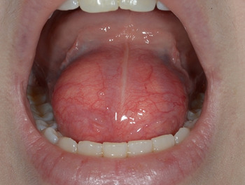
Clinical examination revealed a rather large symmetrical, midline, soft, non-tender and non-pulsatile swelling in the floor of the mouth, with normal covering mucosa (Figure 1). There was mild displacement of the tongue superiorly and posteriorly.
Pre-operative clinical presentation of the dome-shaped, symmetrical sublingual swelling.

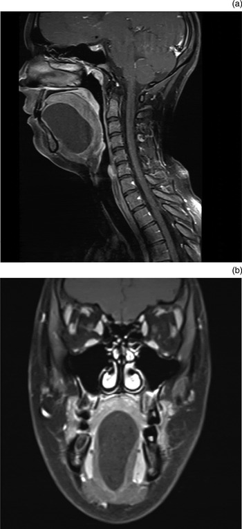
Magnetic resonance imaging (MRI) demonstrated a 6 cm well-defined, thin-walled cystic mass that lay within the sublingual space, with no extensions through the mylohyoid muscle (Figure 2). There was obstruction to the submandibular glands due to a pressure effect on the submandibular papillae. Minor displacement of the epiglottis was also evident.
(a) Sagittal and (b) coronal magnetic resonance imaging scans of the 6 cm well-circumscribed cystic mass, which extends from the sublingual area to the epiglottis level.

The fast growing nature of the cyst and its size raised the suspicion of potential compromise to the airway. Surgical excision was therefore advised.
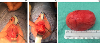
Under general anaesthesia, the patient underwent surgical enucleation of the cyst via an intraoral approach. The cyst was dissected from surrounding tissues en bloc (Figure 3) and the wound was closed primarily.
(a) Intraoral midline incision from the tongue base to the mouth floor, providing adequate access to the lesion. (b) Peri-operative view of the cystic mass. (c) View following removal; the specimen measured approximately 6 × 5 × 3.5 cm.

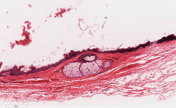
The procedure was uncomplicated and the tongue went back to its original position. Histopathology confirmed the diagnosis of a dermoid cyst (Figure 4).
Histological image of the cyst, showing keratinising squamous epithelium with a distinct granular layer, with a sebaceous gland in the cyst wall. (H&E; 40×).

Discussion
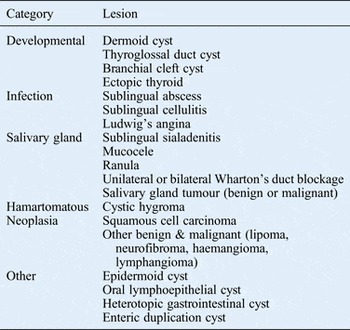
New and Erich found that only 6.94 per cent of dermoid cysts occurred in the head and neck, of which 11 per cent were located in the floor of the mouth.Reference New and Erich6 In our case, the hypothesis of an infection was discarded in light of the clinical presentation, period of evolution and absence of pain. Malignant tumour was ruled out in view of the lesion's clinical aspect and the absence of lymphadenopathy, although the latter is admittedly an imprecise indicator of malignancy. The two most plausible differential diagnoses were that of a ranula or an embryonic abnormality. Table I summarises the differential diagnoses of sublingual or cervical swellings.Reference MacNeil and Moxham7
Differential diagnoses of mouth or neck swellingsReference MacNeil and Moxham7

The pathogenesis of midline cysts of the mouth floor is not well established, with several theories suggested.Reference Menditti, Laino, Ferrara and Baldi5 Meyer divided cysts of the mouth floor into three categories based on histology: dermoid, epidermoid and teratoid.Reference Meyer8 In fact, dermoid cysts occur primarily in the testes and ovaries, and the most common location in the head and neck is the external third of the eyebrow.Reference King, Smith and Burk9
The most recent classification in the literature was outlined by Gordon et al., in 2013.Reference Gordon, Faquin, Lahey and Kaban10 These authors modified Meyer's classification of the cysts,Reference Meyer8 using new terminology, referring to the cysts as congenital germline fusion cysts. Congenital germline fusion cysts have traditionally been referred to as dysontogenic cysts. The following classification is specific to those cysts within the floor of the mouth: (1) epidermoid – stratified squamous epithelium lining with no dermal appendages; (2) dermoid – stratified squamous epithelium lining with dermal appendages, present within associated underlying connective tissue, including sebaceous glands, hair follicles and sweat glands; and (3) teratoid – stratified squamous epithelium lining with elements of all three germ layers, ectoderm, mesoderm and endoderm, in the underlying tissue.
The cysts can be further classified according to their site of development during embryogenesis:Reference Pryor, Lewis, Weaver and Orvidas11 group I develop along the nasolacrimal groove, and give rise to periorbital cysts; group II develop within the prenasal space, giving rise to cysts over the dorsum of the nose; group III develop along the midline of the first and second pharyngeal arches, giving rise to cysts within the sublingual, submental and submandibular regions; and group IV develop in the ventral or dorsal midline, typically in the thyroidal, suprasternal or suboccipital regions.
Three modes of pathogenesis have been described: acquired implantation, congenital teratoma and congenital inclusion.Reference New and Erich6, Reference Pryor, Lewis, Weaver and Orvidas11 However, as highlighted by Gordon et al., a teratoma is a solid neoplasm and should be considered separate to a teratoid variant germline fusion cyst.Reference Gordon, Faquin, Lahey and Kaban10 In view of this, there are only two true modes of pathogenesis: acquired implantation and congenital inclusion.Reference King, Smith and Burk9
Acquired implantation is the method by which a small sample of epithelium is separated and implanted within the underlying tissues, typically following surgery or trauma. This was simulated in a study by Baker and Mitchell by surgically implanting an autogenous sample of epithelium into the deeper connective tissue on the backs of rats.Reference Baker and Mitchell12
Congenital inclusion cysts are formed during embryogenesis, where disruption occurs during the fusion of the embryological components, causing in-folding of the epithelium.Reference Erich and Johnsen13 The mechanism underlying group II (nasal) cysts is described in particular detail within the literature, with interest stemming from the neuroectodermal involvement during development which results in the intracranial extension of some cysts. There have been many proposed theories; however, the most widely accepted theory is that initiated by Grunwald in 1910, and further interpreted by Pratt and Bradley.Reference Bradley14–Reference Rahbar, Shah, Mulliken, Robson, Perez-Atayde and Proctor16
A number of symptoms can be experienced by patients, including dysphagia, dyspnoea, and difficulty masticating and speaking.Reference Hitchin and Smith17, Reference Ohta, Watanabe, Ito, Kubota, Suzuki and Ishida18 Cysts occurring above the mylohyoid muscle tend to displace the tongue superiorly, and those that occur below may cause a double chin appearance extraorally.Reference Mesolella, Cantone, Galli and Iengo19 There have been reports in the literature of dermoid cysts dramatically increasing in size during pregnancy. It has been postulated that this may be caused by an increase in sebum production from the sebaceous glands present in the cyst lining.Reference Boko, Amaglo and Kpemissi20 Mesolella et al. proposed an alternative theory, suggesting that the rapid growth is associated with increased plasma levels of oestrogens and progesterone, which act as growth factors on the cyst.Reference Mesolella, Cantone, Galli and Iengo19 Very large dermoid cysts have been reported in the literature, but these are rare. When they do occur, there may be a risk to the airway, and tracheotomy may even be required as part of management.Reference Boko, Amaglo and Kpemissi20
Radiography is an important adjunct in the diagnosis and management of dermoid cysts. Ultrasound, computed tomography (CT) and MRI have all been reported in the literature. Ultrasound may be helpful in differentiating between solid, vascular and cystic lesions.Reference Jadwani, Misra, Kallianpur and Bansod21 It is generally accepted that CT and MRI imaging allow more precise localisation in comparison to ultrasound. A high signal on T2- and a low signal on T1-weighted imaging typically indicate the presence of cystic structures.Reference Ho and Crean22
Aspiration cytology may be useful in differentiating between types of cysts.Reference Ohta, Watanabe, Ito, Kubota, Suzuki and Ishida18 However, the high-density fluid contents of dermoid cysts do not easily aspirate, limiting the usefulness of this technique.Reference Mesolella, Cantone, Galli and Iengo19 Histological examination is essential to confirm the diagnosis.
Although there are no set rules, the most appropriate time to operate is when symptoms such as dysphagia, dysphonia and dyspnoea are present. Treatment for sublingual dermoid cysts is almost always surgical. The aspiration of cystic contents was previously attempted by Mesolella et al. in order to debulk a large sublingual dermoid cyst; however, this proved unsuccessful because of the high-density fluid content.Reference Mesolella, Cantone, Galli and Iengo19
There is debate in the literature as to the suitability of intraoral and extraoral surgical approaches when removing dermoid cysts. Generally, the size and anatomical location of the cysts are the two most significant considerations.Reference El-Hakim and Alyamani23 It has been stated that an intraoral approach is more suitable for sublingual cysts of up to 6 cm in size, whereas an extraoral approach may be advantageous for those cysts greater than 6 cm.Reference Ohta, Watanabe, Ito, Kubota, Suzuki and Ishida18 There are, however, case reports that detail the removal of very large dermoid cysts carried out by intraoral approach, which claim a cosmetic advantage. Boko et al. reported on the intraoral surgical excision of a dermoid cyst measuring 13 cm in length.Reference Boko, Amaglo and Kpemissi20 McGregor proposed a symphyseal mandibular osteotomy approach for larger lesions, citing restricted surgical access as a limiting factor for conventional intraoral and extraoral approaches.Reference McGregor24 With regards to anatomical location, Longo et al. reported that extraoral incision is mandatory if the cyst is located beneath the mylohyoid muscle.Reference Longo, Maremonti, Mangone, De Maria and Califano25
Prognosis following surgical removal is excellent, with a low incidence of relapse. Malignant changes have been recorded in dermoid cysts, by New and Erich, but not in the mouth floor.Reference New and Erich6 However, a 5 per cent rate of malignant transformation of the teratoid type or oral dermoid cysts has been reported by other authors.Reference Tsirevelou, Papamanthos, Chlopsidis, Zourou and Skoulakis26
• Dermoid cysts in the mouth floor are relatively uncommon developmental lesions
• They tend to be slow growing
• Differential diagnosis is of paramount importance, as recommended surgical techniques vary depending on lesion size and anatomical position
• Prompt diagnosis and treatment can prevent potential airway compromise
Differential diagnosis of cystic lesions in the mouth floor is of paramount importance, as the recommended surgical techniques vary depending on the size and anatomical position of the lesions. The intraoral approach is the preferred treatment modality for those lesions that do not extend below the mylohyoid muscle boundaries; this leads to a satisfactory cosmetic and functional outcome. Extraoral incision is mandatory only if the cyst lies below the mylohyoid muscle.
Acknowledgement
The microscopic view presented is courtesy of Dr Keith Hunter (Reader and Honorary Consultant, Academic Unit of Oral and Maxillofacial Pathology) School of Clinical Dentistry, University of Sheffield, UK.