Introduction
Disrupted connectivity across distributed neural networks has been proposed as a pathophysiological mechanism underlying schizophrenia (SZ) (Andreasen, Reference Andreasen1999; Cannon, Reference Cannon2015; Friston, Reference Friston1998; Stephan, Baldeweg, & Friston, Reference Stephan, Baldeweg and Friston2006; Stephan, Friston, & Frith, Reference Stephan, Friston and Frith2009). These widespread abnormalities can be investigated using resting-state functional magnetic resonance imaging (fMRI)-based connectivity, which measures the temporal synchronization of low-frequency oscillations of the blood oxygen level-dependent (BOLD) signal between brain regions (Biswal, Yetkin, Haughton, & Hyde, Reference Biswal, Yetkin, Haughton and Hyde1995; Raichle, Reference Raichle2011).
While abnormalities have been observed across several functional networks in SZ, dysconnectivity within thalamo–striatal–cortico–cerebellar circuits has been widely replicated (e.g. Anticevic et al., Reference Anticevic, Cole, Repovs, Murray, Brumbaugh, Winkler and Glahn2014; Cheng et al., Reference Cheng, Palaniyappan, Li, Kendrick, Zhang, Luo and Feng2015; Damaraju et al., Reference Damaraju, Allen, Belger, Ford, McEwen, Mathalon and Calhoun2014; Klingner et al., Reference Klingner, Langbein, Dietzek, Smesny, Witte, Sauer and Nenadic2014; Li et al., Reference Li, Wang, Zhang, Rolls, Yang, Palaniyappan and Feng2017; Woodward, Karbasforoushan, & Heckers, Reference Woodward, Karbasforoushan and Heckers2012). Converging evidence has substantiated findings of thalamic hypoconnectivity withprefrontal and cerebellar regions, as well as thalamic hyperconnectivity with sensory regions, suggesting thalamo–striatal–cortico–cerebellar dysconnectivity as a potential biomarker of SZ. Findings that are inconsistent with this pattern [e.g. (Guller, Tononi, & Postle, Reference Guller, Tononi and Postle2012; Wang, Rau, Li, Chen, & Yu, Reference Wang, Rau, Li, Chen and Yu2015)], may stem from heterogeneity in study methodologies and samples common in fMRI research. Further, aspects of thalamic dysconnectivity may relate to positive symptoms, with observations of positive correlations with the thalamus and middle temporal hyperconnectivity and negative correlations with cerebello–thalamic hypoconnectivity (Ferri et al., Reference Ferri, Ford, Roach, Turner, van Erp, Voyvodic and Mathalon2018).
While thalamic dysconnectivity is better characterized in chronic SZ, less is known regarding when these abnormalities emerge in the illness course. Neurodevelopmental models of SZ (e.g. Feinberg, Reference Feinberg1982; Weinberger, Reference Weinberger1987) suggest that abnormalities arise prior to illness onset, stemming from abnormal brain maturation including insufficient pruning of thalamic-sensory connections (Fair et al., Reference Fair, Bathula, Mills, Dias, Blythe, Zhang and Nagel2010; Giraldo-Chica & Woodward, Reference Giraldo-Chica and Woodward2017). However, most investigations of thalamic dysconnectivity in SZ focused on adults with a chronic illness course (e.g. Anticevic et al., Reference Anticevic, Cole, Repovs, Murray, Brumbaugh, Winkler and Glahn2014; Cheng et al., Reference Cheng, Palaniyappan, Li, Kendrick, Zhang, Luo and Feng2015; Damaraju et al., Reference Damaraju, Allen, Belger, Ford, McEwen, Mathalon and Calhoun2014; Klingner et al., Reference Klingner, Langbein, Dietzek, Smesny, Witte, Sauer and Nenadic2014; Li et al., Reference Li, Wang, Zhang, Rolls, Yang, Palaniyappan and Feng2017; Woodward et al., Reference Woodward, Karbasforoushan and Heckers2012). Woodward and Heckers (Reference Woodward and Heckers2016) report thalamic dysconnectivity early in the course of SZ (ESZ), with hypoconnectivity between the thalamus and cerebellum, and hyperconnectivity between the thalamus and somatosensory cortices in individuals <2 years from illness onset. Anticevic et al. (Reference Anticevic, Haut, Murray, Repovs, Yang, Diehl and Cannon2015) report similar abnormalities in a large sample of individuals with psychosis risk syndrome (PRS), with dysconnectivity being more pronounced in those who subsequently developed a psychotic illness. Furthermore, when healthy control (HC) and PRS samples were considered together, there was a negative relationship between positive symptoms and thalamic hypoconnectivity.
These findings suggest that thalamic dysconnectivity patterns observed in chronic SZ may also be present in earlier illness stages. However, with so few studies of thalamic functional connectivity in ESZ and PRS, prior findings warrant replication, as well as the examination of ESZ and PRS samples within the same study. Accordingly, the current study examined whole-brain resting-state thalamic functional connectivity in a sample of 45 individuals meeting PRS criteria, 74 individuals with ESZ and 85 HCs. We also examined five a priori regions of interest (ROIs) that were functionally defined from group differences observed in the multisite North American Prodrome Longitudinal Study (NAPLS-2) cohort in a prior PRS study of thalamic connectivity (Anticevic et al., Reference Anticevic, Haut, Murray, Repovs, Yang, Diehl and Cannon2015). For both whole-brain and ROI-based analyses, we hypothesized that the PRS group would demonstrate increased thalamic connectivity with sensory regions and reduced thalamic connectivity with the cerebellum and prefrontal regions relative to HCs, and that these effects would be more pronounced in the ESZ group. We also examined the relationships between positive symptoms and thalamic dysconnectivity previously observed in chronic SZ (Ferri et al., Reference Ferri, Ford, Roach, Turner, van Erp, Voyvodic and Mathalon2018) and PRS (Anticevic et al., Reference Anticevic, Haut, Murray, Repovs, Yang, Diehl and Cannon2015) samples.
Methods
PRS participants (n = 45) were recruited from the University of California, San Francisco's (UCSF) Path program. PRS participants met Criteria of Psychosis-risk Syndromes (COPS) based on the structured interview for psychosis-risk syndromes (SIPS) (McGlashan, Walsh, & Woods, Reference McGlashan, Walsh and Woods2010; Miller et al., Reference Miller, McGlashan, Rosen, Somjee, Markovich, Stein and Woods2002, Reference Miller, McGlashan, Rosen, Cadenhead, Cannon, Ventura and Woods2003), for at least one of three sub-syndromes: attenuated positive symptom state (APSS; n = 43/45), brief intermittent psychotic state (BIPS n = 0/45), and/or genetic risk and deterioration state (GRDS n = 5/45). Symptom severity in PRS patients was assessed using the scale of prodromal symptoms (SOPS).
ESZ patients (n = 74) within 5 years of illness onset (mean = 1.72 ± 1.35 years) were recruited from the Early Psychosis Clinic at UCSF and the community. Diagnosis of SZ or schizoaffective disorder was confirmed using the Structured Clinical Interview for DSM-IV-TR (SCID) (First, Spitzer, Gibbon, & Williams, Reference First, Spitzer, Gibbon and Williams2002). The scale for the assessment of positive symptoms (SAPS; Andreasen, Reference Andreasen1984) and the scale for the assessment of negative symptoms (SANS; Andreasen, Reference Andreasen1989) were used to assess clinical symptom severity in ESZ. The majority of ESZ patients were prescribed antipsychotic medication (n = 66/74; 89.2%) while none of the PRS participants were (n = 0/45; 0%). After participating in neuroimaging, PRS participants were followed for 24 months or until they transitioned to psychosis. Six PRS individuals transitioned to a psychotic disorder [i.e. met the presence of psychotic symptom (POPS) criteria; Miller et al., Reference Miller, McGlashan, Rosen, Cadenhead, Cannon, Ventura and Woods2003] while 21 were followed for the full 24-month follow-up period and did not transition to psychosis (with the remainder lost to follow-up).
HC participants (n = 85) recruited from the community were screened for Axis I disorders with the SCID-IV-TR (First et al., Reference First, Spitzer, Gibbon and Williams2002), or for HC <16, the Kiddie-Schedule for Affective Disorders and Schizophrenia for school-age children-present and lifetime version (Kaufman et al., Reference Kaufman, Birmaher, Brent, Rao, Flynn, Moreci and Ryan1997).
Participants were 12–35 years old and were excluded for diagnostic and statistical manual of mental disorders (DSM-IV) past-year substance dependence (excepting nicotine), a history of head injury with loss of consciousness, central nervous system disorder, or, for HC participants, a first-degree relative with a psychotic illness. Written informed consent or assent was obtained from study participants under protocols approved by the Institutional Review Board at UCSF. This protocol complies with the ethical standards of national and international committees on human experimentation and with the Helsinki Declaration of 1975, as revised in 2008.
Data acquisition and processing
Resting fMRI data were acquired on a Siemens 3T TIM TRIO scanner at the UCSF Neuroimaging Center using a whole-brain echo-planar imaging (EPI) sequence (6 min): 180 functional images were acquired (32 axial slices, 3.5 mm slice thickness, 1.05 mm inter-slice gap, repetition time (TR) = 2 s, echo time (TE) = 29 ms, flip angle = 75°, field of view (FOV) = 24 cm). Participants were instructed to rest with their eyes open.
Image preprocessing was performed with Statistical Parametric Mapping (SPM8; http://www.fil.ion.ucl.ac.uk/spm/software/spm8/). Motion correction via affine registration realigned all images to the first image using INRIAlign (http://www.fil.ion.ucl.ac.uk/spm/ext/#INRIAlign). Images were slice-time corrected and the mean motion-corrected functional image was normalized to standard neuroanatomical space (Montreal Neurological Institute's MNI-EPI template; http://www.bic.mni.mcgill.ca), resulting in 3 mm3 voxel dimensions. Data were spatially smoothed with a 6 mm full-width half-maximum Gaussian kernel. The artifact detection tools package (ART; http://www.nitrc.org/projects/artifact_detect/) identified outlying volumes based on global image intensity values (Z > 3) and head motion (>2 mm translational movement in the x, y, or z planes, or >0.02° rotation in yaw, pitch, or roll). ACompCor, a regression-based algorithm for denoising BOLD data, was applied (Behzadi, Restom, Liau, & Liu, Reference Behzadi, Restom, Liau and Liu2007). ACompCor performs principal components analysis (PCA) on time-series data by deriving white matter and cerebrospinal fluid (CSF) noise regions from segmented high-resolution T1-weighted anatomical images co-registered to functional data. White matter noise regions were derived from FreeSurfer (http://surfer.nmr.mgh.harvard.edu/) segmentation, while CSF noise regions were derived from SPM8 segmentation. A binary union mask of noise ROIs was co-registered to the mean functional scan. Time-series data for the voxels in the noise masks were then subjected to PCA, and a number of noise components comprising weighted averages of white matter and CSF voxel time-series were identified using a bootstrap procedure (Behzadi et al., Reference Behzadi, Restom, Liau and Liu2007).
The first-level GLM included the mean thalamic seed time-series and seven ART motion parameters, consisting of the temporal derivatives of the six motion parameters as well as a composite measure of total motion across translation and rotation. Regressors were also included for (i) data points identified by the ART toolbox as outliers and (ii) statistically significant (p < 0.05) principal noise components from the ACompCor denoising routine retained from each individual fMRI run.
Data analysis
To control for expected brain maturation across the age range studied and age differences between clinical groups, normal maturation effects were statistically removed from the functional connectivity data. We modeled normal aging effects in the HC group and calculated age-adjusted Z-scores for all subjects based on the HC group age-regression (Fryer et al., Reference Fryer, Woods, Kiehl, Calhoun, Pearlson, Roach and Mathalon2013a, Reference Fryer, Jorgensen, Yetter, Daurignac, Watson, Shanbhag and Mathalon2013b, Reference Fryer, Roach, Wiley, Loewy, Ford and Mathalon2016; Mathalon, Pfefferbaum, Lim, Rosenbloom, & Sullivan, Reference Mathalon, Pfefferbaum, Lim, Rosenbloom and Sullivan2003; Pfefferbaum et al., Reference Pfefferbaum, Lim, Zipursky, Mathalon, Rosenbloom, Lane and Sullivan1992):

Thus, a participant's age-adjusted z-score voxel-wise map reflects the deviation in functional connectivity, expressed in standard deviation units, from that expected for a healthy individual of the same age.
A seed-based connectivity analysis was conducted using the CONN toolbox (Whitfield-Gabrieli & Nieto-Castanon, Reference Whitfield-Gabrieli and Nieto-Castanon2012) (http://www.nitrc.org/projects/conn) with a bilateral seed of the whole thalamus, defined anatomically (Talairach Daemon atlas; Lancaster et al., Reference Lancaster, Woldorff, Parsons, Liotti, Freitas, Rainey and Fox2000). The mean bilateral thalamic seed time-series vector was generated by averaging the time-series data across all seed voxels from the normalized functional images prior to smoothing. Voxel-wise correlation and anti-correlation (i.e. negative correlation) maps were generated for each subject, representing the Pearson's r correlation value between the seed time-series and every voxel in the brain. These r values were transformed to Z-scores via Fisher's transformation, resulting in voxel-wise thalamic functional connectivity Z-score maps for each participant.
Connectivity values were tested for significance between-groups in SPM8 on age-adjusted functional connectivity Z-score maps using a whole-brain false discovery rate (FDR) correction, p < 0.01. Mean connectivity values from FDR-corrected regions showing a significant main effect of Group (HC, ESZ, PRS) were extracted for all participants and followed-up with Tukey-Kramer honest significant difference (HSD) corrected post-hoc tests.
We also examined thalamic connectivity within five a priori ROIs generated from significant between-group differences in thalamic connectivity in an independent sample comparing HCs, PRS individuals who later converted to a psychotic disorder, and PRS individuals who did not (Anticevic et al., Reference Anticevic, Haut, Murray, Repovs, Yang, Diehl and Cannon2015). These ROIs included the left cerebellum, the right lateral PFC, the left sensory/motor cortex, the right sensory/motor cortex, and the anterior cingulate. Pairwise t testing followed-up differences in the thalamic connectivity between groups. Fisher r-to-Z transformed correlation coefficients were HC age-adjusted (z-scored) such that each subject's ROI mean reflects connectivity differences, in standardized units, from the expected connectivity for a healthy subject of the same age.
Scan head motion was compared between the groups via ANOVA by calculating the average motion displacement (Van Dijk, Sabuncu, & Buckner, Reference Van Dijk, Sabuncu and Buckner2012), defined as the root-mean-square (RMS) of the scan-to-scan change in x–y–z translation (in mm) or roll–pitch–yaw rotation (in degrees) parameters. The impact of differences in head motion between the groups was examined by re-running analyses with a covariate for the motion metrics (translational or rotational) that differed between groups. Antipsychotic medication effects within ESZ were examined by correlating chlorpromazine (CPZ) dosage equivalents (Danivas & Venkatasubramanian, Reference Danivas and Venkatasubramanian2013; Woods, Reference Woods2003) with the extracted thalamic connectivity means from each ESZ patient within regions showing a significant Group effect. Non-parametric correlations (Spearman's ρ) were used due to the non-normality of the CPZ variable. Next, means for all clusters showing a significant main effect of Group were extracted and examined between PRS participants who converted to a psychotic disorder within 24 months of study entry (n = 6) v. non-converters (n = 21) who had been followed clinically for at least 24 months.
To examine clinical correlations, several families of tests were conducted guided by past studies in SZ and PRS groups. Our previous chronic SZ study (Ferri et al., Reference Ferri, Ford, Roach, Turner, van Erp, Voyvodic and Mathalon2018), reported thalamic dysconnectivity relationships with positive symptom severity driven by SAPS delusions and hallucinations global ratings. In PRS individuals, prior studies have shown the delusional thought (item P1: Unusual Thought Content; UTC) and suspiciousness (item P2) ratings to most strongly predict transition to psychosis, motivating a focus on these domains (Cannon et al., Reference Cannon, Cadenhead, Cornblatt, Woods, Addington, Walker and Heinssen2008; Perkins et al., Reference Perkins, Jeffries, Cornblatt, Woods, Addington, Bearden and McGlashan2015). In order to interrogate similar domains in ESZ and PRS groups, we included the SAPS persecutory ideas item in ESZ and the SOPS perceptual abnormalities/hallucinations item (item P4) in PRS. Thus, for all ROI clusters showing significant group effects, the ROIs were correlated with SAPS delusions, persecutory ideas, and hallucinations in ESZ, and SOPS UTC, suspiciousness, and SOPS perceptual abnormalities/hallucinations in PRS, with family-wise FDR correction (p < 0.05) applied to control type 1 error.
Lastly, in order to attempt to replicate correlations between connectivity and SOPS positive symptom totals in PRS (Anticevic et al., Reference Anticevic, Haut, Murray, Repovs, Yang, Diehl and Cannon2015), thalamic connectivity values were extracted from the exact ROI clusters reported by Anticevic et al. and summed to derive a separate hyperconnected and hypoconnected distributed ROI summary score. These two summary scores were then correlated with the SOPS positive symptom total score in the PRS sample (using a Bonferroni-adjusted α-level of 0.025). Mean age-adjusted (z-scored) connectivity values were extracted from the left cerebellum (peak MNI coordinate: x = 39, y = −46, z = −47) and the right medial temporal gyrus (MTG; x = 63, y = −1, z = −23) for each ESZ participant to test if the previously observed correlations in chronic SZ between thalamic dysconnectivity to these regions and SAPS global totals (Ferri et al., Reference Ferri, Ford, Roach, Turner, van Erp, Voyvodic and Mathalon2018) would replicate.
Results
Participant demographic and clinical data
The groups did not differ on gender (p = 0.370), handedness (p = 0.145), or average parental SES (p = 0.133). There were no significant group differences in rotational movement (p > 0.29), however there was a significant group effect (p = 0.009) of translational movement, with the HC group moving significantly less than the ESZ group (p = 0.009). There was a trend level group effect of age (p = 0.07), with the HC group being slightly older than the PRS group (p = 0.05). See Table 1 for group means and standard deviations for demographic and clinical variables.
Demographic and clinical data for HC, PRS, and ESZ groups

a Socioeconomic status (SES) measured by the Hollingshead two-factor index (Hollingshead & Redlich, Reference Hollingshead and Redlich1958). Higher Hollingshead scores indicate lower SES. All other assessment measures are scaled such that higher scores reflect greater levels of the measured variable.
Voxel-wise analyses
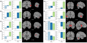
A main effect of Group was detected in eight age-adjusted and FDR-corrected regions. For all regions, the PRS group had mean thalamic connectivity values that numerically fell between those of HC and ESZ patients. Figure 1 and Table 2 depict the thalamic connectivity values and test statistics for pairwise Group follow-up tests for each cluster. Tukey HSD-corrected pairwise follow-up comparisons are reported below for each cluster.
(a) Right middle/superior temporal gyrus (k = 189). This cluster included the right middle and superior temporal gyri. Thalamic connectivity with this region was significantly greater in both ESZ and PRS relative to HC (p's < 0.001) and ESZ was significantly greater than PRS (p = 0.023).
(b) Right middle temporal gyrus 1 (k = 18). This cluster contained voxels primarily from the right middle temporal gyrus with minor extension into the superior temporal gyrus. Thalamic connectivity to this region was significantly greater in ESZ and PRS relative to HC (p's < 0.001), however ESZ was not significantly different from PRS (p = 1.00).
(c) Right middle temporal gyrus 2 (k = 18). This cluster contained voxels primarily from the right middle temporal gyrus with minor extension into the inferior temporal gyrus. Thalamic connectivity with this region was significantly greater in ESZ relative to HC (p < 0.001) and marginally greater relative to PRS (p = 0.057). PRS did not differ from HC (p = 0.180).
(d) Left superior temporal gyrus (k = 37). This cluster contained superior temporal gyrus. Thalamic connectivity with this region was significantly greater in ESZ compared to HC (p < 0.001) and PRS (p = 0.028), however PRS did not significantly differ from HC (p = 0.189).
(e) Left middle temporal gyrus 1 (k = 40). This cluster primarily consisted of voxels from the middle temporal gyrus, but also extended into the middle occipital gyrus. Thalamic connectivity with this region was significantly greater in ESZ relative to both HC and PRS (p's < 0.001), however HC did not differ from PRS (p = 0.878).
(f) Left middle temporal gyrus 2 (k = 432). This cluster primarily included the left middle temporal gyrus, extending into the left superior temporal gyrus. Thalamic connectivity with this region was significantly greater in ESZ relative to HC (p < 0.001) and marginally greater in ESZ compared to PRS (p = 0.057). HC did not differ from PRS (p = 0.180).
(g) Bilateral thalamus (k = 592). This cluster contained both left and right thalamus including the ventral lateral, medial dorsal, ventral anterior, lateral posterior, ventral posterior lateral, ventral posterior medial, and lateral dorsal nuclei. Thalamic connectivity with this region was significantly reduced in both ESZ and PRS relative to HC (p's < 0.001). ESZ was marginally less than PRS (p = 0.088).
(h) Left cerebellum (k = 74). This cluster was primarily in the posterior cerebellum, with extension into the anterior lobe. Thalamic connectivity with the left cerebellum was significantly reduced in both ESZ and PRS relative to HC (p's < 0.001), however ESZ was not significantly different from PRS (p = 0.696). Twenty (23%) of the 74 voxels in this cluster overlapped with the cerebellum ROI from Anticevic et al. (Reference Anticevic, Haut, Murray, Repovs, Yang, Diehl and Cannon2015) study. There were no other overlapping voxels between this whole-brain analysis and the a priori ROI analyses described below.
Bar graphs display age-adjusted mean functional connectivity values by Group for each cluster showing a significant main effect of Group (FDR-corrected 0.010 height threshold). Regions correspond to cluster labels reported in the Results section and Table 2: (a) Right middle/superior temporal gyrus; (b) Right middle temporal gyrus 1; (c) Right middle temporal gyrus 2; (d) Left superior temporal gyrus; (e) Left middle temporal gyrus 1; (f) Left middle temporal gyrus 2; (g) Bilateral thalamus; (h) Left cerebellum.

Brain regions showing significant group differences in thalamic connectivity when comparing age-adjusted z-scores of HC (n = 85), PRS (n = 74), and ESZ (n = 45) groups

ROI analyses
-
Cerebellum. Compared to HC, both PRS (t(201) = −2.223, p = 0.027) and ESZ (t(201) = −2.755, p = 0.006) demonstrated reduced connectivity between the cerebellum and the thalamus, consistent with our whole-brain findings.
-
Right lateral PFC. Compared to HC, thalamic connectivity with right lateral PFC was not significantly reduced in PRS (t(201) = −1.166, p = 0.245), or ESZ (t(201) = −1.794, p = 0.074, with a trend toward an effect in ESZ).
-
Left sensory/motor cortex. Compared to HC, thalamic connectivity to the left sensory/motor cortex was not significantly enhanced in PRS (t(201) = 0.892, p = 0.373), but was significantly enhanced in ESZ (t(201) = 2.361, p = 0.019).
-
Right sensory/motor cortex. Compared to HC, thalamic connectivity with the right sensory/motor cortex was not significantly enhanced in either PRS (t(201) = 0.388, p = 0.669) or ESZ (t(201) = 1.541, p = 0.125).
-
Anterior cingulate cortex (ACC). Thalamic connectivity with the ACC was marginally reduced in PRS compared to HC (t(201) = −1.665, p = 0.097) and significantly reduced in ESZ compared to HC (t(201) = −3.27, p = 0.001).
No significant differences were found between PRS and ESZ for any of the five ROIs (all p's > 0.260). Our results were all in the same direction as Anticevic et al. (Reference Anticevic, Haut, Murray, Repovs, Yang, Diehl and Cannon2015) PRS study.
Motion artifact
Clusters showing a significant main effect of Group were extracted for all subjects, and analyses were repeated adding translational displacement as a covariate. All Group differences reported in the original analysis remained significant (all p's < 0.001). Corrected pairwise comparisons revealed the same pattern of significance, excepting the difference between PRS and ESZ for the left superior temporal gyrus changed from p = 0.028 to 0.053. Further, when dropping the nine ESZ subjects with the most translational movement from analysis, all comparisons of extracted cluster means remain significant (all p's < 0.001), with the same pattern of directionality.
Antipsychotic medication dosage in the ESZ group
Within our ESZ patient sample, CPZ equivalents did not covary with any of the regions showing the main effect of Group (Spearman's ρ < |0.15|; all p's >0.29).
Diagnosis within the ESZ group
Comparison of ESZ subgroups diagnosed with the schizoaffective disorder (n = 23) or SZ (n = 51), showed no significant differences in thalamic dysconnectivity between the diagnostic subgroups (0.741 < p < 0.999).
Psychosis transition within the PRS group
There were no significant differences between individuals who transitioned to a psychotic disorder (n = 6) and those who were clinically followed for 24 months and did not (n = 21) in any of the eight regions showing a main effect of group in thalamic connectivity (all p's > 0.20) or any of five regions from Anticevic et al. (Reference Anticevic, Haut, Murray, Repovs, Yang, Diehl and Cannon2015) (all p's > 0.132).
Correlation of thalamic connectivity with clinical symptoms
No correlations survived adjustment for multiple comparisons within either ESZ or PRS groups for any of the regions examined.
Discussion
These results extend and replicate prior findings by showing thalamic dysconnectivity, in a single study, in both ESZ and PRS. Specifically, voxel-wise analyses demonstrated greater thalamic functional connectivity with middle and superior temporal regions, and reduced connectivity with cerebellar and thalamic regions in ESZ compared to HCs. ROI analyses revealed reduced thalamic connectivity with the cerebellum and the ACC and enhanced connectivity to the left sensory/motor cortex in ESZ compared to HCs. In the PRS group, voxelwise analyses indicated thalamic hyperconnectivity with some, but not all, middle temporal regions, and hypoconnectivity with cerebellar and thalamic regions compared to HCs. Similar to voxel-wise results, ROI analyses also revealed cerebellum hypoconnectivity in PRS patients that was equivalent to ESZ. Overall, these findings converge with the small literatures on ESZ (Woodward & Heckers, Reference Woodward and Heckers2016) and PRS (Anticevic et al., Reference Anticevic, Haut, Murray, Repovs, Yang, Diehl and Cannon2015), and unlike previous studies, were obtained in one setting using identical methods, strengthening evidence that thalamic dysconnectivity patterns associated with chronic SZ are also present in earlier stages of the illness. The brain's intrinsic architecture can be organized into distributed, canonical networks, (Power et al., Reference Power, Cohen, Nelson, Wig, Barnes, Church and Petersen2011; Yeo et al., Reference Yeo, Krienen, Sepulcre, Sabuncu, Lashkari, Hollinshead and Buckner2011), with alterations of many of these well-described functional networks reported in SZ (e.g. Li et al., Reference Li, Wang, Zhang, Rolls, Yang, Palaniyappan and Feng2017). The thalamus is increasingly conceptualized as an integrative hub of functional connectivity, owing to its high degree of global connectivity to many discrete cortical regions; this challenges historical views of the thalamus as a ‘passive’ relay station, recasting the region instead as an actor that integrates information across disparate cortical networks (Hwang, Bertolero, Liu, & D'Esposito, Reference Hwang, Bertolero, Liu and D'Esposito2017). Thus, the thalamic functional connectivity differences observed in our study likely have implications well-beyond discrete regional functioning, through extensive thalamic connectivity with other regions and networks.
Hyperconnectivity with temporal and sensory-motor areas and hypoconnectivity of cerebellar and ACC regions in ESZ patients compared to HCs largely replicate observations of enhanced sensory and reduced cerebellar and prefrontal connectivity in chronic patients (Anticevic et al., Reference Anticevic, Cole, Repovs, Murray, Brumbaugh, Winkler and Glahn2014; Ferri et al., Reference Ferri, Ford, Roach, Turner, van Erp, Voyvodic and Mathalon2018). A prior study in ESZ (Woodward & Heckers, Reference Woodward and Heckers2016) defined six large cortical ROIs and the thalamus in HCs, chronic SZ and ESZ. Any portion of the thalamus showing group differences in connectivity to these six ROIs was then used as a functional thalamic ROI to seed a whole-brain connectivity analysis. Thus, discrepancies between the findings from Woodward and Heckers (Reference Woodward and Heckers2016) and our study may result from methodological differences that include seed definition. Despite these differences, both studies report enhanced thalamic-sensory region covariation in ESZ patients, which together provide evidence suggesting that thalamic dysconnectivity is not a result of the illness chronicity or prolonged medication use, but rather is observed early in the illness.
Thalamic dysconnectivity was also present in PRS, consistent with a previous thalamic seed-based study that reported hyperconnectivity to sensory regions and hypoconnectivity to cerebellar regions and the ACC in PRS (Anticevic et al., Reference Anticevic, Haut, Murray, Repovs, Yang, Diehl and Cannon2015). In the current study, as hypothesized, thalamic dysconnectivity observed in PRS individuals was more pronounced in ESZ, suggesting that thalamic connectivity abnormalities predate illness onset, but may worsen following illness onset (though this speculation warrants further study with a longitudinal design). In our sample, comparisons between PRS participants who transitioned to a psychotic disorder during a 24-month clinical follow-up period v. those who did not were underpowered because only six individuals converted to psychosis. In a larger sample (n = 21 converters) (Anticevic et al., Reference Anticevic, Haut, Murray, Repovs, Yang, Diehl and Cannon2015) individuals who subsequently transitioned to psychosis had a more exaggerated pattern of dysconnectivity at baseline, compared to those who did not. Further study is needed to establish thalamic dysconnectivity as a predictor of psychotic disorder transition. Nevertheless, the current findings suggest that thalamic dysconnectivity is present in PRS, representing a vulnerability marker that may intensify over the illness course. A NAPLS-2 study focused on ‘cross-paradigm’ connectivity to identify state-independent markers of psychosis risk across five paradigms, including resting state. The analysis revealed distributed hyperconnectivity in PRS that was associated with disorganization symptoms and predictive of time to psychosis transition, which suggests that increased cerebello–thalamo–cortical connectivity may be a trait marker of psychosis risk (Cao et al., Reference Cao, Chén, Chung, Forsyth, McEwen, Gee and Cannon2018). The implication of distributed dysconnectivity in the cerebello–thalamo–cortical circuitry in PRS is also consistent with thalamic dysconnectivity observed in functional connectivity studies of PRS seeded fromtemporal (Colibazzi et al., Reference Colibazzi, Yang, Horga, Chao-Gan, Corcoran, Klahr and Peterson2017), cerebellar (Bernard, Orr, & Mittal, Reference Bernard, Orr and Mittal2017), and striatal (Dandash et al., Reference Dandash, Fornito, Lee, Keefe, Chee, Adcock and Harrison2014) regions.
We did not observe significant correlations between thalamic dysconnectivity and positive symptom severity in ESZ or PRS groups. Previously observed correlations of thalamic dysconnectivity with SAPS global scores were obtained in a SZ patient sample with a longer illness duration (17.23 years on average), greater average age (38.73 years), and less variable (s.d. = 5.58) SAPS scores (see Table 1) (Ferri et al., Reference Ferri, Ford, Roach, Turner, van Erp, Voyvodic and Mathalon2018). However, the large chronic SZ sample size (n = 183) in our prior study provided >80% power to detect small magnitude (e.g. |r| > 0.23) correlations. While the ESZ sample in the current study is not small, there was half the power to detect equivalent magnitude correlations to those reported in (Ferri et al., Reference Ferri, Ford, Roach, Turner, van Erp, Voyvodic and Mathalon2018). Differences in the statistical power between the current study and (Anticevic et al., Reference Anticevic, Haut, Murray, Repovs, Yang, Diehl and Cannon2015) are greater; we were not powered to detect the small correlation (r = −0.14) between SOPS positive symptoms and mean hypoconnectivity observed in Anticevic et al.
We note several limitations of this study. First, there was insufficient clinical follow-up data to allow well-powered comparisons of PRS individuals who transitioned to a psychotic illness and those who did not. Next, we note that unlike some prior studies showing thalamo–striatal hypoconnectivity in both chronic SZ (Welsh, Chen, & Taylor, Reference Welsh, Chen and Taylor2010) and PRS (Dandash et al., Reference Dandash, Fornito, Lee, Keefe, Chee, Adcock and Harrison2014) samples, we did not find group differences in connectivity between the thalamus and striatum. This may be a consequence of our voxel-wise analysis and corresponding type 1 error correction strategy which favors detection of larger regional effects over effects in smaller structures such as the striatum.
Another limitation is the antipsychotic medication use in the ESZ group. This limitation is mitigated by similar, albeit attenuated, findings of dysconnectivity in the PRS group, which was antipsychotic medication-free. However, this study was not designed to control for the effects of medication, and we cannot rule out the possibility of undetected medication effects confounding our results in the ESZ group. Widespread modulation of fMRI-based functional connectivity has been reported in association with antipsychotic medication use in first-episode patients with SZ (e.g. Lui et al., Reference Lui, Li, Deng, Jiang, Wu, Tang and Gong2010), although these findings cannot distinguish between direct effects of antipsychotic medication and indirect effects mediated through the clinical improvements produced by medication. It remains an important future research direction to extend our work on thalamic functional connectivity to antipsychotic medication-free ESZ samples.
In conclusion, we replicate previous reports of reduced thalamic connectivity with cerebellar regions and increased connectivity with sensory regions in both individuals at high-risk for psychosis and early illness SZ, but in a single study. Our ROI analyses additionally replicate previous observations of increased connectivity with sensory-motor regions and decreased connectivity with the ACC in early illness SZ. Furthermore, our finding of similar but attenuated thalamic dysconnectivity in high-risk, medication-free PRS adds to a growing literature suggesting that patterns of thalamic connectivity that characterize chronic SZ may predate the onset of psychosis, motivating definitive examination of this question in a longitudinal dataset.
Acknowledgements
Research Supported by the National Institute of Mental Health (NIMH): MH076989 (DHM), MH58262, MH121900 (JMF); MH112189 (AA); Department of Veterans Affairs (VA): CX001028 (SLF), CX0004971 (JMF), Senior Research Career Scientist award to (JMF); NARSAD Independent Investigator Award (AA). Drs Fryer, Ford, and Mathalon are employees of the U.S. Government. The content is solely the responsibility of the authors and does not necessarily represent the views of the Department of Veterans Affairs.
Disclosures
Within the past three years, Dr Mathalon has received compensation as a consultant for Boehringer-Ingelheim, Syndisi, Cadent Therapeutics, and Recognify. . Within the past three years, Dr Anticevic has consulted, held equity and served as a scientific advisory board member for BlackThorn Therapeutics. Drs Ferri, Fryer, Ford, Lowey, Stuart and Mr Roach have no biomedical financial disclosures to declare.
Conflict of interest
None.