




No CrossRef data available.
Article contents
Immune modulation by a high molecular weight fraction from the rat tapeworm Hymenolepis diminuta
Published online by Cambridge University Press: 22 December 2004
Abstract
The host-parasite relationship is exquisitely specific. In exploiting the host niche, a variety of helminth parasites have been shown to directly manipulate their hosts' immune responses. We assessed the ability of a whole-worm extract of Hymenolepis diminuta to modulate immune cell activation. Immune cells isolated from human blood or rodent spleens were activated with the T cell mitogen, concanavalin A (Con A)±H. diminuta extract and cytokine production (i.e. IL-2, -4, -10, -12) and proliferation assessed by ELISA and [3H]thymidine incorporation 24 and 72 h post-treatment, respectively. Co-treatment with the H. diminuta extract (100 μg protein/ml) virtually abolished Con A-induced immune cell proliferation, which was not due to increased apoptosis. Boiling of the worm extract reduced its anti-proliferative effect and fractionation indicated that a >50 kDa component was predominantly responsible for the inhibition of Con A-induced immune cell proliferation. Cytokine determinations revealed that the H. diminuta extract significantly reduced Con A-stimulated IL-2 and IL-4, but enhanced the production of IFNγ, IL-12 and IL-10. The increased IL-12 was due to an LPS contaminant in the extract and a helminth-derived ‘IL-12’-like peptide that bound in the ELISA and Western blots. In contrast, a H. diminuta–derived factor directly stimulated IL-10 production by murine splenocytes, and contaminating LPS synergistically enhanced the production of IL-10. Thus, H. diminuta has the potential to block stimulated T cell proliferation and, by inhibiting IL-4 and promoting IL-10 production, may bias the immune environment towards one of immunoregulation and away from IL-4 dominated T helper 2 type events.
Information
- Type
- Research Article
- Information
- Copyright
- © 2005 Cambridge University Press
INTRODUCTION
The host-parasite relationship is exquisitely complex. Following the elegantly stated ‘Red Queen’ hypothesis (Castrodeza, 1979) the host and parasite play an evolutionary game of leap-frog in which the host evolves to better recognize and neutralize or destroy the parasite, while the parasite evolves mechanisms to evade recognition and subvert any immune response that the host might elicit. Many examples can be cited to illustrate how parasites can affect the behaviour, physiology and immune responses of their hosts. In the case of parasitic helminths, all of the major groups of worms have been shown to either contain or secrete factors that are immunosuppressive, reducing proliferative responses, turning off or skewing cytokine responses, inducing apoptosis or modulating immune cell signal transduction events (Duvaux-Miret et al. 1992; Persat et al. 1996; Allen & MacDonald, 1998; Dematteis et al. 2001; Satoguina et al. 2002; van der Kleij et al. 2002; Lopez-Briones et al. 2003).
Given this ability to modulate their hosts' immune responses, it should be no surprise that one ‘harmonious’ effect of a parasitic infection would be immune modulation that ameliorates other diseases (Desowitz, 1981). In this context we presented data showing that the tapeworm, Hymenolepis diminuta, used both prophylactically and as a treatment, reduced the severity of a murine colitis induced by addition of dextran sodium sulphate to the animals' drinking water (Reardon et al. 2001). Other investigators have likewise presented data in favour of an anti-colitic effect of nematode parasites (Khan et al. 2002; Summers et al. 2003). In addition to other mechanistic possibilities (e.g. affecting the T helper cell cytokine balance), the issue of whether H. diminuta itself has immunoregulatory or immunosuppressive properties is germane to the analysis of the interaction of this worm with permissive and non-permissive hosts. The present study examines this postulate and demonstrates that a crude extract of adult H. diminuta, and soluble products released from the worm, significantly reduce mitogen-induced T cell proliferation, suppress the production of the important T cell growth factor, interleukin (IL)-2, and promote the production of the immunoregulatory cytokine, IL-10.
MATERIALS AND METHODS
Worm extract
H. diminuta was maintained by cyclical passage through flour beetles (T. confusum) and Sprague-Dawley rats. Upon sacrifice, adult worms were flushed from the small intestine with RPMI medium (Invitrogen Canada, Burlington, ON) and rinsed 3 times in warm fresh medium. Worms were either homogenized in 10 ml of PBS medium (60 sec with Polytron homogenizer (Kinematica, Rexdale, ON, Canada)) or incubated in 3 ml of fresh RPMI medium containing 10% (v/v) fetal bovine serum (Sigma Chemical Co. St Louis, MO) at 37 °C for 3 h and the media subsequently collected. Worm extract or culture supernatants were collected and centrifuged (2205 g for 30 min at 4 °C), the supernatant collected, centrifuged again and the supernatant collected into sterile 1·5 ml tubes, the protein content determined using the Bradford assay and the samples were stored at −70 °C. LPS contamination of the samples was determined using the Limulus Amoebocyte lysate assay (Pyrochrome®, Cape Cod Inc., Falmouth, MA) which had a detection level of 0·005 endotoxin units (EU)/ml. Samples used in this study had LPS contamination levels of 3–5 EU/ml.
Fractionation of the H. diminuta extract was achieved using the Vivaspin 500 μl concentrator following the manufacture's instructions (Vivascience AG, Hannover, Germany). Briefly, columns were rinsed with PBS and loaded with 3 mg of extract in 600 μl volume and centrifuged at 4 °C for 30 min at 16050 g, separating the extract in two fractions with ~50 kDa cut-off. Protein concentration of the fractions was determined with the Bio-Rad Bradford assay.
Assessment of immune activity
Immune cell sources
Human peripheral blood mononuclear cells (PMBC) from healthy volunteers taking no medications, and rodent spleen cells were isolated via standard procedures as outlined in previous publications and were suspended in culture media at 106/ml (McKay & Singh, 1997; McKay, Benjamin & Lu, 1998). The human Jurkat T cell line was used in some experiments.
Proliferation
Following isolation, immune cells (105) were seeded in 100 μl of culture medium into 96-well culture plates and either left untreated or treated with the T cell mitogen, concanavalin A (Con A; 2 μg/ml; Sigma Chemical Co.) or the bacterial superantigen, Staphylococcus aureus enterotoxin B (SEB; 1 μg/ml; Sigma Chemical Co. (McKay et al. 1998), which specifically activates T cells. Forty-eight hours later, 1 μCi of [3H]thymidine (Sigma Chemical Co.) was added to each well and after an additional 24 h the cells were harvested onto glass-fibre filter-paper disks and radioactivity determined by scintillation counting and the data recorded as counts per minute (cpm). In additional experiments the mixed immune cell population was fractionated into a monocyte/macrophage depleted population by incubating the cells on 10 cm diameter plastic Petri dishes for 4 h at 37 °C followed by gentle aspiration and retention of the non-adherent lymphoid cells. In another series of experiments the proliferation assay was performed with the addition of 100 units (~20 ng) of recombinant IL-2 (Pierce Biotechnology Inc. Rockford, IL; <1 EU LPS/μg contamination), or the 100 μM of the pan-caspase inhibitor benzyloxycarbonbyl-Val-Ala-Asp (Ome) fluoromethylketone (Z.VAD.FMK; Sigma Chemical Co.) to the culture well at the initiation of the experiment. Trypan blue positivity was checked in parallel immune cell cultures 72 h after exposure to Con A±H. diminuta extract.
In a final series of experiments, male Balb/c mice (10–12 weeks old, Harland Animal Suppliers, Indianapolis, IN) received 1 mg of the H. diminuta extract/100 μl PBS by intraperitoneal (i.p.) injection. Seventy-two hours later the mice were autopsied, splenocytes isolated and Con A-induced proliferative responses compared with those from naïve mice that received PBS only. Handling procedures complied with the Canadian guidelines for animal research.
ELISA
Duo-sets for interferon-γ (IFNγ), IL-2, IL-4, IL-10 and IL-12 were purchased from R&D Systems (Minneapolis, MN) and assays conducted following the manufacture's instructions. Levels of cytokines were determined in medium obtained 24 h after immune cell activation (i.e. 106 immune cells treated with 2 μg/ml Con A). Cytokine determinations were performed in duplicate serial dilutions with assays having a detection limit of 16 pg/ml (i.e. IFNγ, IL-2, and IL-12) or 8 pg/ml (i.e. IL-4 and IL-10).
Immunoblotting for caspase 3 and IL-12
Cells were pelleted and rinsed in cold PBS and then lysed in 300 μl ice-cold lysis buffer (20 mM Tris (pH7·5), 150 mM NaCl, 1 mM EDTA, 1 mM EGTA, 1% Triton X-100, 2·5 mM sodium pyrophosphate, 1 mM Na3VO4) containing a proteinase inhibitor cocktail (Invitrogen) on ice for 30 min, with vortexing at 10-min intervals. Samples were centrifuged (16050 g for 10 min at 4 °C), the supernatant collected and stored at −70 °C. SDS loading buffer (2% SDS, 50 mM Tris, 100 mM DTT, 1% bromophenol blue, 5% glycerol) was added to the samples which were heated to 90–100 °C for 5 min and then 40 μg of protein (i.e. 20–30 μl) was loaded into 10% SDS-PAGE mini-gels (Bio-Rad Laboratories Ltd, Mississauga, ON, Canada), run for 3 h at 100 V, and proteins transferred onto a PVDF membrane (VWR, Mississauga, ON) for 2 h at 100 V at 4 °C. Membranes were blocked with 5% (w/v) non-fat dry milk (in Tris-buffered saline with 0·1% Triton X-100, TBST) and incubated with rabbit polyclonal primary antibodies overnight at 4 °C (anti-caspase-3 (cat. no. 9662; 1[ratio ]1000) or anti-cleaved caspase-3 (cat. no. 9661; 1[ratio ]1000), Cell Signaling Technology, Beverly, MA). Immunoblots were washed thoroughly with TBST and exposed to secondary antibody conjugated to horseradish peroxidase (HRP) for 1 h at room temperature (goat anti-rabbit, 1[ratio ]2000; Santa Cruz Biotechnology Inc., Santa Cruz, CA). The membrane was washed extensively and immunoreactive proteins were visualized using chemiluminescence (ECL, Amersham Pharmacia, Piscataway, NJ) and exposure to Kodak XBL film (Eastman Kodak Company, Rochester, NY).
Samples of H. diminuta were exposed to identical extraction and blotting protocols with the exception that the membrane blocking solution contained 5% BSA. Membranes were probed with a primary rat anti-mouse IL-12/IL-23 p40 antibody (cat. no. DY499; 1[ratio ]200; R&D Systems) and any subsequent immunoreactive proteins detected using a goat anti-rat secondary antibody conjugated to HRP (1 h at room temperature, 1[ratio ]1000; Santa Cruz Biotechnology Inc).
Analyses
Data are presented as mean±standard error of the mean and n values represent the number of animals or human blood donors per experiment. Multiple group analyses were conducted by ANOVA followed by post-hoc pair-wise statistical comparisons using Tukeys or Student's t-test. A level of statistical significance was accepted at P<0·05.
RESULTS
H. diminuta extract blocks mammalian T cell proliferation
Treatment of human PBMC with a crude extract of H. diminuta (H.d.) dose-dependently reduced proliferative responses induced by the T cell mitogen, Con A, or the bacterial superantigen, SEB, that specifically activates T cells by binding to the β-chain of the T cell receptor (Fig.1A). Low doses of the worm extract reduced the stimulated thymidine incorporation by ~50%, while 100 μg/ml reduced proliferation to control, non-stimulated levels. Subsequent experiments focused on Con A only as the immune cell stimulus. When PBMC were depleted of macrophages by plastic plating at 37 °C for 4 h, Con A activation of this immune cell population was blocked by the H. diminuta extract (Fig. 1B (right panel)). Moreover, Con A stimulation of the Jurkat T cell line was inhibited by ~75% by the H. diminuta extract: Con A (25 μg/ml)=46814±2320 vs Con A+H.d. extract (200 μg/ml)=10771±6759* cpm (mean±S.E.M.; n=3; *, P<0·05). The immunosuppressive effect of H. diminuta was also observed with a supernatant collected from worms incubated in RPMI medium for 3 h at 37 °C (Fig. 1B). This H. diminuta supernatant was less effective in blocking the Con A-induced proliferation than the crude extract. Boiling of the worm extract or supernatant to denature the proteins reduced the inhibition of Con A-stimulated PBMC proliferation to 41±8% of the value of non-boiled preparations (n=4).

Fig. 1. Bar graphs showing that co-treatment with a crude extract of Hymenolepis diminuta (H.d.) dose-dependently inhibits concanavalin A (Con A) and Staphylococcus aureus enterotoxin B (SEB) induced human peripheral blood mononuclear cell proliferation (PBMC) (A) n=4 and representative of 3 experiments with Con A and 2 with SEB). (B) H.d. extract alone does not induce proliferation, the anti-proliferative effect is observed with a culture supernatant from H. diminuta (H.d. sup) and the immunosuppressive effect occurs in macrophage-depleted immune cell preparations (mean±S.E.M.; n=4; * and [Dagger], P<0·05 compared to control (cntl) and Con A or SEB only-treated PBMC).
Virtually identical data were obtained with murine splenocytes. H. diminuta extract alone did not alter splenocyte proliferation but completely blocked the effect of Con A (Fig. 2A) and, similarly, rat splenocyte proliferation was reduced to 26±7% of Con A only treated cells (n=4). The inhibition of murine splenocyte proliferation was not overcome by the addition of exogenous IL-2 (100 U) to the cultures: Con A=38830±11620, Con A+H.d. extract=393±44*, Con A+H.d. extract+IL-2=409±60* cpm (n=4; *, P<0·05 compared to Con A). The anti-proliferative effect of the H. diminuta extract was also observed with the H. diminuta supernatant, was reduced by boiling but was not ameliorated by inclusion of polymyxin B to per-absorb contaminating LPS (Fig. 2B).

Fig. 2. (A) Murine splenocyte Con A (2 μg/ml)-induced proliferation is inhibited by the Hymenolepis diminuta extract (100 μg/ml) (n=22). (B) The anti-proliferative effect is observed with H. diminuta supernatant (H.d. sup), is reduced significantly by boiling of the extract but is unaffected by pre-treatment with polymyxin B (PMB, 8 μg/ml – a concentration that blocked the small amount of increased thymidine incorporation induced by LPS only) (n=4). The H. diminuta inhibition of Con A-stimulated thymidine incorporation resided in a >50 KDa fraction of the worm (n=4; panel C) (mean±S.E.B.; * and [Dagger], P<0·05 compared to control (cntl) and time-matched Con A only-treated cells; Ψ, P<0·05 compared to LPS (40 EU) only-treated cells).
Mice that received an i.p. injection of the H. diminuta extract (1 mg/100 μl) displayed significantly reduced splenocyte proliferative responses when challenged in vitro with Con A 72 h later: controls=131110±15343 vs H. diminuta extract=48006±23569* cpm (n=4; *, P<0·005).
Immunoregulatory effect resides in a high molecular weight component of the H. diminuta extract
Gel filtration fractionation revealed that proliferation was only inhibited in splenocyte preparations co-treated with >50 kDa H. diminuta extract (Fig. 2C). Cells treated with the lower molecular weight extract had identical responses to those treated with Con A only. These data support those shown in Fig. 2B indicating that LPS is not the mediator of this effect. Similarly, the ability of the H. diminuta extract to affect cytokine production (see below) resided in the >50 kDa fraction (Table 1).
Table 1. Effect of a high molecular weight component of H. diminuta extract on splenocyte cytokine production (Mean±S.E.M.; n=4 (1 representative experiment of 2).)

Immmunosuppression is not via induction of apoptosis
Western blotting of human PBMC or murine spleen cell extracts 72 h and 24 h, respectively, after exposure to Con A±H. diminuta extract revealed increased cleavage of caspase 3, but this was not different between the two groups (Fig. 3A). Exposure of immune cells to worm extract alone had no effect in this assay. Also, co-treatment with the pan-caspase inhibitor Z-VAD-FMK (100 μM; Sigma Chemical co.) to block apoptosis did not interfere with the H. diminuta extract inhibition of Con A-stimulated PBMC (Fig. 3B) or murine splenocyte proliferation (n=4; data not shown). At the end of some experiments (i.e. 72 h post-treatment) vital staining with trypan blue was conducted. Cells treated with 10 or 50 μg/ml of the worm extract showed no significant increase in trypan blue positivity compared to Con-A only treated cells, while those exposed to the high dose of worm antigen (100 μg/ml) did display a 30% increase in stained cells (n=3). However, given the lack of caspase activation, the data with Z-VAD-FMK and the fact that cytokine levels were examined 24 h post-stimulation, it is unlikely that the cytokine effects described below are solely a consequence decreased immune cell viability.

Fig. 3. Using a caspase 3 antibody that detects the whole molecule (35 kDa) and cleaved forms indicative of activation (19 and 17 kDa), Western blotting revealed approximately equivalent levels of whole and cleaved caspase 3 in extracts from Con A (2 μg/ml)±Hymenolepis diminuta extract (100 μg) treated human PBMC (panel A, n=4). The bar graph in (B) shows that use of the pan-caspase inhibitor Z-VAD-FMK (Z-VAD, 100 μM) did not alter the ability of the worm extract to block Con A-induced PBMC proliferation (mean±S.E.M.; n=4; *, P<0·05 compared to control (cntl)). Similar data were obtained with murine spleen cells (not shown).
Exposure to H. diminuta extract modulates murine spleen cell cytokine production
Stimulated interleukin-2 and IL-4 is reduced
Non-stimulated or H. diminuta extract only treated immune cells had no constitutive expression of either IL-2 or IL-4, the levels of both of which were significantly increased in the medium 24 h after exposure to Con A (Figs 4 and 5). Co-treatment with H. diminuta extract significantly reduced IL-2 production from human PBMC stimulated with Con A or SEB, and Con A stimulated murine splenocytes (Fig. 4). Likewise murine splenocyte IL-4 production induced by Con A was inhibited by H. diminuta extract and culture supernatant from the worm. In the former context, boiling the worm extract significantly blocked the suppression of IL-4 production (Fig. 5).

Fig. 4. Bar graph showing that co-treatment with Hymenolepis diminuta extract (H.d.; 100 μg/ml) reduces human peripheral blood mononuclear cell (PBMC) IL-2 production stimulated by Con A (2 μg/ml) or SEB (1 μg/ml) and Con A-induced murine splenocyte IL-2 (mean±S.E.M.; n=4; *, P<0·05 compared to Con A or SEB only; IL-2 measured 24 h post-treatment).

Fig. 5. Bar graph showing that co-treatment with Hymenolepis diminuta extract (H.d.; 100 μg/ml; n=14) or culture supernatant from the worm (n=4) reduces IL-4 production by Con A (2 μg/ml)-stimulated murine splenocytes. Boiling of the worm extract reduced, but did not ablate, its ability to reduce Con A evoked IL-4 production (mean±S.E.M.; * and [Dagger], P<0·05 compared to Con A and Con A+H.d., respectively; IL-4 measured 24 h post-treatment).
IL-12 and IFNγ production is not blocked by H. diminuta
Murine spleen cells displayed spontaneous IL-12 production and this was significantly increased 24 h after Con A challenge (Fig. 6). In contrast to IL-2 and IL-4 co-treatment with H. diminuta extract appeared to be additive with Con A and in fact splenocytes exposed to the worm extract alone had significantly increased IL-12 production. Analysis of the H. diminuta extract revealed a small LPS contamination (<5 EU/ml) and subsequent experiments with polymyxin B (PBM) reduced both H. diminuta and Con A+H.d. extract evoked IL-12: H.d. only=3015±35, H.d.+PMB=2368±151*, Con A+H.d.=4121±126#, Con A+H.d.+PMB=2436±196* pg/ml (n=4–8; *, P<0·001 compared to H.d. or Con A+H.d.; #, P<0·05 compared to H.d. only). This represented a 22–40% reduction in the stimulated IL-12 production but did not account for the full H. diminuta effect. Worm extract was also found to bind directly in the IL-12 ELISA. Addition of 100 μg of H. diminuta extract gave, on average, ~300 pg IL-12 reading. Complementing this, immunoblots detected an IL-12 immunoreactive IL-12-like protein in the worm extract (Fig. 6 inset). Thus, the IL-12 response from the H. diminuta extract only treated murine splenocytes is likely a combination of LPS effects and detection of the cestode IL-12 like-peptide, with probably only a small amount of IL-12 being induced by a factor derived from the worm.

Fig. 6. Bar graph showing that Hymenolepis diminuta extract (H.d.; 100 μg/ml did not reduce Con A (2 μg/ml)-induced IL-12 production from murine spleen cells (n=6). Worm extract appeared to induce IL-12 (n=14) but this is likely due to a combination of LPS contamination (see text) and a worm homologue that binds in the ELISA and was detectable on Western blots (insert; representative Western blot showing an immunoreactive 40 kDa band in the worm extract (lane 3, arrow) detectable by the anti-IL-12 antibody used in the ELISA; lane 1, culture medium only, lane 2, culture supernatant from H. diminuta) (mean±S.E.M.; * and [Dagger], P<0·05 compared to control and Con A only, respectively; IL-12 measured 24 h post-treatment).
Measurement of stimulated IFNγ production by Con A revealed that co-treatment with the H. diminuta extract actually increased the cytokine level: Con A=30·4±3·3 vs Con A+H.d.=48·3±4·8 ng/ml (n=8; P<0·01).
H. diminuta extract enhances IL-10 production
Murine splenocytes treated with H. diminuta extract, Con A or both agents combined showed a significant increase in IL-10 production (Fig. 7A): supernatant from cultured worms also elicited a direct response from the immune cells (Fig. 7B). The H. diminuta extract did not bind directly in the IL-10 ELISA. Boiling of the worm extract virtually abolished its ability to directly evoke IL-10 from the splenocyes and likewise the effect of the supernatant was reduced by ~70% by boiling (Fig. 7B). Levels of LPS consistent with the contamination of the H. diminuta extract did not directly elicit an IL-10 response, while a 250-fold higher dose resulted in readily detectable amounts of IL-10 and this was not statistically significantly reduced when the LPS was boiled prior to adding to the spleen cell cultures. Finally pre-absorption of LPS with polymyxin B significantly reduced IL-10 induced by the worm extract, suggesting that a protein component from H. diminuta was synergizing with trace amounts of LPS to elicit a considerable IL-10 response (Fig. 7B).

Fig. 7. Exposure to Hymenolepis diminuta extract (H.d.; 100 μg/ml), Con A (2 μg/ml) or both agents evokes increased murine splenocyte IL-10 production (panel A: n=11; *, P<0·05). The direct effect of the worm antigen could be reproduced by supernatant from the worm (H.d. sup.) and was significantly reduced by boiling of the extract or incubation with polymyxin B (PMB, 8 μg/ml) (panel B). Additional controls show that low dose LPS has no significant effect on murine splenocyte IL-10 production and that high doses of LPS (well in excess of the level of contamination in the worm extract) directly elicits IL-10 production that is unaffected by boiling (mean±S.E.M.; n=4–19; *, P<0·05 compared to control; #, P<0·05 compared to H.d. or H.d. sup. only; IL-10 measured 24 h post-treatment; EU, endotoxin units).
DISCUSSION
Intuitively one would accept that successful endoparasites must evade the hosts' immune response aimed at their destruction and/or elimination and, as a corollary of this, that an ability to subvert the hosts' immune responses would be advantageous for the parasite. All of the major groups of parasitic helminth have been shown to be capable of directly modifying their mammalian hosts' immune response (Allen et al. 1998; van der Kleij et al. 2002; Walker et al. 2004). For instance, the cestodes, Taenia solium and Echinococcus multilocularis produce factors that can block mitogen-induced T cell activation and IL-2 production (Persat et al. 1996; Arechavaleta, Molinari & Tato, 1998). In a continuation of the assessment of the interaction of cestodes with their definitive hosts we addressed the issue of whether components from H. diminuta could suppress or modulate immune cell responses. The data presented clearly show that a crude H. diminuta extract and soluble products released from the worm can potently suppress immune cell proliferation and influence cytokine production.
Initial studies revealed that co-treatment with a crude whole-worm extract of H. diminuta dose-dependently reduced the proliferative responses of human peripheral blood mononuclear cells (PBMC) activated by Con A or SEB, both of which stimulate T cells. Likewise the H. diminuta extract blocked ConA induced murine and rat splenocyte proliferation. Similar immunosuppressive events occurred when supernatant from cultured H. diminuta was used, suggesting that secretory/excretory products from the worm, when afforded access to the host immune cells could reduce their activation in vivo – a supposition supported by the reduced Con A responsiveness of splenocytes from mice that received the H. diminuta extract intraperitoneally. Similar events have been observed with other cestodes and in the case of Taenia crassiceps and E. multilocularis, increased CD4+ T cell apoptosis and the production of a macrophage-derived factor were implicated as part of their immunosuppressive activity (Rakha et al. 1991; Lopez-Briones et al. 2003). However, analysis of apoptosis in Con A±H. diminuta extract-treated cells revealed no differences in caspase 3 cleavage and the immunosuppressive effect of the extract was not abrogated by use of a pan-caspase inhibitor. Moreover, the H. diminuta extract was directly affecting T cells since Con-A-induced proliferation of macrophage-reduced immune cell populations or the Jurkat T cell line was inhibited by the extract.
In addition to the blockade of cell proliferation, co-treatment with the H. diminuta extract significantly reduced IL-2 and IL-4 production in response to Con A. At least part of the inhibition of IL-4 production was due to a heat-sensitive molecule as demonstrated by boiling the H. diminuta extract. This inhibition of IL-4 synthesis is intriguing as it suggests that the worm can down-regulate T helper 2 (Th2) responses that are typically considered anti-helminthic (Maizels & Holland, 1998). Also, inhibition of IL-2 production has been cited as a mechanism underlying helminth inhibition of immune cell proliferation (Persat et al. 1996). However, testing this possibility in our system we found that addition of exogenous IL-2 did not prevent the inhibition of Con A-stimulated immune cell proliferation.
It has also been suggested that promotion of a T helper 1-type environment would benefit the helminth parasite by reducing potential anti-worm Th2 immune responses. Our initial observations seemed to support this since the H. diminuta extract alone elicited production of the prototypic Th1 cytokine, IL-12, directly from murine splenocytes. However, neutralization of LPS in the worm extract significantly reduced the production of IL-12 and a component(s) in the extract was found to bind directly in the ELISA. This later finding was confirmed by immunoblotting that revealed an IL-12-like peptide in the H. diminuta extract. This is not an unprecedented observation: T. crassiceps produces an interferon-γ-like molecule (Spolski et al. 2002) as does the nematode, Trichuris muris (Grencis & Entwistle, 1997); Schistosoma mansoni makes structural analogues of mammalian opiates (Leung et al. 1995) and Nippostrongylus brasiliensis has a vasoactive intestinal peptide (VIP)-like molecule with bioactivity similar to mammalian VIP (Foster & Lee, 1995). Thus, it is intriguing to speculate that a H. diminuta derived IL-12 analogue could act as a stimulus to polarize the host immune response towards Th1 events. It is also feasible that the helminth IL-12-like peptide could act as a competitive antagonist for the IL-12 receptor and thus contribute to a general immunosuppressive response. Similarly, we noted that Con A-induced IFNγ production was augmented by co-treatment with the H. diminuta extract. While we did not pursue the mechanism of this event, it is compatible with the IL-12 data and indicates that the worm has the potential to promote Th1 events and certainly, and in contrast to IL-4 production, does not dampen the production of Th1 cytokines.
Assessment of the immunoregulatory potential of H. diminuta revealed that the crude extract or supernatant from the cultured worms elicited significant (i.e. similar to that caused by Con A) IL-10 output from murine splenocytes and, unlike the IL-12 response, the extract did not bind directly in the IL-10 ELISA. Subsequent experiments with the culture supernatant showed that while pre-absorption of contaminating LPS with polymyxin B reduced the production of IL-10, a protein (i.e. boiling sensitive factor) component of the worm extract was also evoking an IL-10 response. Furthermore studies using amounts of LPS that exceeded those measured in the H. diminuta extract revealed only small increases in IL-10 released from murine splenocytes. In the context of the earlier discussion, this could suggest that the worm extract and trace amounts of LPS can synergize to cause IL-10 production from immune cells. Complementing these findings are reports of a S. mansoni-derived lyso-phosphatidylserine evoking IL-10 production (van der Kleij et al. 2002) and the demonstration of an immunosuppressive function of macrophages isolated from nematode-infected mice that can be IL-10-dependent and alternatively IL-10-independent (Gordon, 2003; Maizels & Yazdanbakhsh, 2003). There is much interest in the regulatory/immunosuppressive activity of IL-10 (Mocellin et al. 2003) and it seems reasonable to hypothesize that helminth-derived products which elicit IL-10 could be of benefit to the parasites survival. Moreover, we recently reviewed the case for helminth infection exerting an anti-inflammatory influence that could be beneficial in treating some idiopathic diseases, and the evocation of IL-10 production could be responsible, at least in part, for the anti-colitic effect of helminth infection (Hunter & McKay, 2004).
The ability of H. diminuta to affect the rat gut has been known for over 30 years (Podesta & Mettrick, 1974; Yonge & Webb, 1989), with more recent reports indicating that the worms' presence alters peristalsis, the composition of the microflora and even goblet cell responses (McKay et al. 1990; Dwinell et al. 1997). Short-chain fatty acids from the worm might be expected to affect immune cell activity via altered pH but a series of pilot studies dismissed this possibility (personal observations). Protein denaturation by boiling indicated that inhibition of stimulated proliferation, cytokine production and direct induction of IL-10 by the H. diminuta extract was at least partially due to a heat-sensitive constituent. H. diminuta-derived cGMP was recently shown to affect small intestinal contractile activity (Kroening et al. 2003) and certainly cyclic nucleotides can exert an immunosuppressive influence (Diaz-Granados et al. 2000). However, our preliminary data indicate that the active component of the worm extract is in the >50 kDa fraction. Thus, while further studies are needed to fully characterize the immunoregulatory factor(s) from H. diminuta, the current data point to a peptide with a molecular mass greater than 50 kDa.
Having made the case for H. diminuta immunosuppression and immunoregulation, we must add the caveat that there is an immune response to the worm in the definitive rat host, albeit insufficient to elicit expulsion, and that the worm is spontaneously expelled from mice. Our study does not address this fundamental difference between the permissive and non-permissive hosts, but given the significant worm biomass that develops in the rat we suggest that, once established, direct immunomodulation by worm products may contribute to the maintenance of H. diminuta in its rat host.
In summary, we present data showing that a crude extract and supernatant from cultured H. diminuta are capable of completely blocking mitogen-stimulated human and rodent immune cell proliferation and modulating the cytokine responses of these cells. This immunoregulation is due, in large part, to a helminth-derived factor that occurred in the >50 kDa fraction of the H. diminuta extract. Thus, like other parasitic helminths H. diminuta possesses components with the potential to directly affect its hosts' immune cells, the purification and characterization of which might result in novel, therapeutically useful immunoregulatory agents.
This work was funded by a Crohn's and Colitis Foundation of Canada research operating grant, and in part by a grant from the Canadian Institutes of Health Research (#MT-13421).
References
REFERENCES
ALLEN, J. E. &
MACDONALD, A. S. (
1998).
Profound suppression of cellular proliferation mediated by secretions of nematodes.
Parasite Immunology
20,
241–247.CrossRefGoogle Scholar
ARECHAVALETA, F.,
MOLINARI, J. L. &
TATO, P. (
1998).
A Taenia solium metacestode factor nonspecifically inhibits cytokine production.
Parasitology Research
84,
117–122.Google Scholar
CASTRODEZA, C. (
1979).
Non-progressive evolution, the Red Queen hypothesis and the balance of nature.
Acta Biotheories
28,
11–18.CrossRefGoogle Scholar
DEMATTEIS, S.,
PIROTTO, F.,
MARQUES, J.,
NIETO, A.,
ORN, A. &
BAZ, A. (
2001).
Modulation of the cellular immune response by a carbohydrate rich fraction from Echinococcus granulosus protoscoleces in infected or immunized Balb/c mice.
Parasite Immunology
23,
1–9.CrossRefGoogle Scholar
DESOWITZ, M. (
1981).
Harmonious parasites. In
New Guinea Tapeworms and Jewish Grandmothers. Tales of People and Parasites pp.
179–205.
N.W. Norton & Co.,
London.
DIAZ-GRANADOS, N.,
HOWE, K.,
LU, J. &
McKAY, D. M. (
2000).
Dextran sulphate sodium-induced colonic histopathology, but not altered epithelial ion transport, is reduced by inhibition of phosphodiesterase activity.
American Journal of Pathology
156,
2169–2177.CrossRefGoogle Scholar
DUVAUX-MIRET, O.,
STEFANO, G. B.,
SMITH, E. M.,
DISSOUS, C. &
CAPRON, A. (
1992).
Immunosuppression in the definitive and intermediate hosts of the human parasite Schistosoma mansoni by the release of immunoreactive neuropeptides.
Proceedings of the National Academy of Sciences, USA
89,
778–781.CrossRefGoogle Scholar
DWINELL, M. B.,
BASS, P.,
SCHAEFER, D. M. &
OAKS, J. A. (
1997).
Tapeworm infection decreases intestinal transit and enteric aerobic bacterial populations.
American Journal of Physiology (Gastrointestinal Liver Physiology)
273,
G480–G485.CrossRefGoogle Scholar
FOSTER, N. &
LEE, D. (
1995).
A vasoactive intestinal polypeptide like protein excreted/secreted by Nippostrongylus brasiliensis and its effect on contraction of uninfected rat intestine.
Parasitology
110,
97–104.Google Scholar
GORDON, S. (
2003).
Alternative activation of macrophages.
Nature Reviews Immunology
3,
23–35.CrossRefGoogle Scholar
GRENCIS, R. K. &
ENTWISTLE, G. M. (
1997).
Production of an interferon-gamma homologue by an intestinal nematode: functionally significant or interesting artifact?
Parasitology
115,
S101–S105.Google Scholar
HUNTER, M. M. &
McKAY, D. M. (
2004).
Helminths as therapeutic agents for inflammatory bowel disease.
Alimentary Pharmacology and Therapeutics
19,
167–177.CrossRefGoogle Scholar
KHAN, W. I.,
BLENNERHASSETT, P. A.,
VARGHESE, A. K.,
CHOWDHURY, S. K.,
OMSTED, P.,
DENG, Y. &
COLLINS, S. M. (
2002).
Intestinal nematode infection ameliorates experimental colitis in mice.
Infection and Immunity
71,
5931–5937.CrossRefGoogle Scholar
KROENING, K. D.,
ZIMMERMAN, N. P.,
BASS, P. &
OAKS, J. A. (
2003).
Guanosine 3′,5′-cyclic monophosphate: a tapeworm-secreted signal molecule communicating with the rat host's small intestine.
Journal of Parasitology
89,
1136–1141.CrossRefGoogle Scholar
LEUNG, M. K.,
DISSOUS, C.,
CAPRON, A.,
WOLDEGABER, H.,
DUVAUX-MIRET, O.,
PRYOR, S. &
STEFANO, G. B. (
1995).
Schistosoma mansoni: the presence and potential use of opiate-like substances.
Experimental Parasitology
81,
208–215.CrossRefGoogle Scholar
LOPEZ-BRIONES, S.,
SCIUTTO, E.,
VENTURA, J. L.,
ZENTELLA, A. &
FRAGOSO, G. (
2003).
CD4+ and CD19+ splenocytes undergo apoptosis during an experimental murine infection with Taenia crassiceps.
Parasitology Research
90,
157–163.Google Scholar
MAIZELS, R. M. &
HOLLAND, M. J. (
1998).
Parasite immunology: pathways for expelling intestinal helminths.
Current Opinion in Biology
8,
R711–R714.Google Scholar
MAIZELS, R. M. &
YAZDANBAKHSH, M. (
2003).
Immune regulation by helminth parasites: cellular and molecular mechanisms.
Nature Reviews Immunology
3,
733–744.CrossRefGoogle Scholar
McKAY, D. M.,
BENJAMIN, M. &
LU, J. (
1998).
CD4+ T cells mediate superantigen-induced abnormalities in murine jejunal ion transport.
American Journal of Physiology (Gastrointestinal Liver Physiology)
275,
G29–G38.CrossRefGoogle Scholar
McKAY, D. M.,
HALTON, D. W.,
McCAIGUE, M. D.,
JOHNSTON, C. F.,
FAIRWEATHER, I. &
SHAW, C. (
1990).
Hymenolepis diminuta: Intestinal goblet cell response in male C57 mice.
Experimental Parasitology
71,
9–20.CrossRefGoogle Scholar
McKAY, D. M. &
SINGH, P. K. (
1997).
Superantigen-activation of immune cells evokes epithelial (T84) transport and barrier abnormalities via interferon-γ and tumour necrosis factor-α. Inhibition of increased permeability, but not diminished secretory responses by transforming growth factor β2.
Journal of Immunology
159,
2382–2390.Google Scholar
MOCELLIN, S.,
PANELLI, M. C.,
WANG, E.,
NAGORSEN, D. &
MARINCOLA, F. M. (
2003).
The dual role of IL-10.
Trends in Immunology
24,
36–43.CrossRefGoogle Scholar
PERSAT, F.,
VINCENT, C.,
SCHMITT, D. &
MOJON, M. (
1996).
Inhibition of human peripheral blood mononuclear cell proliferative response by glycosphingolipids from metacestodes of Echinococcus multilocularis.
Infection and Immunity
64,
3682–3687.Google Scholar
PODESTA, R. B. &
METTRICK, D. F. (
1974).
Pathophysiology of cestode infections: effect of Hymenolepis diminuta on oxygen tensions, pH and gastrointestinal function.
International Journal for Parasitology
4,
277–292.CrossRefGoogle Scholar
RAKHA, N. K.,
DIXON, J. B.,
CARTER, S. D.,
CRAIG, P. S.,
JENKINS, P. &
FOLKARD, S. (
1991).
Echinococcus multilocularis antigens modify accessory cell function of macrophages.
Immunology
74,
652–656.Google Scholar
REARDON, C.,
SANCHEZ, A.,
HOGABOAM, C. M. &
McKAY, D. M. (
2001).
Tapeworm infection reduces the ion transport abnormalities induced by dextran sulphate sodium (DSS) colitis.
Infection and Immunity
69,
4417–4423.CrossRefGoogle Scholar
SATOGUINA, J.,
MEMPEL, M.,
LARBI, J.,
BADUSCHE, M.,
LOLIGER, C.,
ADJEI, O.,
GACHELIN, G.,
FLEISCHER, B. &
HOERAUF, A. (
2002).
Antigen-specific T regulatory-1 cells are associated with immunosuppression in a chronic helminth infection (onchocerciasis).
Microbes and Infection
4,
1291–1300.CrossRefGoogle Scholar
SPOLSKI, R. J.,
THOMAS, P. G.,
SEE, E. J.,
MOONEY, K. A. &
KUHN, R. E. (
2002).
Larval Taenia crassiceps secretes a protein with characteristics of murine interferon-gamma.
Parasitology Research
88,
431–438.Google Scholar
SUMMERS, R. W.,
ELLIOT, D. E.,
QADIR, K.,
URBAN, J. F., Jr.,
THOMPSON, R. &
WEINSTOCK, J. V. (
2003).
Trichuris suis seems to be same and possibly effective in the treatment of inflammatory bowel disease.
American Journal of Gastroenterology
98,
2034–2041.CrossRefGoogle Scholar
VAN DER KLEIJ, D.,
LATZ, E.,
BROUWERS, J. F. H. M.,
KRUIZE, Y. C. M.,
SCHMITZ, M.,
KURT-JONES, E. A.,
ESPEVIK, T.,
DE JONG, E. C.,
KAPSENBERG, M. L.,
GOLENBOCK, D. T.,
TIELENS, A. G. M. &
YAZDANBAKHSH, M. (
2002).
A novel host-parasite lipid cross-talk. Schistosomal lyso-phosphatidylserine activates toll-like receptor 2 and affects immune polarization.
Journal of Biological Chemistry
277,
48122–48129.CrossRefGoogle Scholar
WALKER, M.,
BAZ, A.,
DEMATTEIS, S.,
STETTLER, M.,
GOTTSTEIN, B.,
SCHALLER, J. &
HEMPILL, A. (
2004).
Isolation and characterization of a secretory component of Echinococcus multiocularis metacestodes potentially involved in modulating the host-parasite interface.
Infection and Immunity
72,
527–536.CrossRefGoogle Scholar
YONGE, K. A. &
WEBB, R. A. (
1989).
Distribution of histamine in the lumen contents of the small intestine of uninfected and Hymenolepis diminuta-infected rats.
Parasitology Research
76,
162–165.CrossRefGoogle Scholar
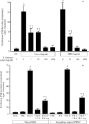
Fig. 1. Bar graphs showing that co-treatment with a crude extract of Hymenolepis diminuta (H.d.) dose-dependently inhibits concanavalin A (Con A) and Staphylococcus aureus enterotoxin B (SEB) induced human peripheral blood mononuclear cell proliferation (PBMC) (A) n=4 and representative of 3 experiments with Con A and 2 with SEB). (B) H.d. extract alone does not induce proliferation, the anti-proliferative effect is observed with a culture supernatant from H. diminuta (H.d. sup) and the immunosuppressive effect occurs in macrophage-depleted immune cell preparations (mean±S.E.M.; n=4; * and [Dagger], P<0·05 compared to control (cntl) and Con A or SEB only-treated PBMC).
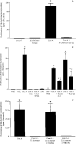
Fig. 2. (A) Murine splenocyte Con A (2 μg/ml)-induced proliferation is inhibited by the Hymenolepis diminuta extract (100 μg/ml) (n=22). (B) The anti-proliferative effect is observed with H. diminuta supernatant (H.d. sup), is reduced significantly by boiling of the extract but is unaffected by pre-treatment with polymyxin B (PMB, 8 μg/ml – a concentration that blocked the small amount of increased thymidine incorporation induced by LPS only) (n=4). The H. diminuta inhibition of Con A-stimulated thymidine incorporation resided in a >50 KDa fraction of the worm (n=4; panel C) (mean±S.E.B.; * and [Dagger], P<0·05 compared to control (cntl) and time-matched Con A only-treated cells; Ψ, P<0·05 compared to LPS (40 EU) only-treated cells).

Table 1. Effect of a high molecular weight component of H. diminuta extract on splenocyte cytokine production
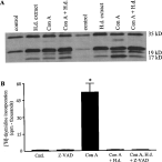
Fig. 3. Using a caspase 3 antibody that detects the whole molecule (35 kDa) and cleaved forms indicative of activation (19 and 17 kDa), Western blotting revealed approximately equivalent levels of whole and cleaved caspase 3 in extracts from Con A (2 μg/ml)±Hymenolepis diminuta extract (100 μg) treated human PBMC (panel A, n=4). The bar graph in (B) shows that use of the pan-caspase inhibitor Z-VAD-FMK (Z-VAD, 100 μM) did not alter the ability of the worm extract to block Con A-induced PBMC proliferation (mean±S.E.M.; n=4; *, P<0·05 compared to control (cntl)). Similar data were obtained with murine spleen cells (not shown).
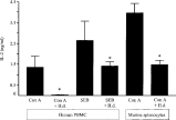
Fig. 4. Bar graph showing that co-treatment with Hymenolepis diminuta extract (H.d.; 100 μg/ml) reduces human peripheral blood mononuclear cell (PBMC) IL-2 production stimulated by Con A (2 μg/ml) or SEB (1 μg/ml) and Con A-induced murine splenocyte IL-2 (mean±S.E.M.; n=4; *, P<0·05 compared to Con A or SEB only; IL-2 measured 24 h post-treatment).
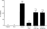
Fig. 5. Bar graph showing that co-treatment with Hymenolepis diminuta extract (H.d.; 100 μg/ml; n=14) or culture supernatant from the worm (n=4) reduces IL-4 production by Con A (2 μg/ml)-stimulated murine splenocytes. Boiling of the worm extract reduced, but did not ablate, its ability to reduce Con A evoked IL-4 production (mean±S.E.M.; * and [Dagger], P<0·05 compared to Con A and Con A+H.d., respectively; IL-4 measured 24 h post-treatment).
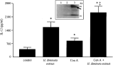
Fig. 6. Bar graph showing that Hymenolepis diminuta extract (H.d.; 100 μg/ml did not reduce Con A (2 μg/ml)-induced IL-12 production from murine spleen cells (n=6). Worm extract appeared to induce IL-12 (n=14) but this is likely due to a combination of LPS contamination (see text) and a worm homologue that binds in the ELISA and was detectable on Western blots (insert; representative Western blot showing an immunoreactive 40 kDa band in the worm extract (lane 3, arrow) detectable by the anti-IL-12 antibody used in the ELISA; lane 1, culture medium only, lane 2, culture supernatant from H. diminuta) (mean±S.E.M.; * and [Dagger], P<0·05 compared to control and Con A only, respectively; IL-12 measured 24 h post-treatment).

Fig. 7. Exposure to Hymenolepis diminuta extract (H.d.; 100 μg/ml), Con A (2 μg/ml) or both agents evokes increased murine splenocyte IL-10 production (panel A: n=11; *, P<0·05). The direct effect of the worm antigen could be reproduced by supernatant from the worm (H.d. sup.) and was significantly reduced by boiling of the extract or incubation with polymyxin B (PMB, 8 μg/ml) (panel B). Additional controls show that low dose LPS has no significant effect on murine splenocyte IL-10 production and that high doses of LPS (well in excess of the level of contamination in the worm extract) directly elicits IL-10 production that is unaffected by boiling (mean±S.E.M.; n=4–19; *, P<0·05 compared to control; #, P<0·05 compared to H.d. or H.d. sup. only; IL-10 measured 24 h post-treatment; EU, endotoxin units).
 You have
Access
- 24
- Cited by