Congenital cardiac fibroma is the second most common benign primary cardiac tumour in children after rhabdomyoma.Reference Marx and Moran 1 Some patients are asymptomatic but others are prone to drug-resistant ventricular arrhythmia from infancy, which is often life-threatening.Reference Marx and Moran 1 In such cases, subtotal tumour resection is performed for two reasons. First, total resection is frequently difficult to achieve because of the relatively thin ventricular wall and indeterminate tumour border. Second, mid-term surgical outcomes appear optimal.Reference Padalino, Vida and Boccuzzo 2 , Reference Miyake, Del Nido and Alexander 3 However, no electrophysiological data have shown that subtotal resection can solely prevent further ventricular arrhythmia. Here, we report a case of congenital cardiac fibroma in an infant treated with partial resection and implantable cardioverter-defibrillator implantation. The infant did not experience cardiovascular events since the surgery, except one episode of inappropriate shock. However, impending compression of the left coronary artery by the coil lead was suspected in coronary angiography findings later in the clinical course. We performed an electrophysiological study 5 years postoperatively and confirmed complete elimination of the arrhythmogenic substrate.
Case report
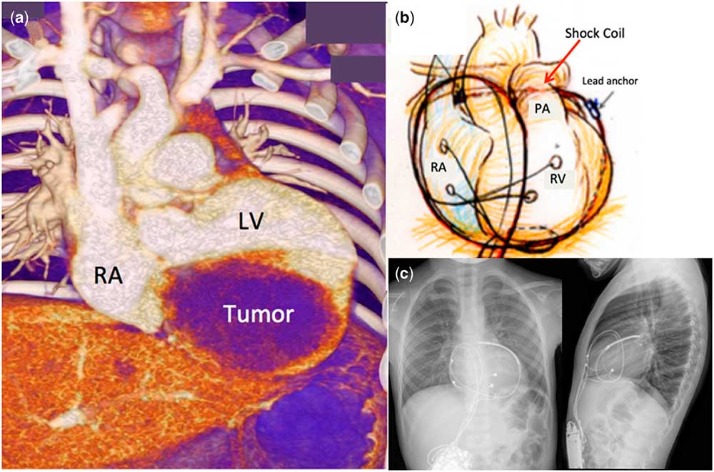
A 4-month-old male infant suspected with a large cardiac fibroma was admitted to our emergency department because of ventricular fibrillation and aborted cardiac death. His past medical history was not significant except failure to thrive. An echocardiography and contrast CT scan revealed a large cardiac tumour (35×28 mm) originating from the left ventricular inferior wall (Fig 1a). He was closely observed at our outpatient clinic and was admitted after several episodes of ventricular fibrillation and resuscitation at 4 months of age. Medical treatment, including maximal amiodarone and propranolol doses, was ineffective, and he underwent subtotal tumour resection and implantable cardioverter-defibrillator (Fortify Assura ICDTM; St Jude Medical, St Paul, Minnesota, United States of America) implantation via a pericardial approach at 19 months of age. We decided to place the device because he had experienced six episodes of ventricular fibrillation, and there was no consensus as to whether partial resection alone is sufficient to prevent further events. Intraoperatively, a coil lead was placed through the transverse pericardial sinus and sewn to the posterior pericardial wall (Fig 1b). Biopsy indicated that the tumour was a cardiac fibroma.
(a) Contrast CT scan (3D reconstructed) showing the tumour originating from the left ventricular inferior wall. (b) A coil lead was placed through the transverse pericardial sinus and sewn to the posterior pericardial wall. (c) Post-operative chest X-ray (two-direction).

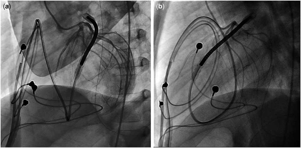
The patient experienced one episode of inappropriate shock because of sinus tachycardia 1 month after implantation, but no other cardiac events occurred. Antiarrhythmic medications were discontinued 2 years postoperatively. A follow-up coronary angiography performed 5 years postoperatively showed that the left circumflex artery was compressed by the shock lead (Fig 2). Although there were no signs or symptoms of acute coronary syndrome, impending compression of the coronary artery by the coil lead was suspected. The lead was thought to be pulled down as his thoraces grew longitudinally. Electrophysiology studies were performed to evaluate the possibility of device removal. A strict right ventricular stimulation programme under isoproterenol infusion did not elicit ventricular tachycardia, and a thorough investigation of the left ventricular endocardium revealed neither late potential nor fractionated potential. He underwent removal of the implantable cardioverter-defibrillator and leads at 7 years of age without complications. There was no postoperative change in electrocardiogram findings and he has been asymptomatic after the operation.
Left anterior oblique view (a) and lateral view (b) of left coronary angiography showing the left circumflex artery compressed by a coil lead.

Discussion
The present case provides two clinical messages. First, subtotal tumour resection can solely eliminate arrhythmogenic substrates. Second, implantable cardioverter-defibrillator leads, particularly coil leads implanted via a pericardial approach in infants, may cause complications later during childhood.
The efficacy of subtotal tumour resection
It is well known that mid-term outcomes of subtotal tumour resection are optimal.Reference Padalino, Vida and Boccuzzo 2 , Reference Miyake, Del Nido and Alexander 3 Miyake et al reported that 16 of 25 patients with congenital cardiac fibroma with ventricular tachycardia who underwent total or subtotal resection had no major complications.Reference Miyake, Del Nido and Alexander 3 They reported a lack of clinically significant arrhythmias in all cases over a mean follow-up of 6 years. In addition, they reported seven recent cases (including non-fibroma cases) of electrophysiology studies with negative ventricular stimulation. Subsequent clinical questions involve the location of the arrhythmogenic substrate (inside or outside the tumour), its electroanatomical character, and its elimination via surgery.
Recently, Sakamoto et al reported that intraoperative electroanatomical mapping of the epicardium can reveal a fractionated potential or late diastolic potential, which are considered ventricular arrhythmia substrates.Reference Sakamoto, Shibata and Murata 4 They reported potentials recorded near the border between tumors and normal tissue. A plausible cause is normal myocardial tension, and debulking may be sufficient to prevent further recurrence of fatal ventricular arrhythmia even if a residual tumour exists.
Our case supports the above possibility. Clinically evident cardiovascular events never relapsed in our case after subtotal tumour resection, and there was no evidence of even subclinical ventricular arrhythmias. Further, electrophysiology studies, including ventricular stimulation, and thorough investigation of the left ventricular endocardium proved the elimination of the arrhythmogenic substrate.
Tissue hypoxia and mechanoelectrical coupling may explain how a focal passive stretch of myocardium generates arrhythmogenesis at the cellular and molecular level. Stretched cardiac myocytes may be exposed to relatively scarce oxygen supply because of stretching of nearby microvasculature. The other possibility is mechanoelectrical coupling. According to Orini et al, acute mechanical stress activates specific channels allowing various cations (including calcium) to cross the cell membrane, causing action potential shortening and diastolic depolarisation that may lead to ventricular arrhythmias.Reference Orini, Nanda and Yates 5
Adverse effects of implantable cardioverter-defibrillator implantation
Among various surgical techniques for implantable cardioverter-defibrillator implantation in children and infants, pericardial approach is a widely accepted method in which a coil lead is placed through the transverse sinus under fluoroscopic guidance.Reference Suzuki, Motohashi and Matsumoto 6 Late complications, including lead fracture, buckling leads, constrictive pericarditis, and potential risk of cardiac strangulation, must be considered.Reference Suzuki, Motohashi and Matsumoto 6 In our case, a coil lead was pulled down with increase in the patient’s height and was found to be on the verge of coronary artery compromise.
Such complications may not be prevented until subcutaneous implantable cardioverter-defibrillators can be used in infants. Clinicians must be aware of the risks of placing leads in growing children and repeatedly assess complications via CT scan or other methods.
In conclusion, subtotal tumour resection can eliminate arrhythmogenic substrates whereas implantable cardioverter-defibrillator leads, particularly coil leads implanted via a pericardial approach in infants, can cause complications later during childhood. Avoiding device implantation in such patients may be an option, but further investigation is warranted.
Acknowledgements
The authors are deeply grateful to Dr Kazuhiro Takahashi for contribution of this work on account of decision-making and early management of ICD implantation.
Financial Support
The authors received no specific grant from any funding agency, or from commercial or not-for-profit sectors.
Conflicts of Interest
None.
Ethical Standards
The authors assert that this report complies with the ethical standards of the Helsinki convention, and consent for this publication has been granted by the patient’s family.
Supplementary material
To view supplementary material for this article, please visit https://doi.org/10.1017/S1047951118001750