Implications
A greater number of low birth-weight piglets is observed in hyperprolific sows. Muscles of these light piglets had generally less myofibers, a trait that is mainly fixed before birth and influences postnatal growth. Maternal dietary strategies have been largely unsuccessful to restore a normal muscle development in intrauterine growth-restricted piglets. Restricted feeding followed by overfeeding leads to compensatory muscle growth during refeeding, but its effects on fetal muscle development when applied to pregnant sows had not been investigated. Current results show that this feeding regimen did not allow light fetuses for recovery of normal skeletal muscle phenotype.
Introduction
Skeletal muscles are fundamental components of the body as they ensure force, movement and locomotion, maintain posture, and participate to resting energy expenditure and whole-body insulin sensitivity (Brown, Reference Brown2014 for a review). The mass and functions of skeletal muscle are largely determined by the number, size and type of its constitutive myofibers. In mammals, the formation of myofibers takes place in utero, with the appearance of two or three successive waves of precursor cells (myoblasts), which constitute the origin of the different waves of muscle fibers (Rehfeldt et al., Reference Rehfeldt, Te Pas, Wimmers, Brameld, Nissen, Berri, Valente, Power, Picard, Stickland and Oksbjerg2011 for a review). The total number of muscle fibers is considered to be set at 90 days of gestation in the pig (birth at 114 days), or at least, is definitely fixed in the first weeks after birth (Bérard et al., Reference Bérard, Kalbe, Lösel, Tuchscherer and Rehfeldt2011). Therefore, environmental insults or stimuli during the prenatal period may have immediate and long-lasting consequences on muscle characteristics (Rehfeldt et al., Reference Rehfeldt, Te Pas, Wimmers, Brameld, Nissen, Berri, Valente, Power, Picard, Stickland and Oksbjerg2011). The natural variation in birth weights within porcine litters, which results from uterine crowding limiting nutrient availability (Foxcroft et al., Reference Foxcroft, Dixon, Novak, Putman, Town and Vinsky2006), has been notably considered to clarify the effects of in utero nutrition on muscle development. Small for gestational age pigs exhibit a lower number of muscle fibers (Wigmore and Stickland, Reference Wigmore and Stickland1983), and a lower ratio of secondary to primary myofiber populations when compared with their normal weight littermates at birth (Bérard et al., Reference Bérard, Pardo, Béthaz, Kreuzer and Bee2010). However, the developmental time course of myosin heavy chain (MyHC) isoforms, that are important genes in myogenesis as they code for proteins of the contractile apparatus, were only slightly affected by variations in piglet birth weights (Bérard et al., Reference Bérard, Pardo, Béthaz, Kreuzer and Bee2010). Altogether, the underlying regulatory mechanisms accounting for these changes remain then to be clarified. Different nutritional interventions with the aim of optimizing in utero environment have been also undertaken to prevent intrauterine growth restriction and (or) restore normal myofiber development in light piglets. To date, these maternal feeding strategies have been largely unsuccessful to improve muscle traits of the offspring. For instance, excessive protein intake in pregnant gilts had no benefits on muscle weight of newborn piglets, nor did it affect the expression of myogenic regulatory factors and MyHC isoforms in skeletal muscles (Rehfeldt et al., Reference Rehfeldt, Lefaucheur, Block, Stabenow, Pfuhl, Otten, Metges and Kalbe2012). Increasing feed and energy intake of sows (over the recommended levels) during specific periods of gestation had either no effects on total fiber number and the ratio of secondary to primary fibers (Markham et al., Reference Markham, Latorre, Lawlor, Ashton, McNamara, Natter, Rowlerson and Stickland2009) or even this decreased muscle weight in the progeny (Nissen et al., Reference Nissen, Danielsen, Jorgensen and Oksbjerg2003; Cerisuelo et al., Reference Cerisuelo, Baucells, Gasa, Coma, Carrión, Chapinal and Sala2009). In offspring from sows overfed during the 50 to 80 days of gestation and fed back to a standard level during the last third of gestation, no difference for MYOG, a regulatory gene involved in myogenesis, was shown but a higher expression level of IGF1 acting on postnatal growth was demonstrated in treated pigs at market weight (McNamara et al., Reference McNamara, Giblin, Markham, Stickland, Berry, O’Reilly, Lynch, Kerry and Lawlor2011). A reverse strategy may be to induce catch-up growth mechanisms during the last trimester of gestation after an initial period of restricted maternal feeding. Recently, Farmer et al. (Reference Farmer, Palin and Martel-Kennes2014) have shown that maternal diet deprivation during the first 70 days of gestation and subsequent feed over-allowance until farrowing led to lower maternal body and fat gains during the restrictive period, but accelerated gains from day 70 to day 108 of gestation. The present study, which was a subset of the aforementioned trial, aimed to determine the effects of this experimental feeding strategy on muscle development of light and medium-weight fetuses just before birth.
Material and methods
Animals and sample collection
Sows were reared and cared according to a recommended code of practice of Agriculture and Agri-Food Canada and experimental procedures were approved by an institutional animal care committee of the Dairy and Swine Research and Development Centre of Agriculture and Agri-Food Canada in Sherbrooke (Canada). Sows used in this study were part of a larger experiment previously described by Farmer et al. (Reference Farmer, Palin and Martel-Kennes2014) where 136 Yorkshire×Landrace gilts were bred via artificial insemination with pooled semen from Duroc boars. Sows were divided into the following two groups: (1) the control group (CTL) receiving a conventional diet during gestation (n=59) and (2) the treated group (TRT) undergoing feed restriction between insemination and 10 weeks of gestation followed by overfeeding up to farrowing (n=56). Feed allowance was fixed at 2.27 kg/day throughout gestation for the two groups. The TRT regimen provided 70% (restriction diet) and 115% (over-allowance diet), respectively, of the CP and digestible energy contents provided by the CTL regimen. The difference in energy and protein intakes was due to different compositions of diets, with the restriction diet containing more fiber (mainly as oat hulls) to dilute the nutrients compared with the CTL and compensatory diets; the over-allowance diet contained more fat, and more protein from soybean meal than the CTL diet. A detailed composition of the diets together with reproductive performance of sows can be found in Farmer et al. (Reference Farmer, Palin and Martel-Kennes2014). In the present study, a subset of 14 first-parity sows (n=7 per dietary group) was used. These sows were sacrificed at day 110 of gestation. After bleeding, their uteri were immediately removed and each uterine horn was opened. Fetuses were counted, weighed and sexed. Fetuses were categorized as normal weight (NWT) when their weight was within 1 s.d. unit of the average weight of the whole litter, and as low (LWT) when their weight was at least 1.5 s.d. units below the average weight of the whole litter. The longissimus dorsi muscle (LM) was collected from one NWT and one LWT fetus in each litter. Samples were then frozen in liquid nitrogen, stored at −70°C and sent under dry ice to INRA UMR Pegase (Saint Gilles, France) for analyses.
Extraction of total RNA
Approximately 40 mg of fetal muscle tissues were homogenized with a stainless steel ball mill (Qiagen Tissue-Lyse, Courtaboeuf, France; twice 2 min at 30 Hz) in the presence of Trizol® (Invitrogen, Saint-Aubin, France). Total RNA was extracted and purified using a silica-membrane technology following the manufacturer’s instructions (Nucleospin RNA II kit; Macherey Nagel, Hoerdt, France), and then, it was quantified using a NanoDrop ND-1000 spectrophotometer (Thermo Scientific, Illkirch, France). The integrity of RNA was assessed using the Agilent RNA 6000 Nano kit on an Agilent 2100 Bioanalyzer (Agilent Technologies, Massy, France). Samples had RNA Integrity Number >7 and A260/280 ratio >1.8.
Complementary DNA synthesis and quantitative real-time PCR amplifications
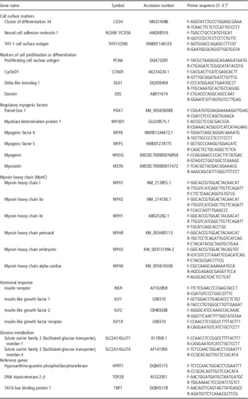
Reverse transcription was performed from 1 µg of total RNA using a High Capacity cDNA Reverse Transcription Kit (Applied Biosystems, Foster City, CA, USA) and random hexamers. The focus was made on candidate genes known to be important in skeletal muscle development, such as markers of tissue-resident stem cells, myogenic regulatory factors, MyHC isoforms and IGFs, as well as on actors of glucose uptake and energy use. Specific primers and TaqMan probes (Table 1) were defined from porcine sequences using the Primer Express software (Version 3.0, Applied Biosystems). To avoid amplification of genomic DNA, primers located on different exons were preferred. Expression levels of genes coding for the different MyHC isoforms were quantified by TaqMan technology qPCR, as previously described (Lefaucheur et al., Reference Lefaucheur, Milan, Ecolan and Le Callenec2004). Expression levels of other selected genes were measured using the SybrGreen technology, as previously described (Gondret et al., Reference Gondret, Père, Tacher, Daré, Trefeu, Le Huërou-Luron and Louveau2013). All reactions were performed on a StepOnePlusTM Real Time PCR system (Applied Biosystems). Specificity of the amplification products was checked by dissociation curves analysis. For each primer pair, the amplification efficiency of qPCR reaction was determined using calibration curves generated with six decreasing concentrations of cDNA from pooled muscle samples (from 12.5 to 12.5E-3 ng RNA). Three reference genes, HPRT1 (hypoxanthine-guanine transferase PhospoRibosyl), TBP1 (TATA box binding protein 1) and TOP2B (DNA topoisomerase 2-beta), were identified as the most stable ones by the GeNorm algorithm (http://medgen.ugent.be/~jvdesomp/genorm/). For each sample, the normalized expression (N) of each gene was thus calculated according to the following formula: N=E −ΔCq (sample−calibrator)/NF, where E is obtained from the slope of the calibration curve, Cq is the quantification cycle, and calibrator is a pool of all muscle samples, and NF is the normalization factor calculated from the three reference genes.
Primer sequences used for qPCR amplifications of studied genes in pig skeletal muscle tissue

1 F=forward primer; R=reverse primer; P=TaqMan probe.
MyHC electrophoresis
The relative abundance of the different MyHC isoforms was also determined at the protein level in the LM of fetuses. Approximately 100 mg of muscle tissue was homogenized with a stainless steel ball mill (Qiagen Tissue-Lyse) in the presence of Laemmli (Sigma, Saint-Quentin Fallavier, France; S-3401). Proteins were extracted by heating the mixture at 100°C for 6 min, and the homogenate was centrifuged at 15 000×g for 15 min at 20°C. Protein concentration in the supernatant was determined using the RC DC Protein Assay kit (BioRad Laboratories, Hercules, CA, USA) and bovine serum albumin as a standard. The different isotypes of MyHC were separated using a standard one-dimensional sodium dodecyl sulfate–polyacrylamide gel electrophoresis (SDS–PAGE; Lefaucheur et al., Reference Lefaucheur, Ecolan, Lossec, Gabillard, Butler-Browne and Herpin2001). Separative gels stained by colloidal Coomassie Brillant Blue G250 were scanned with the ImageQuant LAS 4000 imager and analyzed using the ImageQuant TL 7.0 image analysis software (GE Healthcare; Vélizy Villacoublay; France). Bands corresponding to the different MyHC isoforms (Figure 1a) were identified according to Lefaucheur et al. (Reference Lefaucheur, Ecolan, Lossec, Gabillard, Butler-Browne and Herpin2001). The proportion of the different MyHC isoforms was expressed as percentages of the sum of MyHCs within a lane.
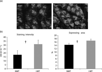
Immunocytochemical localization of DLK1/Pref1 protein. Slides were obtained from longissimus muscles sampled at 110 days of gestation in light (LWT) or medium-weight (NWT) littermates of sows fed a control diet. (a) Representative photos for one fetus in each fetal weight group, together with (b) semi-quantification of the signals in each weight group, are shown. Values are means with their s.e.m. Staining intensity (P=0.08) and percent of expressing area (P=0.06) tended be the highest in the small fetuses.

Immunocytochemistry
Transverse sections (8 µm thick) were obtained in muscle samples of fetuses from CTL sows only (n=7 per group), using a cryostat (2800 Frigocut N; Reichert-Jung, Heidelberg, Germany). These sections were mounted on silane-coated slides for enhanced adhesion. Immunocytochemistry was used to investigate the protein expression of the delta-like 1 homolog (PREF1/DLK1), as previously described (Gondret et al., Reference Gondret, Perruchot, Tacher, Bérard and Bee2011). A goat anti-human PREF1/DLK antibody (1 : 25; R&D systems, Minneapolis, MN, USA) was used as primary antibody, and a fluoresceine-conjugated goat anti-mouse IgG (Jackson, Interchim Montluçon, France) was used as secondary antibody. Qualitative staining analyses were performed on a self-developed plugin for ImageJ (ImageJ 1.43: National Institutes of Health, Bethesda, MD, USA), and representative images obtained in small and medium-weight fetuses are shown.
Statistics
The data for mRNA levels of selected genes and relative protein abundance of MyHC isoforms were submitted to ANOVA using the SAS software (v9.2; SAS Institute, Cary, NC, USA). The MIXED procedure was used, with fetal weight category (NWT and LWT), maternal nutrition groups (CTL and TRT) and their interaction as main effects, and sow within feeding groups as a random effect. Because there were almost no significant interactions (P>0.10) between maternal dietary regimen and fetal weight category for analyzed traits, data are generally presented as the least squares means for the effect of fetal weight categories, and then, the effects of maternal dietary treatments. In the rare occasions where there was a significant interaction, the data are shown for the four experimental groups.
Within CTL sows, immunohistochemistry measurements were analyzed with the nonparametric method (NPAR1WAY procedure of SAS) to determine the effect of fetal weight categories (low or normal weights). Means within each weight categories and P-values of Wilcoxon tests for the two groups of samples are then indicated. Differences between groups were considered significant if P<0.05, and as tendencies if 0.05<P⩽0.10.
Results
Irrespective of maternal dietary treatments, BW of LWT fetuses was 32% lower than average weight of NWT fetuses (824±140 v. 1218±192 g, respectively, P<0.001) at 110 days of gestation. Maternal dietary treatment had no effect on the mean weight of fetuses in litters (CTL: 1028±260 g, TRT: 1015±268 g, P=0.85). In addition, there was no treatment effect (P=0.25) on the weight reduction of LWT relative to NWT fetuses.
Stem cell markers
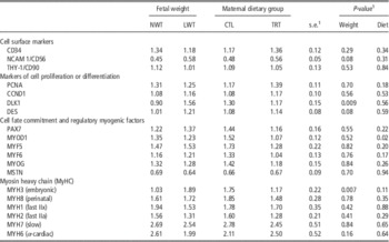
The expression levels of stem cell surface markers CD34 (involved in hematopoietic stem cell proliferation) and Thy-1/CD90 (a mesenchymal stem cell marker) were similar in LM muscles of LWT and NWT fetuses (Table 2). In contrast, the expression of the precursor cell marker NCAM1/CD56 (involved in cell-matrix interactions during development) tended to be greater (29%, P=0.08) in the LM muscle of LWT fetuses compared with NWT fetuses. Expression levels of these genes in fetal muscles were not influenced by maternal dietary treatments.
Effects of fetal weight categories and maternal dietary regimen during gestation on the expression levels of genes involved in muscle cell development in pig fetuses on day 110 of gestation

1 Data are least square means for the effects of fetal weight categories (LWT: low v. NWT: normal) and of maternal dietary strategies (TRT: restricted and subsequent overfeed allowance; CTL: control). Fetuses were sampled in n=7 sows per dietary group. There were no interaction (P>0.10) between fetal weight and maternal diets on gene expression levels. s.e., pooled standard errors of the LSmeans.
Cell proliferative capacity
Expression levels of proliferating cell nuclear antigen (PCNA) involved in mitotic cell cycle and of cyclin D1 (CCND1) contributing to the temporal coordination of mitotic events, did not differ between muscles of light and medium-weight fetuses. On the contrary, the mRNA level of the delta-like 1 homolog (DLK1/Pref1) involved in the negative control of early cell differentiation, was 73% greater (P<0.01) in LWT fetuses than in NWT fetuses. Maternal dietary treatment did not affect expression level of DLK1 in fetal muscles.
In an attempt to check expression changes between fetal weight categories for DLK1 at the protein level, immunocytochemical analyses were also performed on muscle tissue slides within fetuses of CTL sows (Figure 1). Staining intensity and percentage expressing area for DLK1/PREF1 protein tended to be slightly higher in the smallest fetuses than in their medium littermates (P<0.10).
Cell fate commitment and myogenic differentiation
The satellite cell marker PAX7 involved in the regulation of cell fate commitment, as well as myostatin (MSTN), a negative regulator of cell growth, were expressed at similar levels in the LM muscle of all fetuses (Table 2). The myogenic regulators MYOD1, MYF5, MYOG and MYF6/MRF4 were not affected by fetal weight categories, but mRNA level for MYOD1 was lower in fetuses from TRT sows (−30%; P=0.02) than in fetuses from CTL sows.
Myofibrils organization and contractile function
Differences in fetal weights between littermates at 110 days of gestation had affected the expression levels of genes playing important roles in organization of the myofibrils. The DES gene, coding for the muscle-specific member of the intermediate filament family desmin, tended to have a greater expression level (+20%; P=0.08) in LM muscles from LWT than NWT fetuses, whatever the maternal dietary groups. Among the six different transcripts of MyHC isoforms detected at this gestational stage, the expression level of the embryonic isoform MYH3 was 64% greater (P<0.01) in LWT fetuses than in NWT fetuses (Table 2). Consequently, the ratio of perinatal (MYH8) to embryonic (MYH3) isoforms was lower in LWT than in NWT fetuses (1.44 v. 2.31; P=0.04, respectively). In addition, the ratio of the sum of adult fast MyHC (MYH1+MYH2) to developmental (MHY3+MYH8) isoforms was 31% lower in LWT than in NWT fetuses (0.43 v. 0.62; P<0.01, respectively). Maternal diets during gestation had no effects on MyHC expression levels in fetal muscle.
MyCH isoforms protein separation
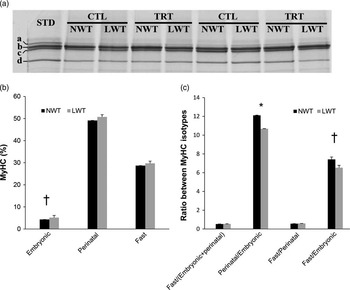
A complementary study using gradient SDS–PAGE was performed to put evidence of MyHC distribution at the protein level. At this stage, the most prominent isoform was the perinatal MyHC (representing about 50% of the total MyHC) followed in decreasing order, by the sum of adult fast MyHC, the sum of type I+α-cardiac MyHC, and the embryonic MyHC (Figure 2). Similarly to what was found at the molecular level, a greater abundance of the embryonic MyHC protein was observed in LWT fetuses than in NWT fetuses (5.3% v. 4.3%, P=0.05). The perinatal-to-embryonic MyHC protein ratio was also lower (P<0.05) in LWT fetuses than in NWT fetuses. In addition, the ratio of adult fast to developmental MyHC isoforms tended to be lower (P=0.09) in LWT fetuses compared with NWT fetuses.
Myosin heavy chain (MyHC) electrophoresis of the pig longissimus muscle on day 110 of gestation. (a) Representative sodium dodecyl sulfate–polyacrylamide gel electrophoresis (SDS–PAGE) of MyHC profiles. Bands correspond to MyHC embryonic (a, emb), perinatal (b, peri), adult fast IIa+IIx+IIb (c, fast) and adult I+α-cardiac (d, slow) isotypes. STD: a mixture of the different muscle samples including all MyHC isotypes used as a reference. (b) Calculated proportions of developmental MyHC and adult fast MyHC in small weight (LWT) or normal weight (NWT) fetuses in sows fed control diet (CTL) or submitted to restricted/over-allowance of feed (TRT) during gestation. Because there were no interactions between fetal weight categories and maternal strategies, least-square means in LWT and NWT are shown. (c) Ratio between MyHC isoforms in LWT and NWT fetuses. Black bars, NWT; gray bars, LWT. *P<0.05; † P<0.10.

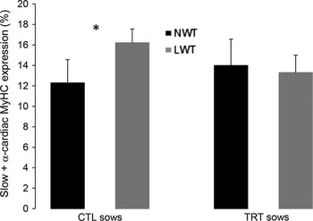
Maternal dietary treatment per se did not affect MyHC isotype proportions. However, there was a trend (P=0.07) for an interaction between maternal diet and fetal weight categories. Indeed, a higher proportion of MyHC slow protein was present in LWT than NWT fetuses (16.2% v. 12.3%, P=0.05) within the CTL sow group, while there was no difference between fetal weight categories within the TRT sow group (Figure 3).
Percentage of slow +α-cardiac myosin heavy chain (MyHC) in the longissimus muscle of fetuses sampled on day 110 of gestation. One low (LWT) and one normal weight (NWT) fetuses were collected within each litter from sows that were either fed a control diet (CTL) or submitted to feed restriction followed by overfeeding during gestation (TRT, treated; n=7 sows per dietary group). The distribution of MyHC isotypes was studied by sodium dodecyl sulfate–polyacrylamide gel electrophoresis (SDS–PAGE). Maternal diet and fetal weight categories interacted on the relative proportion of the slow-type MyHC protein. Black bars, NWT; gray bars, LWT. *P=0.05.

Glucose–insulin axis and IGFs
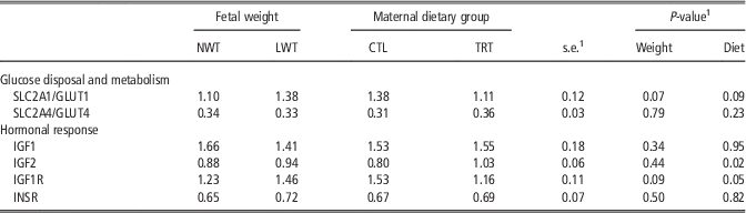
The mRNA level of the facilitated glucose transporter SLC2A1/GLUT1 tended to be higher (+25%; P=0.06) in LWT fetuses compared with NWT fetuses, but it tended to be reduced (−20%, P<0.10) in TRT fetuses than in CTL fetuses (Table 3). The insulin-related glucose transport GLUT4 and the insulin receptor (INSR) gene did not differ between fetal weight categories or maternal dietary groups. The expression level of IGF1 receptor (IGF1R) tended to be higher in the LWT fetuses compared with the NWT fetuses (+18%; P=0.09), while it was lower (−24%, P=0.02) in TRT fetuses compared with CTL fetuses, whatever their weight categories. Expression levels of IGF1 and IGF2 did not differ between weight categories, but IGF2 was upregulated (+29%, P=0.02) in LM muscle of TRT fetuses compared with CTL fetuses.
Effects of fetal weight categories and maternal dietary regimen during gestation on the expression levels of genes involved in muscle glucose uptake and insulin–IGF axis in pig fetuses on day 110 of gestation

1 There were no interactions (P>0.10) between fetal weight and maternal diets on gene expression levels. Fetuses were sampled in n=7 sows per dietary group. Therefore, data are shown as least square means for the effects of fetal weight categories (LWT: low v. NWT: normal) and of maternal dietary strategies (TRT: restricted and subsequently overfed during gestation; CTL: control). s.e., pooled standard errors of the LSmeans.
Discussion
Spontaneous growth restriction altered early myogenic events, and this could not be prevented by maternal feed restriction/overfeed allowance in gestation
The present study shows that spontaneous variations in fetal weight in both treated and control sows were associated with subtle differences in expression levels of some genes involved in muscle cell development, which are likely indicative of retarded myofiber differentiation. Indeed, the LM muscle of small fetuses at day 110 of gestation exhibited the highest expression of DLK1, a gene regulating differentiation of several cell types. This gene, which is highly expressed in embryonic muscle, is sharply down-regulated during postnatal development in porcine muscles (Oczkowicz et al., Reference Oczkowicz, Piestrzyska-Kajtoch, Piórkowska, Rejduch and Rózycki2010) and almost completely abolished in human and mouse adult muscle (Andersen et al., Reference Andersen, Schrøder and Jensen2008). Altogether time-related decrease in DLK1 expression level seems to be a mandatory step for normal myogenesis (White et al., Reference White, Vuocolo, McDonagh, Grounds, Harper, Cockett and Tellam2008). In support to our findings in skeletal muscle of intrauterine growth-restricted fetuses, it has been shown that circulating levels of soluble DLK1 are higher in small-for-gestational age babies at birth (de Zegher et al., Reference de Zegher, Díaz, Sebastiani, Martín-Ancel, Sánchez-Infantes, López-Bermejo and Ibáñez2012). In the present study, NCAM1/CD56, a gene encoding a cell surface marker in precursor cells carrying a high myogenic potential (Perruchot et al., Reference Perruchot, Lefaucheur, Barreau, Casteilla and Louveau2013), also tended to be higher in the lightest fetuses. This may be related to the fact that some DLK1 positive cells located below the basal lamina co-express NCAM1/CD56 (Peault et al., Reference Peault, Rudnicki, Torrente, Cossu, Tremblay, Partridge, Gussoni, Kunkel and Huard2007). In addition, it has been shown that porcine proliferating progenitor cells also express desmin (Wilschut et al., Reference Wilschut, Jaksani, Van Den Dolder, Haagsman and Roelen2008), which tended to be upregulated in the LWT fetuses compared with NWT fetuses in the present study. Finally, the smallest fetuses at day 110 of gestation exhibited a higher expression of embryonic MyHC, one of the developmental types of MyHCs that predominate during proliferation and differentiation of porcine muscular cells (Perruchot et al., Reference Perruchot, Ecolan, Sorensen, Oksbjerg and Lefaucheur2012). Because most MyHCs are synthesized upon terminal differentiation of muscle cells, a higher ratio of the embryonic MyHC to adult fast MyHC is then considered as a good indicator of muscle immature phenotype in newborn piglets (Rehfeldt et al., Reference Rehfeldt, Lefaucheur, Block, Stabenow, Pfuhl, Otten, Metges and Kalbe2012). In the present study, the ratios of perinatal to embryonic MyHCs and of adults to developmental MyHC isoforms were generally lower in LWT than in NWT fetuses. Taken together, the present data argue for retarded muscle development in small porcine fetuses compared with their normal weight littermates just before birth. These changes likely account for the lower number of myofibers previously demonstrated in intrauterine growth retarded pigs (Wigmore and Stickland, Reference Wigmore and Stickland1983). In the present study, the relative proportion of slow MyHC protein isoform was, however, higher in LWT fetuses than in NWT fetuses of control sows. Because the MyHC slow isotype is predominant in primary fibers, this suggests delayed formation of secondary myofibers rather than of primary fibers in the smallest fetuses. In support, a lower secondary/primary fiber number ratio has been reported in some muscles of piglets having suffered from intrauterine crowding compared with their normal progeny (Bérard et al., Reference Bérard, Pardo, Béthaz, Kreuzer and Bee2010). Early maternal diet deprivation followed by feed over-allowance during gestation did not modify expression levels of stem cell markers, developmental myogenic signals and MyHC isoforms (except MyHC I percentage) in fetal muscles. This suggests that this maternal feeding strategy was mainly ineffective in improving muscle development of experimental fetuses.
Myogenic regulatory factors did not differ between fetal weight categories
In the present study, other genes considered in literature as important for myogenic events, such as the myogenic regulatory factors (Oksbjerg et al., Reference Oksbjerg, Gondret and Vestergaard2004) and MSTN, a negative regulator of myogenesis (Patruno et al., Reference Patruno, Caliaro, Maccatrozzo, Sacchetto, Martinello, Toniolo, Reggiani and Mascarello2008), did not differ between light and medium-weight fetuses at day 110 of gestation. In agreement, another study has also reported greater expression of the embryonic MyHC isoform and reduced muscle weight in small newborn piglets, without any differences in expression levels of MYF5, MYF6, MYOD and MYOG in the LM (Rehfeldt et al., Reference Rehfeldt, Lefaucheur, Block, Stabenow, Pfuhl, Otten, Metges and Kalbe2012). On the contrary, a greater expression of MSTN has been demonstrated in semitendinosus muscle of intrauterine growth-restricted pig fetuses compared with their largest littermates at 61 to 89 days of gestation (Karunaratne et al., Reference Karunaratne, Bayol, Ashton, Simbi and Stickland2009). The difference between the above-mentioned study and our results may be explained by the fact that expression values of these regulatory myogenic factors are likely age- and muscle- (and muscle region) dependent. In support to this assumption, it has been shown that MSTN and MYOD1 mRNA levels are expressed more in porcine LM at 30 and 70 days of gestation than at birth (Patruno et al., Reference Patruno, Caliaro, Maccatrozzo, Sacchetto, Martinello, Toniolo, Reggiani and Mascarello2008). In the present study, a lower expression of MYOD1 was, however, observed in LM of fetuses from treated sows compared with controls at day 110 of gestation. This study was a subset of a larger experiment, showing a tendency for piglet birth weight to be lower in the TRT group than in the CTL group (Farmer et al., Reference Farmer, Palin and Martel-Kennes2014). The lower expression of MYOD1 in the subset of TRT offspring considered just before birth may be thus related to the lower development of piglets undergoing this interventional maternal strategy.
IGF signaling in fetal muscle could be altered by maternal feeding strategies
Metabolic actors are also important for proper functioning of skeletal muscles at birth and in later life. In the present study, we suggested that both spontaneous variation in fetal weight and maternal nutrition can alter glucose disposal and IGFs in fetal pig muscle. Indeed, the facilitative glucose transporter SLC2A1/GLUT1 tended to have a greater expression level in LWT than in NWT fetuses. The expression of GLUT1 predominates during fetal and early postnatal life (Gaster et al., Reference Gaster, Handberg, Beck-Nielsen and Schroeder2000), and then decreases gradually during later growth (Santalucia et al., Reference Santalucia, Camps, Castelló, Muñoz, Nuel, Testar, Palacin and Zorzano1992). Therefore, the greater expression of GLUT1 in muscle of LWT fetuses compared with NWT fetuses suggests a delayed muscle metabolic maturation in growth-restricted fetuses. In addition, although IGFs expression in muscle did not differ between fetal weight categories, IGF1R was expressed more in the lightest fetuses at day 110 of gestation. The biological functions of both IGF-I and IGF-II are primarily mediated by IGF1R, and transcript level for IGF1R declines in the last part of gestation in normal pig fetuses (Louveau et al., Reference Louveau, Combes, Cochard and Bonneau1996). Therefore, this observation argues again for lower metabolic development of muscle of growth-retarded fetuses. Importantly, feed restriction followed by overfeed allowance of sows during gestation resulted in decreased levels of IGF1R mRNA but upregulated IGF2 in muscle of treated fetuses when compared with fetuses of control maternal regimen. This suggests an altered susceptibility of fetal muscle to IGFs signaling in offspring of sows undergoing dietary restriction followed by over-allowance of feed during gestation.
Conclusions
The present study shows that small porcine fetuses exhibited a delay in skeletal muscle maturation at the end of gestation. Restricted feeding followed by feed over allowance of gestating sows as a nutritional strategy to trigger catch-up growth during the realimentation period, had only little effects on fetal muscle development and physiology. Altogether, this feeding strategy did not allow for small fetuses to recover a normal muscle phenotype.
Acknowledgments
The authors gratefully acknowledge C. Tréfeu, L. Finot and D. Beaudry for their skillful technical assistance, as well as the staff at the CRF and COOP Fédérée research farms, especially I. Cormier and A. St-Cyr, and the staff of the AAFC Swine Complex for slaughter of the animals. Sincere thanks are extended to La COOP Fédérée (St-Romuald, QC, Canada) and INRA for their financial and technical supports.