Introduction
The use of magnetic micro- and nanoparticles in biological and biomedical applications goes back nearly 100 years. In the early 20th century, Heilbronn and Seifritz used magnetic forces on iron and nickel microparticles embedded within slime molds and cellular cytoplasm to probe their rheological properties. Reference Heilbrunn1,Reference Seifritz2 More than 60 years ago, Nobel Laureate Francis Crick, along with Arthur Hughes, expanded on these studies using well-characterized iron oxide microparticles. Reference Crick and Hughes3 In the decades since, the use of magnetic micro- and nanoparticles in biomedical applications has undergone rapid growth, with exponential growth in both publications and citations in this field since the early 2000s. Research in this area is growing at a remarkable pace. Novel applications that exploit the most fundamental principle of magnetism and magnetic particles—action at a distance—are being developed that have the potential to revolutionize fields as diverse as cancer therapy, gene therapy, developmental biology, and tissue engineering.
Most magnetic particles used in biomedical applications are superparamagnetic iron oxides, particularly for in vivo applications. Iron oxide is well tolerated by living organisms; in fact, virtually all living organisms, including humans, store iron in the form of iron oxide. However, aggregations of magnetic particles circulating within the bloodstream of a human or animal can have catastrophic consequences, as the clusters can block the flow of blood. By coating superparamagnetic iron oxides with biocompatible polymers such as dextran or polyethylene-glycol or synthesizing multiple superparamagnetic crystallites within a polymer matrix, it is possible to prevent particle aggregation (and the associated potential for embolism formation when the particles are introduced intravenously) as well as attenuate any immune response, facilitating longer circulation times. Here we explore both established and emerging biomedical applications of mesoscale magnetic particles.
Magnetic cell separation
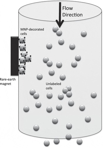
One of the most widely used applications for mesoscale magnetic particles is magnetic cell separation to isolate a specific cell population from a heterogeneous sample. Cells are labeled with antibody-conjugated magnetic particles targeted to specific proteins or peptides (antigens) displayed on the surface of the desired cell population. Magnetically labeled cells can then be separated from the rest of the (non-labeled) cell population using a high gradient magnetic separator ( Figure 1 ).
Principles of magnetic cell separation. Cells in suspension decorated with magnetic micro- or nanoparticles are attracted to the magnet as they pass through the column. Unlabeled cells pass through without being captured. After the sample has cleared the column, the magnet is removed, and the supernatant is washed out with the target cells. Note: MNP, magnetic nanoparticle.

In 1990, Miltenyi et al. Reference Miltenyi, Muller, Weichel and Radbruch4 introduced a magnetic cell separation system that employed fluorescently labeled, antibody-conjugated magnetic microparticles in combination with a separation column filled with steel wool. A static magnetic field magnetizes the steel wool, enabling trapping of the magnetically labeled cells. With the magnetic field in place, unlabeled cells are washed through the column. After removing the magnetic field, the labeled cells are released from the steel wool in a simple washing step, allowing for efficient, rapid cell separation with excellent cell viability.
In order to address challenges, such as low target antigen expression levels on the surface of some cells, McCloskey et al. Reference McCloskey, Chalmers and Zborowski5 modeled three different scenarios to enhance the magnetophoretic mobility (the movement of magnetic particles, and the cells to which they are attached, along a field gradient). It was found that microparticles (particles with hydrodynamic diameters of more than 1 µm) improve the magnetophoretic mobility of cells by several orders of magnitude in comparison to nanoparticles (<100 nm). However, when dealing with cells expressing low numbers of antigens, indirect antibody labeling is recommended. This is done by grafting multiple polyclonal antibodies (antibodies that bind multiple sites on the cell surface antigen) onto magnetic nanoparticles. Reference McCloskey, Chalmers, Moore and Zborowski6
MRI contrast enhancement
The first routinely used clinical application of magnetic nanoparticles was as negative contrast agents in magnetic resonance imaging (MRI). Field inhomogeneities introduced by the presence of the magnetic particles promote rapid dephasing of protons in nearby water molecules, resulting in negative contrast in T2-weighted MR images. Reference Carroll, Huffstetler, Miles, Goff, Davis, Riffle, House, Woodward and St Pierre7,Reference Pankhurst, Connolly, Jones and Dobson8
It is worth noting that despite their potential, superparamagnetic iron oxide nanoparticles (SPIONs) are not routinely used for most clinical MRI examinations. This is primarily because SPIONs are negative contrast agents, making clinical diagnosis somewhat more difficult. In addition, negative contrast may also arise from metal deposits, calcification, and bleeding, further confusing diagnosis.
Hence, while until recently, several brands of SPION MRI contrast agents were available, most of them have been discontinued to give way for ultrasmall iron oxide nanoparticles, such as Supravist and gadolinium-based contrast agents, such as Magnevist, which require lower doses.
Nevertheless, there are currently exciting developments with SPIONs in MRI. Recently, Carroll et al. demonstrated that magnetite nanoparticles coated with co-polyethers, when “clustered” (MBIClusters), improved their T2 relaxivities by a factor of 1.7 in comparison to the commercial contrast agent Feridex. Reference Carroll, Huffstetler, Miles, Goff, Davis, Riffle, House, Woodward and St Pierre7 This was taken a step further when the same group showed that the loading of these MBIClusters with the antibiotic gentamicin did not affect their high MRI sensitivity, highlighting the “theranostic” (combined diagnostic and therapeutic) potential of these particles by combining imaging with drug delivery. Reference Pothayee, Balasubramaniam, Pothayee, Jain, Hu, Lin, Davis, Sriranganathan, Koretsky and Riffle9
Magnetic drug targeting
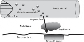
In the late 1970s, Widder et al. introduced the concept of magnetic drug delivery using mesoscale magnetic particles as carriers of chemotherapy compounds ( Figure 2 ). Reference Widder, Senyei and Scarpelli10 The group conjugated the cytotoxic drug “adriamycin” (better known as doxorubicin) to magnetic microspheres and, using a high-gradient magnetic field, targeted them to a specific site in a rat model. This resulted in a localized drug concentration that, if achieved through systemic intravenous delivery, would require a 100-fold higher dose of the free drug. Reference Widder, Senyei and Scarpelli10
Principles of magnetic targeting for drug delivery. Magnetic nanoparticles carrying a therapeutic agent (not to scale) are pulled from circulation by a high gradient magnetic field positioned at the target site (such as a tumor). The magnetic nanoparticles collect at the target site and exit the vasculature via an enhanced permeability and retention effect.
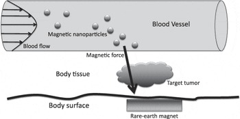
Although these first results were promising and there has been progress with the loading and release of drugs from magnetic particles (e.g., through the use of thermoresponsive polymers Reference Pothayee, Balasubramaniam, Pothayee, Jain, Hu, Lin, Davis, Sriranganathan, Koretsky and Riffle9 ), magnetic drug delivery has yet to fulfill its early potential. This is primarily due to the fact that the magnetic field strength and gradient decrease exponentially with distance from the magnet, making targeting sites deep within tissue difficult. Also, the hydrodynamic drag on the particles in rapidly flowing blood competes with the magnetic force. In addition, ballistic effects of proteins and cells traveling within the blood make particle capture more difficult.
One of the main requirements of magnetic nanoparticles (MNPs) for magnetic targeting is that they are very small (<100–200 nm hydrodynamic diameter), so the iron oxide cores are superparamagnetic, and the MNP/composite particle can evade the reticuloendothelial system—the body’s immune surveillance system. With current magnetic materials being used for these applications (primarily iron oxides), this small size means that the magnetic moment of the particle—which is composed of both iron oxide and diamagnetic polymer matrix—is not sufficiently large to experience a magnetic force larger than the hydrodynamic drag on the individual particles in most cases.
Several groups are investigating techniques for overcoming these issues, such as implanting magnetic meshes near the target site, Reference Kempe and Kempe11,Reference Yellen, Forbes, Halverson, Fridman, Barbee, Chorny, Levy and Friedman12 using novel, high-gradient magnet arrays such as Halbach—so-called “one-sided flux”—arrays, Reference Kyrtatos, Lehtolainen, Junemann-Ramirez, Garcia-Prieto, Price, Martin, Gadian, Pankhurst and Lythgoe13 using MNP-doped cells as carriers in order to generate a larger force while eliminating any immune response, Reference Muthana, Scott, Farrow, Morrow, Murdoch, Grubb, Brown, Dobson and Lewis14 and a concept called “magnetic pushing” based on the geometric interaction of multiple field sources to create null field points. Reference Sarwar, Nemirovski and Shapiro15
Nanomagnetic gene transfection
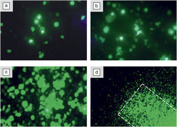
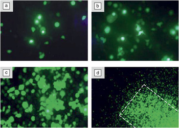
An important and useful biomedical application of MNPs that had its genesis in the principles of magnetic drug targeting is nanomagnetic transfection (non-viral introduction of exogenous genes into a cell) for gene delivery. Magnetic microparticles were first used by our group in 2000 to deliver genes into cells by linking a viral vector to the particles. By placing a NdFeB magnet beneath a culture dish, a microparticle/virus complex was targeted to a specific area of cells growing in the culture, resulting in enhanced transduction of those cells, while non-targeted cells showed little evidence of fluorescent reporter gene expression ( Figure 3 ). Reference Mah, Fraites, Zolotukhin, Song, Flotte, Dobson, Batich and Byrne16,Reference Mah, Zolotukhin, Fraites, Dobson, Batich and Byrne17
In vitro transduction of green fluorescent protein (GFP) reporter genes into C12S cells. The C12S cells were transduced using microparticle/virus complexes. (a) Control 1: Non-magnetic, polystyrene microsphere-conjugated rAAV encoding GFP. (b) Control 2: Magnetic microsphere conjugated-rAAV encoding GFP with no magnet. (c–d) Magnet-assisted transduction of magnetic microsphere conjugated-rAAV encoding GFP (magnet placement indicated by white dotted line). Reprinted with permission from Reference 16. © 2002 Nature Publishing Group.
More recently, in vitro transfection efficiency and protein expression have been further enhanced for both DNA and small-interfering RNA delivery by mechanical stimulation through the use of oscillating magnet arrays and pulsed electromagnets. Reference Kamau, Hassa, Steitz, Petri-Fink, Hofmann, Hofmann-Amtenbrink, von Rechenberg and Hottiger18–Reference McBain, Griesenbach, Xenariou, Keramane, Batich, Alton and Dobson20 In this case, MNP/biomolecule complexes are introduced into cell cultures. Arrays of NdFeB magnets placed below the cell culture plate are attached to microprocessor controlled stepper motor/slide assemblies. These slide assemblies oscillate along the slide axis, essentially “dragging” the particles over the surface of the cells. Mechanical stimulation of the cell surface promotes endocytosis (the uptake of materials into the cell via membrane engulfment) of the MNP/biomolecule complex, improving uptake. Reference Lim, Clements and Dobson21 This motion also appears to increase protein yield, perhaps via enhanced activation of the cell’s transcription machinery.
Another approach for gene delivery combines nanomagnetic transfection with ultrasound and nanoparticle loaded microbubbles. Reference Stride, Porter, Prieto and Pankhurst22 This technique is interesting not only for transfection, but also potentially for drug delivery and imaging. Depending on the design, nanoparticles or drugs can be incorporated within the bubble coating or be loaded into the bubble itself, depending on the acoustic field. Different applied physical effects, such as microjetting, may improve drug delivery, opening an array of possible theranostic applications. Reference Owen, Pankhurst and Stride23,Reference Stride and Coussios24
Magnetic fluid hyperthermia
The concept of magnetic fluid hyperthermia (MFH) was first proposed by Gilchrist Reference Gilchrist, Medal, Shorey, Hanselman, Parrott and Taylor25 in 1957 and relies on coupling magnetic nanoparticles to a radio frequency (RF) magnetic field, generally at frequencies between 100 KHz and 1 MHz, to generate heat at a targeted site within the body. As the human body is primarily transparent to magnetic fields, energy can be delivered to MNPs targeted to tumors within the body via MFH. This energy can be dissipated as heat via two mechanisms—(1) frictional heating due to Brownian rotation of magnetically blocked particles within the tissue, and (2) Néel relaxation, or phase lag of the electron spin orientations, in superparamagnetic particles as they attempt to follow the changing field vector (there are other possible sources such as eddy currents and hysteretic heating, but these are negligible in comparison). Reference Carroll, Huffstetler, Miles, Goff, Davis, Riffle, House, Woodward and St Pierre7 Therefore, the heating potential of a nanoparticle suspension is strongly dependent on magnetic particle core size and composition, as well as its hydrodynamic diameter and size distribution, in addition to the magnetic field strength and frequency. Reference Rosensweig26 By heating tumor cells above ∼42°C for a period of time, a process known generally as “hyperthermia,” cellular apoptosis (programmed cell death), or necrosis may be induced. Additionally, the heated cells are more susceptible to chemotherapy and radiotherapy in this stressed state.
Although MFH has been used as complementary therapy in cancer treatment, and there are a number of studies that indicate synergistic effects when hyperthermia is applied in combination with chemo- and radiation therapy, it was not until the end of the last decade that the first clinical MFH trials were started. Reference Johannsen, Gneveckow, Taymoorian, Thiesen, Waldofner, Scholz, Jung, Jordan, Wust and Loening27–Reference Maier-Hauff, Ulrich, Nestler, Niehoff, Wust, Thiesen, Orawa, Budach and Jordan29 Since then, the MFH community has focused primarily on improvements in the heating potential of magnetic nanoparticles. To date, the most significant heating has been achieved by iron oxide nanocubes, with a specific absorption rate of up to 2,452 W/g at 520 kHz and 29 kA/m. Reference Guardia, Di Corato, Lartigue, Wilhelm, Espinosa, Garcia-Hernandez, Gazeau, Manna and Pellegrino30 Additionally, magnetically more favorable core materials have also been investigated, such as CoFe2O4-coated MnFe2O4. Reference Lee, Jang, Choi, Moon, Noh, Kim, Kim, Kim, Park and Cheon31 However, toxicity of these particles may be an issue for in vivo applications.
Despite these successes, the biological consequences of MFH are not properly understood. Following the original experiments by Gilchrist, it was assumed that a macroscopic increase in temperature (hyperthermia) would be required to enable cell death. Reference Rabin32 However, there is a growing body of evidence indicating the presence of highly localized heating effects affecting cells in the absence of a detectable, macroscopic temperature increase. Reference Kozissnik, Bohorquez, Dobson and Rinaldi33
Nanomagnetic actuation of cell surface receptors
Targeted, remote magnetic manipulation of specific cell surface receptors is a relatively recent development with potential implications for tissue engineering and regenerative medicine, developmental biology, and cancer therapy. The technique, which grew out of work on magnetic twisting cytometry Reference Valberg and Butler34,Reference Wang, Butler and Ingber35 and magnetic microparticle-based cell membrane stretching experiments, Reference Glogauer and Ferrier36,Reference Glogauer, Ferrier and Mcculloch37 is based on coupling magnetic nanoparticles to specific cell surface receptors via antibodies or other receptor-specific targeting molecules (such as arginine-glycine-aspartic acid targeting integrin receptors).
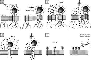
It is possible to induce conformational changes in cell surface receptor proteins by “pulling” on magnetic nanoparticles attached to them ( Figure 4 ). This “activates” biochemical signaling pathways within the cell, inducing a response in the cell that may be, for example, the production of a specific type of tissue matrix, differentiation of a stem cell down a specific lineage, initiation of cell division, or activation of apoptosis pathways to kill the cell. Targeted nanomagnetic actuation of specific cell surface receptors was first demonstrated by our group in 2002, and later expanded to target and magnetically activate specific cell signaling pathways both for tissue matrix production and to remotely activate and control stem cell differentiation both in vitro and in vivo. Reference Cartmell, Dobson, Verschueren and El Haj38–Reference Sura, Zhang, Lees, Zghoul, Wolbank, van Griensven, Redl, El Haj and Dobson42
Magnetic actuation of cell surface receptors. (a) Magnetic twisting: Large (micron-sized) magnetic particles coated with arginine-glycine-aspartic acid (RGD) molecules attach to integrin receptors on the cell membrane. The receptors are linked to actin filaments (black lines). A magnetizing pulse gives the particle a remanent magnetization (B = magnetic field vector). A torque is then applied to the particle via a “twisting field.” The force required to twist the particle is related to the mechanical properties of the actin filaments. (b) Mechanosensitive (MS) ion-channel activation: Mesoscale magnetic particles are bound to the integrin receptors and, upon the application of a high-gradient magnetic field, the particles are pulled toward the field, deforming the cell membrane and activating nearby MS ion channels. (c) Targeted ion-channel activation: Magnetic nanoparticles are attached to an ion channel via an antibody. Upon activation of a high-gradient magnetic field source, the ion channel is forced open. (d) Receptor clustering: Magnetic nanoparticles are bound to receptor complexes. In the absence of a magnetic field, the receptors are spaced along the membrane surface. When a high-gradient field is applied with a magnetic needle, the receptors are pulled toward the field source, initiating receptor clustering that can, for example, trigger intracellular signaling. Reproduced with permission from Reference 39. © 2008 Nature Publishing Group.

Recently, the technique has been adapted to manipulate signaling pathways that are activated via receptor clustering. Many so-called “death receptors” are activated via clustering, opening up the potential for remotely triggering apoptosis pathways in cancer cells. Mannix et al. first demonstrated magnetically triggered receptor clustering by coating superparamagnetic nanoparticles with monovalent ligands targeting transmembrane receptors to activate specific cell signaling pathways. Reference Mannix, Kumar, Cassiola, Montoya-Zavala, Feinstein, Prentiss and Ingber43 The application of a high gradient magnetic field via a magnetic needle induced clustering between the nanoparticles and therefore between the bound receptors, directly stimulating a signaling cascade within the cell. In a more recent article, Cho et al. adapted this idea in vivo and demonstrated the initiation of apoptosis in the tail region of zebrafish embryos by targeting and clustering tumor necrosis factor-related apoptosis inducing ligands. Reference Cho, Lee, Son, Lee, Yoo, Kim, Park, Shin and Cheon44
Concluding remarks
The use of mesoscale magnetic particles for biomedical applications is growing rapidly. Though few of these applications have reached the clinic, the potential is great, particularly as iron oxides are found naturally in the body. Current research focuses not only on the development of novel particles with enhanced biocompatibility and magnetic properties but also on applications that, through the coupling of particles in vivo to external fields, open up potential for magnetically actuated remote control of human physiology.
Currently, there are several challenges that must be overcome before mesocale magnetic particles can realize their full biomedical potential. Enhanced magnetic materials with higher moments and zero- or near-zero-remanance ground states are required for generating higher forces in order to develop more effective in vivo transducers and targeting agents. Improved biocompatibility and biomolecular surface functionalization for cell targeting will enable longer circulation times and improved activation of cell signaling pathways to control cell behavior. Finally, it is necessary to develop models and experiments that improve our understanding of the interactions of mesoscale magnetic structures with the complex biological environment in vivo and how magnetic energy can be deposited and transduced to activate and control biological or physiological processes.