Transposition of the great arteries represents one of the more common congenital cardiac lesions that necessitates infant surgical repair. Echocardiography is the most important diagnostic tool in the initial diagnosis, detailed surgical planning, and late follow-up of this lesion. The diagnosis of transposition of the great arteries is generally not difficult for those with some training in congenital heart disease. However, a detailed understanding of variations and coexisting lesions that alter the management is essential to perform and interpret the echocardiogram correctly. The present manuscript will review these features and identify the pitfalls that may occur in the imaging of this lesion.
Imaging the great vessel relationship
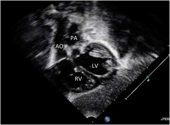
One of the key features of imaging in any congenital cardiac lesion is to clearly define the great vessel relationship. Nearly all imaging planes can provide direct or inferential evidence of abnormal great vessel relationships. The subcostal imaging planes are particularly valuable. A posterior-to-anterior sweep demonstrates the mitral valve in continuity with the pulmonary artery. The bifurcation of the pulmonary artery is usually easy to recognise. In addition, when colour Doppler interrogation is undertaken the flow through the ductus arteriosus can also be helpful in identifying the pulmonary artery. The sagittal imaging plane typically demonstrates the parallel nature of the great vessels, with the aorta being anterior to the pulmonary artery (Fig 1).
Subcostal sagittal view demonstrating the aorta (Ao) anterior to the pulmonary artery (PA). LV = left ventricle; RV = right ventricle.

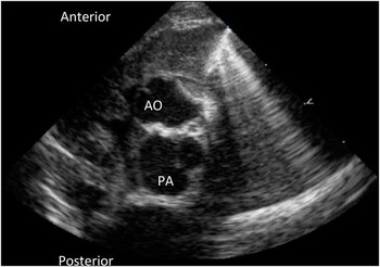
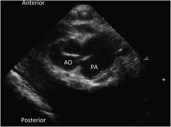
The parasternal imaging planes are also quite helpful in confirming the diagnosis of transposition of the great arteries. The parasternal long-axis plane demonstrates the relationship of the valve with the mitral valve. The short-axis plane allows one to recognise both semilunar valves en face (Fig 2). This plane confirms the abnormal great vessel relationship and also provides key anatomic features that will guide the surgery. In the case of ventriculoarterial discordance, the aorta is described in terms of its relationship with the pulmonary artery. By Van Praagh nomenclature, these positions can include “D” to the right, “A” directly anterior, or “L” to the left.Reference Van 1 The more widely used classification scheme favoured by Anderson et alReference Crupi, Anderson, Ho, Lincoln and Buckley 2 would favour descriptive terms to convey the anatomic relationship of the aorta with the pulmonary artery. In nearly all cases of ventriculoarterial discordance, the anatomic repair would include a LeCompte manoeuvre, where the branch pulmonary arteries are draped anterior to the aorta. However, there can be some cases where the aorta sits parallel or even slightly posterior to the right (Fig 3). Occasionally, the anatomic repair results in the branch pulmonary arteries passing posterior to the aorta. Hence, appropriate description of the semilunar relationship is needed.
Parasternal short-axis image of the semilunar valves demonstrating the aorta (Ao) to be anterior and to the right of the pulmonary artery (PA).

Parasternal short-axis image side-by-side relationship of the aorta (Ao) and pulmonary artery (PA).

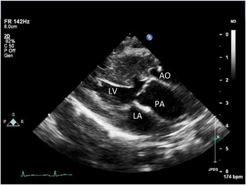
The imaging planes that best delineate the great vessel arrangement are also critical to define abnormalities of the semilunar valves or the ventricular outflow tracts. Such abnormalities almost always occur in association with a ventricular septal defect. When a ventricular septal defect is present, there may be some overlap between the diagnosis of double-outlet right ventricle and transposition of the great arteries with ventricular septal defect. Although controversy exists, a reasonable rule of thumb would be to ascribe the pulmonary artery to the ventricle based on the proportion of the valve that overlies a given ventricle. Hence, if more than 50% of the pulmonary valve is related to the left ventricle, one would use the term transposition of the great arteries with ventricular septal defect (Fig 4).
Parasternal long-axis image of transposition of the great arteries (TGA) with ventricular septal defect. Note that the pulmonary artery (PA) is predominantly committed to the left ventricle (LV). AO = aorta; LA = left atrium.

All forms of ventricular septal defect may be seen in transposition of the great arteries, the most clinically relevant being perimembranous. High-quality two-dimensional imaging is particularly important as the pressure in both ventricles is often equal or nearly so at the time of neonatal imaging, as many of these children receive prostaglandin therapy to maintain patency of the arterial duct. Similarly, because ventricular pressure may be equal, additional ventricular septal defects may not be appreciated when one relies on colour Doppler imaging alone. Care must be taken to interrogate the entire septum sweeps in the subcostal and parasternal short-axis planes. Echocardiography certainly plays a role in guiding the decision to attempt the surgical closure of the ventricular septal defect at the time of the arterial switch procedure. In isolated ventricular septal defects, the decision to pursue surgical closure is determined by many factors including signs and symptoms of cardiac failure. The decision to surgically close the transposition of the great arteries with ventricular septal defect is related primarily to the size and type of the defect. In general, small ventricular septal defects (<3 mm in diameter) do not require surgical closure. However, the decision depends on a variety of features including the ease of access and visualisation by the surgeon.
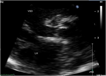
When a ventricular septal defect is present, this may result in outflow tract obstruction. Most commonly, this is left ventricular outflow tract obstruction or sub-pulmonary stenosis. This feature is best appreciated in parasternal long-axis imaging (Fig 5). In almost all of these cases, the pulmonary valve may have abnormalities such as doming leaflets, annular hypoplasia, or supravalvar stenosis. It is important for the echocardiographer to provide measurements of all elements of the left ventricular outflow tract. The surgeon can employ several strategies to address the case of transposition of the great arteries with ventricular septal defect and pulmonary stenosis. Aortic translocation or the Nikaidoh procedure is well suited for severe outflow tract obstruction.Reference Nikaidoh 3 This approach has largely supplanted the Rastelli-type approach. In other cases, arterial switch and ventricular septal defect closure can be performed even if there is valvar pulmonary stenosis. In general, pulmonary valve annulus diameter plotted as a Z score (using aortic valve normative data) of > −2.5 should be sufficient to consider an arterial switch procedure.Reference Sohn, Brizard, Cochrane, Wilkinson, Mas and Karl 4
Parasternal long-axis image of transposition of the great arteries (TGA) with ventricular septal defect and sub-pulmonary stenosis with a small pulmonary valve. LA = left atrium; PA = pulmonary artery; VSD = ventriclular septal defect.

Evaluation of the aortic arch in transposition of the great arteries is also an important component of the evaluation. Coarctation of the aorta occurs in 5–10% of cases of transposition of the great arteries.Reference Serraf, Lacour-Gayet and Bruniaux 5 Suprasternal imaging is the best imaging plane. Owing to the fact that the aortic root is more anterior in D-transposition of the great arteries, the orientation of the aortic arch is less acute than is seen in the normal heart. This means that the origin of the ductus arteriosus may give the impression of narrowing of the isthmus even when coarctation of the aorta is not present. Two-dimensional measurements can be helpful in making the diagnosis of coarctation of the aorta. As the arterial duct may be maintained patent with prostaglandin therapy, Doppler findings may not be particularly valuable in this determination.
Imaging of the atrial septum in D-transposition of the great arteries is relatively straightforward. Subcostal imaging planes should be used for this purpose. These images can be of value in clinical management vis-à-vis atrial septostomy. The approach to atrial septostomy varies among institutions. For those centres that use atrial septostomy selectively, the echocardiogram can provide supporting evidence for the premise that the lack of atrial level mixing is responsible for hypoxaemia. An absent or small (<5 cm) foramen ovale would support this assessment. In addition, pulse width Doppler can provide information regarding a mean gradient across the atrial septum. In many centres, echocardiography is used to guide atrial septostomy at the bedside. This has been shown to be successful and safe.Reference Beitzke, Stein and Suppan 6 Lastly, echocardiography can determine the effectiveness of the balloon atrial septostomy.
Assessment of the coronary arteries
The primary technical challenge in the arterial switch procedure relates to translocation of the coronary arteries. This is due to the small calibre of the vessels, but also due to the numerous anatomic variations and the possibility of an intramural course. Several guiding principles can be considered when imaging the coronary arteries.Reference Gremmels, Tacy, Brook and Silverman 7 First, multiple imaging planes are required to completely image the coronary arteries.
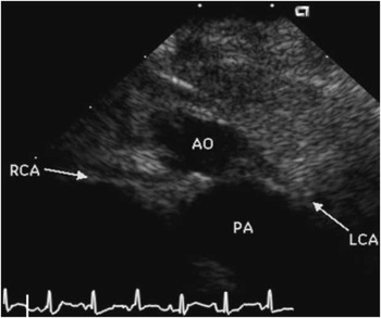
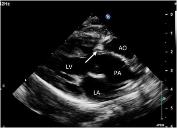
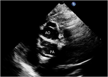
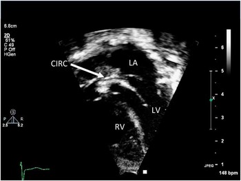
The echocardiographic views that are most useful for imaging the coronary arteries include the parasternal short-axis, the apical four-chamber, and the left oblique subcostal views obtained by rotating the transducer approximately 45° clockwise from the long-axis view of the left ventricle, and a parasternal long-axis view angled to the left.Reference Pasquini, Sanders and Parness 8 The parasternal short-axis view at the level of the aortic root usually displays the origins of the coronary arteries from the right and left posterior sinuses of Valsalva (Fig 6). However, the origin of the right coronary artery is not always well visualised in this plane. As such, a parasternal long-axis view through the aortic root may provide the best visualisation of the origin of the right coronary artery (Fig 7). A counterclockwise rotation from the parasternal short-axis view allows the right coronary artery to be followed to the right atrioventricular groove. The course of the left coronary artery can be demonstrated by angling leftward from a parasternal long-axis view through the aortic root. When the scan plane is tangential to the left side of the aortic root, the origin and proximal portion of the left coronary artery are displayed in cross-section. Further angulation to the left follows the left coronary artery to its bifurcation into the left anterior descending and left circumflex coronary arteries (Fig 8). The apical four-chamber and left oblique subxiphoid views are useful for locating the left circumflex coronary when it arises from the right coronary artery or for locating the left coronary artery in most cases of single right coronary artery. In such cases, the circumflex coronary artery passes posterior to the pulmonary artery (Fig 9).
Parasternal short-axis image demonstrating the origin of the left coronary artery (LCA) and right coronary artery (RCA) from the aorta (Ao). PA = pulmonary artery.

Parasternal long-axis demonstrates the origin of the right coronary artery (RCA) from the aorta (Ao). LA = left atrium; LV = left ventricle; PA = pulmonary artery.

Parasternal short-axis image demonstrating the origin of the left coronary artery (LCA) into the left anterior descending (LAD) and circumflex coronary artery (Cx). AO = aorta; CIRC = circumflex; LA = left atrium; PA = pulmonary artery.

Apical image demonstrating the circumflex coronary artery (CIRC) passing posterior to the pulmonary annulus. LA = left atrium; LV = left ventricle; RV = rightventricle.

The identification of intramural coronary artery poses the greatest challenge for the echocardiographer. The intramural course limits the surgeon's ability to mobilise the coronary arteries and in some series has been associated with a higher risk of mortality. The features that usually suggest an intramural course include a course that passes between the semilunar valves.Reference Pasquini, Parness, Colan, Wernovsky, Mayer and Sanders 9 Typically, the origin of the coronary artery is close to the commissure of the aortic valve (Fig 10). In addition, the intramural coronary artery may have a “high takeoff” arising further from the aortic valve annulus than usual.
Parasternal short-axis image demonstrating the intramural course of the left coronary artery (LCA) passing between the aorta (Ao) and pulmonary artery (PA).

Commissural alignment is also a feature that must be analysed. In the majority of the cases of D-transposition of the great arteries, the commissures of the pulmonary valve and aortic valve align. However, in some cases there may be an offset of up to 60° (Figs 11a and b). This malalignment of the commissures can complicate translocation of the coronary arteries, as the surgeon needs to translocate the “coronary button” to a location that is away from the commissures.
( a ) Parasternal short-axis image demonstrating alignment of the commissures of the aortic (Ao) valve and pulmonary artery (PA) valve. ( b ) Parasternal short-axis image demonstrating malalignment of the commissures of the Ao valve and PA valve.

Echocardiographic assessment of the child who presents “late”
Occasionally, the child with transposition of the great arteries may present after the 1st week of life. If there is a sizable ventricular septal defect present, then both ventricles have been exposed to systemic pressure and the arterial switch can be safely undertaken. However, if the ventricular septal defect is small or there is no ventricular septal defect, the left ventricle may have been exposed to systolic pressures much lower than the systolic arterial blood pressure (Fig 12). In such cases, the echocardiographer will need to examine the suitability of the left ventricle to support systemic circulation. Some studies have shown that the left ventricular mass can be helpful in predicting which patients can proceed to arterial switch without banding. Using M-mode measurements, the left ventricular mass can be estimated.Reference Lacour-Gayet, Piot and Zoghbi 10 It should be recognised that not all centres base their decision to undertake a “late” arterial switch on echocardiographic measurements.
Parasternal short-axis view demonstrating bowing of the ventricular septum towards the left ventricle (LV) suggesting elevated right ventricular (RV) pressure relative to LV.

Post-operative assessment
Fortunately, following anatomic repair of D-transposition of the great arteries, significant residual lesions are relatively uncommon. The echocardiographer, however, needs to be aware of a number of post-operative complications that may manifest soon after the arterial switch procedure, or in some cases many years later. As with numerous other congenital cardiac lesions, residual ventricular septal defects may occur. In general, defects <4 mm in diameter are unlikely to result in a significant shunt.Reference Rychik, Norwood and Chin 11 Residual defects may be found either adjacent to the site of patch closure or less commonly from a second previously unrecognised defect.
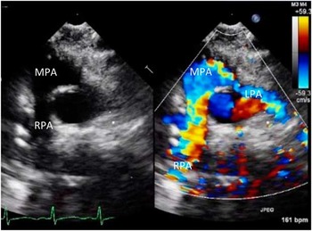
Owing to the fact that the branch pulmonary arteries are draped anterior to the aorta with the LeCompte manoeuvre, branch pulmonary artery stenosis is a known complication. In order to best visualise the branch pulmonary arteries, following the arterial switch and high parasternal short-axis image is particularly valuable (Fig 13). General peak Doppler gradients of <25 mmHg are well tolerated and do not require further intervention.
High parasternal short-axis view demonstrating the branch pulmonary arteries anterior to the aorta (Ao) following the arterial switch with LeCompte manoeuvre. LPA = left pulmonary artery; MPA = main pulmonary artery; RPA = right pulmonary artery.

Rarely, the anastomic site of the great vessels may be narrowed. This can result in either supravalvar pulmonary stenosis or supravalvar aortic stenosis. This complication is likely to be identified immediately following surgery. A number of imaging planes can help identify this lesion. Although parasternal long- and short-axis views are most valuable in demonstrating the two-dimensional narrowing, numerous other transducer positions are best at obtaining the most accurate Doppler gradients.
Acute problems with the coronary arteries, although very rare, can be catastrophic. In most such cases, there are numerous other features that would raise the suspicion of such a complication. These features include electrocardiographic changes, arrhythmias, or concerns raised directly by the surgeon regarding the coronary implantation. It is valuable to use both two-dimensional and Doppler interrogation of the coronary arteries. Colour Doppler of the coronaries should be performed with a low Nyquist setting and a small colour Doppler sector in order to optimise frame rate. It should be recognised that immediately following surgery the acoustic windows for the parasternal planes can be challenging, and satisfactory colour Doppler interrogation of the coronary arteries is not always possible. As such, it is also important to examine myocardial contractility, particularly wall-motion abnormalities. As acute coronary complications are likely to impact either the entire right or left coronary system, one would expect significant regional myocardial dysfunction. Identification of acute coronary complications needs to be made in a timely manner, as the low cardiac output is likely to result from this lesion.
Late lesions and complications
Coronary artery complications can occur months or years after an arterial switch procedure. The identification of these lesions is particularly challenging for a number of reasons. Acoustic windows may limit both two-dimensional and Doppler imaging of the proximal coronary arteries. In addition, the coronary circulation may develop collateral circulation and thus overt wall-motion abnormalities may be absent. Several studies have suggested that routine screening echocardiography may be insufficient to diagnose such complications.Reference Taylor, Dymarkowski and Hamaekers 12 Some studies have stated that alternative imaging modalities such as coronary angiography or computed tomography be performed routinely on these patients as they enter adolescence. Recognising these challenges, the echocardiographer should still perform a careful interrogation of the coronary arteries whenever follow-up echocardiography is performed. Increasingly, stress echocardiography is being used to identify coronary perfusion abnormalities in school-aged children and adolescents following arterial switch.Reference Hui, Chau, Leung, Chiu and Cheung 13 The diagnostic yield of this modality remains to be determined.
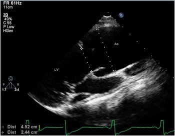
An additional complication that is being increasingly recognised following the arterial switch procedure is aortic insufficiency and aortic root dilation. With transposition of the great arteries, the pulmonary artery arises from the left ventricle. Following the arterial switch procedure, the pulmonary valve and pulmonary root serve as the aortic valve and aortic root.Reference Hourihan, Colan, Wernovsky, Maheswari, Mayer and Sanders 14 Some investigators have used the term “neo-aorta”. Following the arterial switch, the aortic root is predisposed to dilation (Fig 14), and thus the aortic valve may develop insufficiency. Therefore, follow-up echocardiographic studies should focus on the aortic valve function and aortic root dimensions. Guidelines recently published by the American Society of Echocardiography suggest that measurements of the aortic root should be made in end-systole using a parasternal long-axis imaging plane.Reference Lopez, Colan and Frommelt 15 These measurements are then typically expressed as a Z score. Although there are published guidelines of when to offer surgery for aortic root dilation in Marfan syndrome, it is not known whether there is a specific aortic root dimension or Z score that justifies pre-emptive surgical intervention following the arterial switch.
Parasternal long-axis view demonstrating dilation of the aortic (Ao) root. LV = left ventricle.

Summary
The remarkable improvement in mortality and morbidity for surgical repair of D-transposition of the great arteries remains one of the greatest successes in congenital heart care. Echocardiography has contributed significantly to this success. Adequate pre-operative imaging is essential to surgical planning especially with respect to delineation of coronary artery anatomy. Post-operative assessment needs to focus on several key features including the coronary arteries. Echocardiography is an essential element of the periodic follow-up of a child or adult with repaired D-transposition of the great arteries.