Introduction
Mucopolysaccharidoses are a group of inherited metabolic disorders. Currently, seven distinct clinical types of mucopolysaccharidoses, with different prevalence rates, have been described.Reference Muenzer1 Children with mucopolysaccharidoses generally appear normal at birth, but develop a variety of disease signs and symptoms resulting from the abnormal degradation and excessive storage of glycosaminoglycans, including dermatan sulphate, keratan sulphate, heparan sulphate and chondroitin sulphate, depending on the type of mucopolysaccharidosis. Typical somatic symptoms and signs include facial dysmorphology, hepatosplenomegaly, joint contractures, skeletal deformities, visual impairment, hernias and valvular heart disease. Neurological findings in mucopolysaccharidoses include hydrocephalus, carpal tunnel syndrome, optic nerve compression, and spinal cord compression and cervical myelopathy. A substantial proportion of patients with mucopolysaccharidoses also experience cognitive impairment, learning difficulties and behavioural problems.Reference Young and Harper2 Otolaryngological manifestations of mucopolysaccharidoses include recurrent middle-ear effusion, chronic upper airway infection, upper airway obstruction, macroglossia and voice disorders.Reference Mendelsohn, Harmatz, Bodamer, Burton, Giugliani and Jones3
Mucopolysaccharidoses patients undergo surgical procedures quite frequently before the diagnosis is made.Reference Keilmann, Nakarat, Bruce, Molter and Malm4 According to an international patient registry for patients with mucopolysaccharidosis II (Hunter Outcome Survey), ventilation tubes were inserted in 50 per cent of patients (at a median age of 3.5 years), adenoidectomy was performed in 47 per cent of patients (median age, 3.5 years) and tonsillectomy was conducted in 35 per cent of patients (median age, 4.8 years).Reference Keilmann and Malm5 As stated by Walker et al., in many patients the narrowing of the upper airways leads to a higher resistance of the airways, and in severe cases it can lead to stridor, respiratory distress and obstructive sleep apnoea. It is often not possible to ventilate or intubate the patients; the temporary airway obstruction can cause negative pressure (potentially obstructive) and pulmonary oedema.Reference Walker, Belani, Braunlin, Bruce, Hack and Harmatz6
Mostly after induction or extubation, a complete airway obstruction can cause profound hypoxaemia and cardiac arrest. Post-intubation problems such as stridor, lower airway collapse or infection, or the need for reintubation or tracheostomy, may occur. One paper reports that 22 patients out of 487 with mucopolysaccharidosis II had a tracheotomy.Reference Keilmann and Malm5
There is currently no universal examination protocol useful in determining the mucosal alterations of the larynx and hypopharynx as observed on rigid or flexible endoscopy performed in patients without general anaesthesia. The Cormack–Lehane classification, used to describe the width of the larynx as observed during intubation, has proved to be insufficient, as it only describes the visibility of the vocal folds.Reference Cormack and Lehane7 The use of a standardised classification protocol could be useful to assess the risk of intubation and extubation, and could provide information about the natural course of the disease and the efficacy of enzyme replacement therapy.
Materials and methods
The described procedure was designed as part of the examinations conducted to monitor the course of mucopolysaccharidosis otorhinolaryngological manifestations. These examinations included inspection of the head and neck, microscopy of the ear, rhinoscopy, assessment of the maximal distance of the teeth (ideally with a special curved ruler), inspection of the mouth and oropharynx, and audiometry. All procedures contributing to this work complied with the ethical standards of the relevant national and institutional guidelines on human experimentation, and with the Helsinki Declaration of 1975 (as revised in 2008). Informed consent was obtained from all patients in the study.
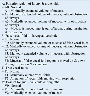
We developed a classification system in which the alterations of mucosa in the posterior region of the larynx and the arytenoids (‘A0’ – ‘A4’), the false vocal folds (‘F0’ – ‘F4’), the true vocal folds (‘T0’ – ‘T2’), and the vallecula and epiglottis (‘V0’ – ‘V2’) are described according to rigid or flexible endoscopic findings of the upper airways. Detailed information is provided in Table I. Examples from different patients are shown in Figure 1.
Examples demonstrating the classification of mucosal alterations in: the posterior region of the larynx and the arytenoids (‘A0’–‘A4’), the false vocal folds (‘F0’–‘F4’), and the vallecula and epiglottis (‘V0’–‘V2’).

Classification of mucosal alterations of larynx and hypopharynx in mucopolysaccharidoses patients
Three physicians who specialised in laryngology used this classification, in a blinded manner, for: 11 patients with mucopolysaccharidosis I (7 females, aged 7–35 years, with a mean age of 22.6 years), 15 patients with mucopolysaccharidosis II (0 females, 5–45 years, mean age of 20.8 years), 13 patients with mucopolysaccharidosis IV (4 females, 4–25 years, mean age of 13.0 years) and 16 patients with mucopolysaccharidosis VI (8 females, 6–44 years, mean age of 23.25 years). Fifteen patients were examined prior to the start of enzyme replacement therapy and several times during the therapy.
The intraclass correlation was calculated for the three examiners. The results of the patients with mucopolysaccharidosis II were compared to all others as we hypothesised that patients with mucopolysaccharidosis II were the most severely affected. The Mann–Whitney U test was used for this comparison. Using a correlation analysis, we examined whether the mucosal alterations were age-dependent. Finally, we compared the results of the new classification system to those of two classification systems established in anaesthesiology to judge the risk of intubation, using a correlation analysis.Reference Cormack and Lehane7, Reference Mallampati, Gatt, Gugino, Desai, Waraksa and Freiberger8
Results
About half of all patients tolerated rigid or flexible endoscopy during the routine examinations. Fifty-two per cent of the examinations were carried out using a flexible endoscope under local anaesthesia of the nose, and 48 per cent of the examinations were carried out using a rigid laryngoscope transorally, without local anaesthesia in most cases (mucopolysaccharidosis I, 48 and 52 per cent respectively; mucopolysaccharidosis II, 60 and 40 per cent; mucopolysaccharidosis IV, 73 and 27 per cent; and mucopolysaccharidosis VI, 30 and 70 per cent).
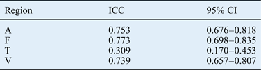
The intraclass correlation coefficients for the alterations of mucosa were: 0.75 (95 per cent confidence interval (CI) = 0.68–0.82) in the posterior region of the larynx and the arytenoids, 0.77 (95 per cent CI = 0.70–0.84) for the false vocal folds, 0.31 (95 per cent CI = 0.17–0.45) for the true vocal folds, and 0.74 (95 per cent CI = 0.66–0.81) for the vallecula and epiglottis.
As seen in Figure 2, the mucosal alterations of the posterior region of the larynx and the arytenoids, and the false vocal folds, were classified as more severe in patients with mucopolysaccharidosis II, compared to those with mucopolysaccharidoses I, IV and VI who had comparable alterations.
Classification of the mucosal alterations in: (a) the posterior region of the larynx and the arytenoids (‘A0’–‘A4’), (b) the vallecula and epiglottis (‘V0’–‘V2’), (c) the false vocal folds (‘F0’–‘F4’), and (d) the true vocal folds (‘T0’–‘T2’), in patients with mucopolysaccharidoses I, II, IV and VI.
Using the Mann–Whitney U test, a significant difference between patients with mucopolysaccharidosis II and the other mucopolysaccharidoses patients was observed for the alterations of mucosa in: the posterior region of the larynx and the arytenoids (p = 0.012), the false vocal folds (p = 0.009), and the vallecula and epiglottis (p = 0.036), but not for the true vocal folds.
As seen in Table II, the alterations of mucosa in the posterior region of the larynx and the arytenoids (‘A’) were correlated with all other endoscopic classifications (‘F’, ‘T’ and ‘V’), and with the classifications of Mallampati and Cormack–Lehane. In the correlation analysis, there was no dependency on age (data not shown).
Correlations between parameters
ICC = intraclass correlation coefficients; CI = confidence interval; A = posterior region of the larynx and the arytenoids; F = false vocal folds; T = true vocal folds; V = vallecula and epiglottis
In 15 patients who received enzyme replacement therapy (who were followed longitudinally), gradual improvements, deteriorations or irregular developments were observed (Figure 3).
Classification of the mucosal alterations in: (a) the posterior region of the larynx and the arytenoids (‘A0’–‘A4’), and (b) the false vocal folds (‘F0’–‘F4’), in patients with mucopolysaccharidoses I, II and VI who received enzyme replacement therapy (‘ERT’). Pt no. = patient number
Examples of the assessment findings of one patient with mucopolysaccharidosis II are shown in Figure 4. Mucosa of the posterior region of the larynx and the arytenoids, and the false vocal folds, was only minimally enlarged before the start of enzyme replacement therapy. Four months after the start of enzyme replacement therapy, the mucosa was markedly extended, and it moved into and out of the larynx during inspiration and expiration.
Endoscopic pictures of the same patient: (a) before the start of enzyme replacement therapy; (b & c) 4 months after the start of enzyme replacement therapy ((b) during inspiration (mucosa is moved into the larynx) and (c) during expiration); (d) after 8 months of enzyme replacement therapy; and (e) after 14 months of enzyme replacement therapy. A = posterior region of the larynx and the arytenoids; F = false vocal folds; T = true vocal folds
Discussion
Semenza and Pyeritz reported on the respiratory complications of mucopolysaccharidoses in 1988.Reference Semenza and Pyeritz9 The authors reviewed 10 previous studies with regard to 32 patients with mucosal alterations of the pharynx and larynx. For their own investigation, 21 patients were chosen retrospectively; these patients had a history of respiratory problems or had undergone a thorough evaluation of their respiratory system. Supraglottic narrowing was diagnosed by direct laryngoscopy or computed tomography. In a 25-year-old male with mucopolysaccharidosis II, they found large floppy arytenoids, unevenly deformed vocal folds and an ‘infantile epiglottis’. In a 54-year-old male with mucopolysaccharidosis IV, a tracheotomy was performed and laser therapy undertaken to remove redundant aryepiglottic tissue. The authors suggested that respiratory involvement was responsible for a high degree of morbidity and mortality. Belani et al. found that it was easier to see the vocal folds during laryngoscopy in children when they were younger and smaller.Reference Belani, Krivit, Carpenter, Braunlin, Buckley and Liao10 Pre-operative obstructive breathing was associated with a significantly higher incidence of post-extubation obstruction.
The first comprehensive review of otorhinolaryngological manifestations of mucopolysaccharidoses was reported by Simmons et al.Reference Simmons, Bruce, Penney, Wraith and Rothera11 They gave a summary of the ENT features of mucopolysaccharidoses I, II, III, IV and VI, and described adenotonsillar hypertrophy as almost universal in this group of patients. Within the frame of progressive airway compromise, they proposed that the depositions may also be in the walls of the pharynx and the larynx. In addition, the authors reported several cases with festoons of excessive tissue associated with substance deposition over the arytenoid cartilages and the aryepiglottic folds. In extreme cases, these were seen to collapse into the laryngeal inlet and cause severe airway compromise. The authors described performing laser excision of the excess tissue in some cases. Simmons et al. concluded that these conditions, and other systemic effects of the disease, must be considered when surgery under general anaesthesia is indicated.Reference Simmons, Bruce, Penney, Wraith and Rothera11
Walker et al. and Berger et al. emphasised the importance of mucosal alterations of the hypopharynx and larynx for airway management, and respiratory and sleep disorders, in mucopolysaccharidoses patients.Reference Walker, Darowski, Morris and Wrath12, Reference Berger, Fagondes, Giugliani, Hardy, Lee and McArdle13
In our study, about half of the patients tolerated an endoscopy during routine examinations performed partially under local anaesthesia, without sedation or general anaesthesia. The alterations of mucosa usually increase over a lifetime. In older patients with predominantly physical alterations, endoscopy was tolerated well. At least in these patients, a regular standardised assessment of the alterations seems to be reasonable, preferably before an emergency situation occurs.
Comparing the mucosal alterations found in the four regions, the most severe alterations were in the posterior region of the larynx and the arytenoids, and in the region of the false vocal folds. In some patients, the true vocal folds could not be classified because the false vocal folds impeded this. In the patients in whom the vocal folds could be classified, the highest category (‘T2’) was never reached.
Comparing the patients with mucopolysaccharidoses I, II, IV and VI, those with mucopolysaccharidosis II demonstrated the most severe mucosal alterations. This is in accordance with the experience of Walker et al., who reported about 34 children who underwent general anaesthesia for 110 procedures.Reference Walker, Belani, Braunlin, Bruce, Hack and Harmatz6 They found the highest incidence of difficult intubation in patients with mucopolysaccharidosis II. During laryngoscopy, the vocal folds were visible in only 19 out of 55 cases of mucopolysaccharidosis II.
The classification system used in this study is suitable to describe the natural course of the disease and possible treatment effects. In addition to symptomatic treatment, bone marrow transplantation and enzyme replacement therapy are also used. Bone marrow transplantation has been employed in patients with mucopolysaccharidoses since the 1980s. Yeung et al. reported 14 patients who underwent bone marrow transplantation.Reference Yeung, Cowan, Horn and Rosbe14 Eleven of the 13 children who had symptoms of upper airway obstruction documented during clinical examination, and who underwent serial pulmonary function testing before bone marrow transplantation, showed marked improvement in these symptoms based on the results of subsequent pulmonary function testing and clinical history, within weeks to months after transplantation.
Currently, enzyme replacement therapy is available for mucopolysaccharidoses I, II and VI. Harmatz et al. conducted a study on enzyme replacement therapy in mucopolysaccharidosis VI and found a reduction of sleep apnoea in those patients receiving treatment who had no tracheostomy.Reference Harmatz, Whitley, Waber, Pais, Steiner and Plecko15 They explained this as an improvement in upper airway obstruction at week 48.
Treatment efficacy was evaluated in a phase II/III clinical study of enzyme replacement therapy in patients with mucopolysaccharidosis II using a composite endpoint consisting of distance walked in 6 minutes and percentage of predicted forced vital capacity.Reference Muenzer, Wraith, Beck, Giugliani, Harmatz and Eng16 The authors reported a clear clinical benefit, which was primarily reflected in an improvement in the 6-minute walk test distance rather than an improvement in the forced vital capacity percentage. They suggested that the smaller effect on pulmonary function may represent difficulties in accurately determining forced vital capacity percentage in patients with mucopolysaccharidosis II, because the formulae for predicted forced vital capacity assume both normal growth and the ability to accurately measure height. Mucopolysaccharidosis II patients under the age of nine years are typically of normal stature, whereas older mucopolysaccharidoses patients demonstrate a lower height than normal subjects of the same age. In such cases, the forced vital capacity percentage is misleadingly high. However, most of the patients experienced an improvement in joint flexibility; the authors assumed that they were able to stand up straighter, leading to an increased height and subsequent increase in predicted forced vital capacity.
• Mucopolysaccharidoses patients often develop mucosal alterations of the larynx and hypopharynx
• These alterations can lead to respiratory distress, obstructive sleep apnoea and post-intubation problems
• The alterations can be documented using video endoscopy, with both rigid and flexible endoscopes
• A classification system for the alterations was developed and utilised in 55 mucopolysaccharidoses patients
• The most severe alterations were in the posterior larynx and false vocal fold regions
• Alterations were most severe in patients with mucopolysaccharidosis II
With regard to the 15 patients followed up longitudinally, before and after the start of enzyme replacement therapy, we saw no clear trend. In each patient, the therapy may ameliorate the symptoms while the natural course of the disease may lead to a deterioration of symptoms.
In some patients with mucopolysaccharidosis II, who were reassessed six months after starting enzyme replacement therapy, their classification worsened. When studying the videos of the patients with a temporal worsening, we observed redundant mucosa relapsing into the larynx, moving with the airflow and even causing noises. This could be explained by a partial depletion of the deposits, leading to excessive mucosa for some time. In patients with extremely excessive tissue, laser surgery may be indicated.Reference Simmons, Bruce, Penney, Wraith and Rothera11 Surgery was not used on our patients as the alterations mostly proved to be transient after enzyme replacement therapy, and improved after some months.
Conclusion
Quantification of the mucosal alterations was possible using video endoscopy, with both a rigid and flexible endoscope (depending on the patients’ needs). The new classification system is superior to the Cormack–Lehane classification system for patients with mucopolysaccharidoses. Regular monitoring of the abovementioned parameters can provide information about the disease's natural course and the efficacy of enzyme replacement therapy.