In the 1970s, a soil sample collected from the Vai Atare region of Rapa Nui (also known as Easter Island) in the Pacific Ocean was found to contain a bacterial strain of Streptomyces hygroscopicus producing a compound with potent antifungal activity (Ref. Reference Vezina, Kudelski and Sehgal1). The purified antibiotic, named rapamycin after the location where it was discovered, was characterised as a lipophilic macrolidic lactone – practically insoluble in water but soluble in most organic solvents. In addition to antifungal activity, rapamycin was found to have impressive immunosuppressive qualities (Refs Reference McAlpine2, Reference Sehgal3). The National Cancer Institute (NCI) identified rapamycin as a potent, mainly cytostatic agent against many types of human cancer (Refs Reference Douros and Suffness4, Reference Faivre, Kroemer and Raymond5). However, because of its low solubility in water and the fact that the patent for the drug had expired, the development of rapamycin as an antitumour agent was delayed considerably (Refs Reference Faivre, Kroemer and Raymond5, Reference Hidalgo6).
Intensive investigation of the mechanism of action of rapamycin led to the discovery of an evolutionarily conserved pathway that integrates signals arising from growth factors, nutrients and energy status. This pathway regulates metabolism, growth and proliferation in normal cells and plays a central role in cell proliferation, survival and drug resistance in many human cancers (Refs Reference Harris and Lawrence7, Reference Tee and Blenis8, Reference Bjornsti and Houghton9). The core of this pathway corresponds to the target of rapamycin (TOR), discovered originally through two mutations conferring resistance to the growth inhibitory effect of rapamycin in the budding yeast (Saccharomyces cerevisae) (Refs Reference Heitman, Movva and Hall10, Reference Kunz11, Reference Helliwell12, Reference Chen13). All eukaryotes studied to date contain a TOR gene. There are two yeast genes (TOR1 and TOR2); the mammalian homologue, named mammalian target of rapamycin (MTOR or FRAP1), is a single gene (Refs Reference Sabatini14, Reference Brown15, Reference Sabers16).
The core of the mTOR pathway: mammalian target of rapamycin complexes (mTORCs)
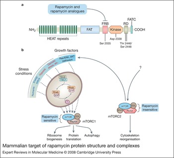
The MTOR gene maps to chromosome 1p36.2 in the human genome and encodes a 289 kDa protein that contains a kinase catalytic domain similar to that found in phosphoinositide 3-kinase (PI3K) (Refs Reference Brunn17, Reference Burnett18, Reference Cardenas and Heitman19). However, unlike PI3K, mTOR does not have lipid kinase activity and instead functions as a Ser/Thr protein kinase (Refs Reference Brunn17, 18). mTOR is the founding member of the PI3K-related kinase (PI3KK) family, which also includes DNA-dependent protein kinase, ATM (ataxia telangiectasia mutated), ATR (ATM- and Rad3-related) and SMG-1 (‘human homologue of suppressor with morphogenetic effect on genitalia 1’) (Ref. Reference Abraham20). In addition to the catalytic domain, the mTOR molecule contains 20 HEAT (Huntington elongation factor 1A–protein phosphatase 2A subunit–TOR) repeats that are crucial for protein–protein interactions, FAT (FRAP–ATM–TRAPP2) and FATC (FAT C-terminal) domains characteristic of the PI3KK family, a putative autoinhibitory repressor domain (RD) and an FKB12 rapamycin-binding domain (FRB). The FRB is the immediate target of rapamycin and rapamycin analogues (Fig. 1a) (Refs Reference Helliwell12, Reference Andrade and Bork21, Reference Bosotti, Isacchi and Sonnhammer22, Reference Peterson23, Reference Takahashi24, Reference Sekulic25, Reference Cafferkey26).
Mammalian target of rapamycin protein structure and complexes. (a) The human mammalian target of rapamycin (mTOR) gene encodes a protein of 2549 amino acids. Point mutation at the catalytic domain changing Asp2338 to Ala eliminates mTOR kinase activity. The FAT and FATC domains, flanking the catalytic domain are believed to be necessary for the appropriate folding or organisation of the catalytic domain and perhaps interact. The FRB region, just upstream of the catalytic domain, is characteristic of mTOR and the only region for which definite structural information exists. Mutation of Ser2035 to Arg in this region abolishes the binding of the FKBP12–rapamycin complex. The 20 HEAT repeats at the N-terminus, each ~39 amino acids long, contain several conserved hydrophobic residues. It is believed that each repeat forms a pair of antiparallel α-helices that stack in parallel with other repeats forming a large surface for protein–protein interactions. Between the catalytic and the FATC domain there is a poorly understood regulatory region containing two phosphorylation sites: Thr2446 and Ser2448. Deletion of this region results in activation of mTOR. Recently it was shown that both sites are phosphorylated by the downstream target of mTOR, S6K1, and not by AKT as it was initially believed. However, the functional significance of this regulatory loop between mTOR and S6K1 remains elusive. (b) There are at least two different mTOR protein complexes: mTORC1 and mTORC2. Most of the known functions of mTOR, and particularly the regulatory effect on protein translation and autophagy, are attributable to mTORC1 and can be inhibited by rapamycin. The two other proteins of the complex, GβL and Raptor, have no catalytic activity. The human gene for Raptor encodes a protein 1335 amino acids long, which contains a unique N-terminal region, three HEAT motifs and seven WD40 repeats. GβL protein, which is 260 amino acids long, consists almost entirely of seven WD40 repeats, is similar in sequence to the β subunits of heterotrimeric G proteins and interacts with the kinase domain of mTOR. Collectively, mTOR, GβL and Raptor have 23 HEAT and 14 WT40 motifs, increasing the potential of protein–protein interaction. No definite model exists for mTORC1 function; however, it seems that Raptor acts as adaptor that presents substrates to mTOR, and that Raptor and GβL both modulate mTOR activity. Many of the signals that regulate mTORC1 activity concentrate on the TSC1–TSC2 complex. For example, activated AKT or ERK pathways result in phosphorylation of TSC2 and inactivation of TSC1–TSC2. TSC1–TSC2, when active, inactivates the small protein Rheb, promoting the hydrolysis of GTP bound to Rheb. The protein that catalyses the opposite step remains elusive. Rheb seems to regulate mTOR indirectly, through FKBP38, a member of the FK506-binding protein (FKBP) family. FKBP38 binds to mTOR and inhibits its activity. Rheb interacts directly with FKBP38 and prevents its association with mTOR in a GTP-dependent manner. The second complex – mTORC2 – is known to influence cytoskeleton organisation and cell polarity and contains the evolutionary conserved protein Rictor (instead of Raptor), SIN1 and protor-1. SIN1 exists in at least three different isoforms defining three different mTORC2 complexes with different target specificities. However, all of these isoforms contribute to AKT activation, creating a regulatory loop for the activation of mTORC1. Insulin regulates the activity of two of these mTORC2 variations. However, the exact mechanism of mTOR2 activation and whether it is upstream of mTORC1 remains unclear. mTORC2 function is not generally inhibited by rapamycin. Abbreviations: FAT, FRAP–ATM–TRAPP2; FATC, FAT C-terminal; HEAT, Huntington elongation factor 1A–protein phosphatase 2A subunit–TOR; FKBP12, FK507-binding protein 12; FRB, FKBP-rapamycin binding; mTORC1/2, mammalian target of rapamycin complex 1/2.

mTOR functions as part of a larger multiprotein signalling complex. Recent studies show that mTOR participates in at least two different signalling complexes – mTORC1 and mTORC2 – which have different functions (Fig. 1b) (Ref. Reference Loewith27). mTORC1, which is sensitive to rapamycin, is currently believed to have three components: mTOR, Raptor (regulatory-associated protein of mTOR; an adaptor protein approximately half the size of mTOR) and GβL (G protein β-subunit-like protein, also known as mLST8; a 36 kDa protein believed to be important for sensing upstream signals) (Refs Reference Kim28, Reference Hara29). Both Raptor and GβL are evolutionary conserved. However, the exact relationship between these components and their functions are not completely understood. It is believed that upstream signals associated with growth factors, energy status, nutrient availability and various conditions of stress, such as hypoxia, reactive oxygen species, DNA damage and hyperosmotic environment regulate the strength of association between mTOR and Raptor and, in this way, the recruitment of various downstream substrates of mTOR (Refs Reference Kim30, Reference Sarbassov31). In turn, activation of mTORC1 promotes ribosome biogenesis and the rates of translation and protein synthesis. Activation of mTORC1 also inhibits autophagy, influencing metabolic mass, cell proliferation and cell survival (Ref. Reference Sarbassov, Ali and Sabatini32). All these functions are inhibited by rapamycin or rapamycin analogues (Ref. Reference Kim30).
mTORC2, which is generally rapamycin insensitive, contains at least four components: mTOR, GβL, Rictor (rapamycin-independent companion of mTOR; a ~200 kDa adaptor protein) and mSIN1 (mammalian stress-activated protein kinase interacting protein 1) (Refs Reference Sarbassov33, Reference Jacinto34, Reference Jacinto35, Reference Frias36). Very recently a fifth component of undetermined significance, named protor-1, was discovered (reviewed in Ref. Reference Guertin and Sabatini161). mTORC2 is regulated by unknown upstream signals, and the exact mechanisms of mTORC2 activation are poorly understood. However, it is believed that various growth factors contribute to mTORC2 activation. Compared with mTORC1, the function of mTORC2 is less well studied, but it is known that mTORC2 activity regulates cytoskeleton organisation and promotes activation of AKT (also known as protein kinase B) (Refs Reference Sarbassov31, Reference Sarbassov33, Reference Jacinto34). Since AKT is an important upstream regulator for the activation of mTORC1, an autoregulatory loop between the two complexes seems to exist (Refs Reference Sarbassov31, Reference Sarbassov, Ali and Sabatini32).
Downstream effectors of mTOR
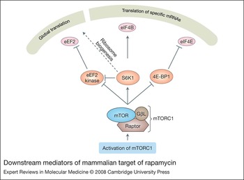
Although the regulation of protein production has been studied mainly at the genetic and transcriptional levels, it is becoming clear that regulation of translation is equally important (Ref. Reference Ruggero and Pandolfi37). mTORC1 acts mostly at this level, regulating the rate of mRNA translation and ribosome biosynthesis. Two critical mTORC1 downstream effectors of translation are the eukaryotic initiation factor 4E (eIF4E) binding protein 1 (4E-BP1) and p70 ribosomal protein S6 kinase (S6K1) (Fig. 2) (Refs Reference Beretta38, Reference Price39, Reference Chung40, Reference Hay and Sonenberg41).
Downstream mediators of mammalian target of rapamycin. 4E-BP1, S6K1 and eEF2 kinase are among the crucial downstream mediators of mammalian target of rapamycin (mTOR) that orchestrate the response of the translational machinery to various stress signals and growth factors. In the inactive state, 4E-BP1 blocks eIF4E from binding the cap structure of mRNA, and the translation elongation factor eEF2 is phosphorylated and inactivated by the corresponding eEF2 kinase. In the active state, activated mTORC1 phosphorylates and neutralises 4E-BP1, so that eIF4E can bind the cap structure of mRNA. In addition, S6K1, activated by mTORC1, phosphorylates and activates the eIF4B RNA helicase. Activated S6K1 phosphorylates and inhibits eEF2 kinase so that the underphosphorylated and active eEF2 can contribute to the elongation step of translation. Finally, activation of mTORC1 enhances ribosome biogenesis through many intermediate steps generally promoting the rate of translation. Abbreviations: 4E-BP1, 4E-binding protein-1; eEF2, eukaryotic elongation factor 2; eIF-4E, eukaryotic initiator factor 4E; mTORC, mammalian target of rapamycin complex; S6K1, p70 ribosomal protein S6 kinase.

The rate of translation of most eukaryotic mRNAs is dependent on the binding of initiator factors (eIF) to the cap (m7GpppN – where N is any nucleotide) structure at the 5′ untranslated mRNA terminus (Ref. Reference Gingras, Raught and Sonenberg42). The eIF4E 5′CAP-binding subunit of the eIF4F initiator complex is recruited and neutralised by 4E-BP1. Direct or indirect phosphorylation of 4E-BP1 by activated mTORC1 disrupts the association between 4E-BP1 and eIF4E, facilitating the 5′CAP-binding function of eIF4E and thereby promoting translation (Fig. 2) (Refs Reference Gingras, Raught and Sonenberg42, Reference Gingras, Raught and Sonenberg43, Reference Wang and Proud44).
S6K1, which is phosphorylated and activated by mTORC1 and PDK1, was previously thought to play a critical role in the translation of mRNAs with the TOP (terminal oligopyrimidine) sequence at the 5′ untranslated region, among which are many that encode components of the translational machinery (Ref. Reference Meyuhas45). However, more recent studies have shown that S6K1 (and the similar S6K2), as well as the phosphorylation status of its well-known target, ribosomal protein S6, are dispensable for 5′TOP mRNA translation, although mTORC1 activation remains crucial (Refs Reference Pende46, Reference Tang47, Reference Stolovich48). Recent studies have established two more connections between mTORC1-mediated S6K1 activation and translation (Ref. Reference Peterson and Sabatini49). First, activated S6K1 phosphorylates the RNA helicase eIF4B, promoting the association of eIF4B with the eIF3 scaffold component of the initiator complex, and the association of eIF3 with the eIF4G scaffold component of the eIF4F initiator complex (Refs Reference Holz50, Reference Shahbazian51). Second, activated S6K1 phosphorylates and inactivates the eukaryotic elongation factor 2 (eEF2) kinase, resulting in underphosphorylated and functional eEF2. The latter is important for the promotion of the elongation steps of translation (Fig. 2) (Refs Reference Browne and Proud52, Reference Wang53).
Regulation of ribosomal biogenesis is a central mechanism for the control of global levels of translation and cell growth in eukaryotes, and is tightly regulated by mTORC1 (Ref. Reference Mayer and Grummt54). Activation of mTORC1 promotes the biosynthesis of ribosomal RNAs and proteins by activating transcription, mediated by RNA polymerase (Pol) I, Pol II and Pol III in the nucleus and accelerating the translation of 5′TOP mRNA into ribosomal proteins in the cytoplasm (Ref. Reference Mayer and Grummt54). Although the exact mechanisms for the regulation 5′TOP mRNA in the cytoplasm remain elusive, it seems likely that enhancement of transcription in the nucleus by Pol I is mediated partly by S6K1, which activates and increases the availability of TIF-IA (transcription initiator factor IA) and UBF (upstream binding factor), essential cofactors of Pol I (Refs Reference Mayer55, Reference Hannan56).
Autophagy, more specifically macroautophagy, the process by which cellular components including organelles are catabolised and recycled, is an important survival mechanism that is tightly regulated by mTORC1 (Ref. Reference Shintani and Klionsky57). The exact mechanisms by which activated mTORC1 inhibits autophagy are poorly understood, although it seems probable that negative regulation of a homologue of ATG1 kinase that mediates early activation steps of the autophagic process is involved (Refs Reference Kamada58, Reference Mordier59).
Downstream targets of mTORC2 that mediate cytoskeleton reorganisation seem to include the small GTPases Rho and Rac, and the protein kinase PKCα (Refs Reference Sarbassov33, Reference Jacinto34). However, knowledge about the pathway downstream of mTORC2 is limited.
Upstream regulators of mTOR
Pivotal to the understanding of upstream regulation of mTOR activity was the discovery of the importance of TSC1 and TSC2 (tuberous sclerosis complex 1 and 2, also known as amartin and tuberin, respectively) (Refs Reference Tapon60, Reference Gao61). Lack of TSC1 or TSC2 expression results in overactivation of mTOR (Refs Reference Goncharova62, Reference Kwiatkowski63). The heterodimer of TSC1/TSC2 is a GTPase-activating protein (GAP) for the small GTP-binding protein Rheb (RAS homologue enriched in brain), which catalyses the hydrolysis of Rheb-bound GTP and promotes the conversion of Rheb-GTP to Rheb-GDP (Refs Reference Saucedo64, Reference Stocker65, Reference Zhang66, Reference Tee67). In turn, Rheb-GTP activates mTOR kinase (Ref. Reference Long68). Signals regulating mTORC1 activity including those associated with growth factors, energy status and various stressful stimuli (e.g. hypoxia) target the TSC1/TSC2 heterodimer (Fig. 1B) (Refs Reference Sarbassov, Ali and Sabatini32, Reference Hay and Sonenberg41).
Two of the most important mTORC1-regulating pathways are the AKT and the Ras–ERK (extracellular-signal-regulated kinase)/MAPK (mitogen-activated protein kinase) pathways emanating from cell membrane growth-factor-activated tyrosine kinase or seven-transmembrane receptors (Refs Reference Cantley69, Reference Johnson and Lapadat70, Reference Roux and Blenis71). A critical step for AKT activation is phosphorylation at the D3 position of the inositol ring of the plasma membrane lipid phosphatidylinositol 4,5-bisphosphate [PtdIns(Reference Douros and Suffness4,Reference Faivre, Kroemer and Raymond5)P 2] by PI3K. PI3K is activated by interaction with activated receptor tyrosine kinases (RTKs) and/or associated adaptors, such as those of the IRS family. Phosphorylation of PtdIns(Reference Douros and Suffness4,Reference Faivre, Kroemer and Raymond5)P 2 produces PtdIns(Reference Sehgal3,Reference Douros and Suffness4,Reference Faivre, Kroemer and Raymond5)P 3, which offers plasma membrane docking sites for proteins that contain the pleckstrin homology (PH) domain, such as AKT and PDK1 (phosphoinositide-dependent kinase). In this way, PtdIns(Reference Sehgal3,Reference Douros and Suffness4,Reference Faivre, Kroemer and Raymond5)P 3-associated AKT is phosphorylated in the catalytic domain by the proximal PtdIns(Reference Sehgal3,Reference Douros and Suffness4,Reference Faivre, Kroemer and Raymond5)P 3-associated PDK1 and becomes activated (Ref. Reference Cantley69). However, full activation of AKT requires a second phosphorylation at the conserved C-terminal hydrophobic motif by a second ‘PDK2’ kinase. A recent study has shown that this ‘PDK2’ is, in fact, mTOR kinase nucleated at mTORC2 (Ref. Reference Sarbassov31). Activated AKT, in turn, phosphorylates TSC2 and inactivates the TSC1/TSC2 heterodimer, neutralising the inhibitory activity of TSC1/TSC2 on mTORC1 (Refs Reference Gao61, Reference Inoki72, Reference Potter, Pedraza and Xu73).
In the Ras–ERK pathway, activated RTKs, through adaptors, recruit guanine-nucleotide exchange factors, such as SOS (son of sevenless), which stimulate the exchange of GDP for GTP on the GTPase Ras, allowing the activation of downstream effectors such as the MAPKKK Raf. It has been shown that downstream effectors of the Ras–ERK pathway – ERK (a MAPK) and the MAPK-activated protein kinase (MP) p90 ribosomal S6 kinase – can phosphorylate TSC2 and inactivate the TSC1/TSC2 heterodimer (Refs Reference Ma74, Reference Ballif75, Reference Roux76).
The AMP-activated protein kinase (AMPK) has a central place in the energy-status-associated mTOR signalling pathway (Ref. Reference Wullschleger, Loewith and Hall77). AMPK is a Ser/Thr kinase that senses the AMP:ATP ratio and is activated by conditions that impinge on the energy balance of the cell, thereby increasing the relative AMP concentration. Fully activated AMPK, for which the additional step of its phosphorylation by the Ser/Thr kinase LBK1 is necessary, is able to phosphorylate TSC2, inactivating TSC1/TSC2 (Refs Reference Inoki, Zhu and Guan78, Reference Corradetti79, Reference Shaw80). AMPK regulation of mTORC1 is also noteworthy because it has been shown to be used by DNA-damage-response signals mediated by activated p53 (Ref. Reference Feng81).
When there is a scarcity of nutrients (particularly amino acids leucine and arginine), the activity of the mTORC1 is low; nutrient abundance boosts mTORC1 activity (Ref. Reference Wullschleger, Loewith and Hall77). Where the exact nutrient input signal is localised within the mTOR regulatory network is controversial. There are contradictory data placing the input above the level of TSC1/TSC2, or lower, at Rheb, or even at the level of mTORC1. Recently, it was shown that hVps34, a class III PI3K (totally unrelated to the PI3K mentioned in the AKT pathway), can be activated by amino acids and inhibited by AMPK (Refs Reference Byfield, Murray and Backer82, Reference Nobukuni83). However, the exact mechanism by which hVps34 regulates mTORC1 activity, and whether this occurs in parallel with the TSC–Rheb–mTORC1 pathway, remains uncertain.
In addition, the links between many stress signals, including those associated with hyperosmotic conditions, overproduction of reactive oxygen species, mechanical stressors and the mTOR regulatory network is generally poorly understood (Ref. Reference Reiling and Sabatini84). Interestingly, the hypoxia input that culminates in mTORC1 downregulation was shown recently to be above the TSC1/TSC2 level and to depend on a hypoxia-induced 34 kDa protein, Redd1/RTP801 (Refs Reference Brugarolas85, Reference Reiling and Hafen86). However, the exact mechanism by which Redd1 leads to TSC1/TSC2 inhibition remains unknown.
mTOR is a major integrator of oncogenic pathways
The importance of the mTOR pathway for human cancer may be better appreciated after realising that almost every member of the mTOR regulatory network behaves as an oncogene or tumour suppressor gene. Although mutations of the MTOR gene itself have not been described in cancers to date, genetic alterations or deregulated expression of RTKs, RAS, ERK, PI3K, PTEN, AKT, TSC1/TSC2, 4EBP-1, eIF4E and S6K1 have been identified in many types of cancer (Refs Reference Faivre, Kroemer and Raymond5, Reference Shaw and Cantley87, Reference Cully88). In addition, germline genetic alterations of PTEN, TSC1/TSC2, NF1 and LBK1 are the cause of various tumour-prone syndromes, each one characterised by a specific spectrum of neoplasms (Refs Reference Faivre, Kroemer and Raymond5, Reference Shaw and Cantley87, Reference Cully88).
The mechanisms by which overactivation of the mTOR pathway promotes the oncogenic phenotype are under intense investigation and are not fully elucidated. However, it seems that both deregulation of autophagy and protein translation by mTOR play major roles. On the one hand, suppression of autophagy (also a consequence of mTOR activation) by inactivation of beclin – an important downstream autophagy regulator – is oncogenic in animal models (Refs Reference Liang89, Reference Yue90). On the other hand, the translation of many proteins that promote cell-cycle progression, including cyclins and other oncogenes (e.g. Myc), are promoted by mTOR activation (Ref. Reference Averous and Proud91). Accordingly, the mTOR downstream regulator of translation eIF4E has been shown to be oncogenic in many studies including animal models of lymphoma (Refs Reference Ruggero92, Reference Wendel93).
Clinical aspects of mTOR signalling in lymphomas
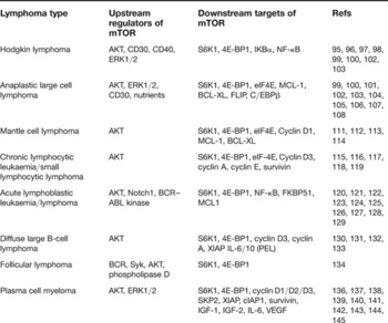
The therapy of patients with haematological malignancies, including lymphomas, is in flux because efforts are focusing on the development of biologically targeted therapies, as opposed to the traditional, and mostly empirical, chemotherapeutical approaches. Here we summarise the findings of recent studies regarding the activation status, regulation and downstream targets of mTOR signalling in lymphomas (Table 1), as well as clinical efforts to exploit mTOR inhibition as part of clinical trials of lymphoma patients.
mTOR signalling in lymphomas

Abbreviations: AKT, akt murine thymoma viral oncogene homologue; BCL-XL, long isoform of the BCL-2-related gene; BCR, B-cell receptor; BCR–ABL, fusion product of breakpoint cluster region gene on chromosome 22 and Abelson kinase; C/EBPβ; CCAAT enhancer binding protein β; cIAP1, cellular inhibitor of apoptosis 1; 4E-BP1, 4E-binding protein 1; eIF-4E, eukaryotic initiator factor 4E; ERK, extracellular-signal-regulated kinase; FLIP, FLICE-inhibitory protein; IGF, insulin-like growth factor; IL, interleukin; MCL-1, myeloid cell leukaemia 1; NF, nuclear factor; PEL, pleural effusion lymphoma; S6K1, p70 ribosomal protein S6 kinase; SKP2, S-phase kinase-associated protein 2; Syk, spleen tyrosine kinase; VEGF, vascular endothelial growth factor; XIAP, X-linked inhibitor of apoptosis.
Hodgkin lymphoma
Hodgkin lymphoma (HL), previously known as Hodgkin's disease, represents ~30% of all lymphoma cases worldwide. HL is further divided into nodular lymphocyte predominant and classical types, with the latter representing ~94% of all cases. Classical HL is typically composed of a small number (often <1%) of large neoplastic cells, known as Hodgkin/Reed–Sternberg cells (HRS), within a reactive cellular background (Ref. Reference Jaffe94). As mTOR activity and its downstream effector, S6K1, are known to be important determinants of cell size, it is not surprising that the mTOR pathway is activated in HL cell lines and primary tumours (Refs Reference Montagne95, Reference Stocker and Hafen96, Reference Fingar97). Expression of activated mTOR, S6K1 and phosphorylated 4E-BP1 proteins in primary HRS cells has been reported in most HL tumours by using immunohistochemistry (Ref. Reference Isaacson98). Treatment of HRS cells with rapamycin or the rapamycin analogue RAD0001 results in G1–S cell-cycle arrest, but not apoptosis (Ref. Reference Jundt99). However, rapamycin was shown to enhance the cytotoxicity of a chemotherapeutic agent (doxorubicin) against HRS cells (Ref. Reference Jundt99). Also, RAD0001 has an antitumour effect in HL murine xenograft models. The growth-inhibitory effect of RAD001 on HRS cells is accompanied by decreased NF-κB activity – an important survival factor for HRS cells, mediated by stabilisation of IκBα – as a result of translational regulation (Ref. Reference Jundt99). The exact mechanism of mTOR activation in HRS cells is not completely understood. It seems that AKT, an upstream regulator of mTOR, plays an important role. Activated (phosphorylated) AKT was found to be expressed by 64% of primary HL tumours in one study, and by most cases in another (Refs Reference Georgakis100, Reference Dutton101). Furthermore, activation of tumour necrosis factor (TNF) receptors, including CD40, CD30 and RANK, which are characteristic of HRS cells, seems to contribute to AKT activation (Ref. Reference Georgakis100). ERK1/2, another important upstream regulator of mTOR, is also activated in HRS cells (Refs Reference Zheng102, Reference Watanabe103). However, the functional significance of this ERK–mTOR link in HL is unknown.
Anaplastic large cell lymphoma
Activation of the mTOR pathway has been shown in several types of non-Hodgkin lymphoma (NHL). Anaplastic large-cell lymphoma (ALCL) is a type of NHL of T- or null-cell lineage that represents approximately 2% of NHLs in North America and Europe (Ref. Reference Jaffe94). A large subset of ALCL cases, more frequent in children and young adults, is characterised by constitutive overactivation of anaplastic lymphoma kinase (ALK) as a result of the t(2;Reference Faivre, Kroemer and Raymond5)(p23;q35) or other chromosomal abnormalities involving the ALK gene (Ref. Reference Jaffe94).
ALK+ ALCL cell lines and most tumours (as well as ALK− tumours to a lesser degree) express activated constituents of the mTOR pathway, including phosphorylated 4E-BP1, p70S6K1 and mTOR, and elevated levels of eIF4E (Ref. Reference Vega104). Rapamycin treatment or downregulation of mTOR by MTOR-specific siRNA was shown to induce G1–S cell-cycle arrest and apoptosis of ALK+ ALCL cells, accompanied by downregulation of anti-apoptotic molecules, such as MCL-1, BCL-XL and FLIP, and cell-cycle-promoting proteins, including cyclin D2. Rapamycin was also shown to enhance the cytotoxicity of chemotherapeutic agents in ALK+ ALCL cells (Ref. Reference Vega104). Similar results were obtained using the rapamycin analogue RAD0001, which was also shown to have antitumour activity in ALCL xenograft animal models (Ref. Reference Jundt99). Interestingly, downregulation of a truncated isoform of the transcription factor CCAAT enhancer-binding protein β (CEBPβ) as a result of RAD001 treatment was shown to be essential for antitumour activity in one study (Ref. Reference Jundt99). AKT and ERK activation, as well as nutrient availability, contribute to mTOR activation in ALCL cells (Refs Reference Vega104, Reference Marzec105). Activation of both AKT and ERK has been shown by immunohistochemistry in most ALCL tumours (Refs Reference Watanabe103, Reference Rassidakis106). ALK has been causally linked to AKT and ERK activation in ALK+ tumours (Refs Reference Bai107, Reference Marzec108, Reference Gu109, Reference Pulford, Morris and Turturro110). By contrast, the cause of AKT, ERK and consequently mTOR activation in ALK− ALCL tumours is less well understood. However, TNF-receptor signalling, including CD30 and AP-1 transcription factor activity, may play an important role (Ref. Reference Watanabe103).
Mantle cell lymphoma
Mantle cell lymphoma (MCL) is a type of B-cell NHL characterised by t(11;Reference Sabatini14)(q13;q32) and cyclin D1 overexpression (Ref. Reference Jaffe94). These tumours represent approximately 6% of all NHLs in North America and Europe. Because patients with MCL are usually elderly and have poor long-term survival, attempts to develop novel and biological targeted therapies are of particular importance.
Recent studies have shown that the mTOR pathway is activated in MCL cell lines and a substantial subset of MCL tumours (Ref. Reference Peponi111). Inhibition of the mTOR pathway by rapamycin or rapamycin analogues resulted mainly in cell-cycle arrest of MCL cells (Refs Reference Peponi111, Reference Hipp112, Reference Haritunians113). In addition, rapamycin or its analogues enhanced the cytotoxic effect of chemotherapeutic agents, such as doxorubicin, vincristine or rituximab, which are traditional components of MCL treatment regimens (Ref. Reference Haritunians113). In addition, total mTOR inhibition using MTOR-specific siRNA induced cell-cycle arrest and apoptosis of MCL cells accompanied by downregulation of cyclin D1 and anti-apoptotic proteins, including MCL-1 and BCL-XL (Ref. Reference Peponi111). Interestingly, downregulation of eIF4E, a downstream target of mTOR, recapitulated most of the effects of mTOR inhibition, underlining the importance of the regulation of translational machinery for the biological effects of mTOR inhibition in MCL cells (Ref. Reference Peponi111). Activation of AKT seems to contribute to mTOR activation in MCL cells, and specific inhibition of AKT induces cell-cycle arrest and apoptosis of MCL cells through mTOR downregulation, as well as involving targets that are independent of the mTOR pathway, including mdm2, p27, FRKHL-1, BAD and GSK-3β (Ref. Reference Rudelius114). The cause of AKT activation in MCL, observed more frequently in the clinically aggressive blastoid variant of MCL, is not well understood. PTEN, a known AKT inhibitor, is expressed in most cases of MCL. Furthermore, activating mutations of PI3K have not been detected (Ref. Reference Rudelius114). It seems that other factors, in addition to AKT, contribute to mTOR activation in MCL (Ref. Reference Peponi111).
Chronic lymphocytic leukaemia/small lymphocytic lymphoma
Chronic lymphocytic leukaemia/small lymphocytic lymphoma (CLL/SLL) is a B-cell NHL that can present as either leukaemia or lymphoma. CLL/SLL is the most common type of human leukaemia and represents approximately 6% of all lymphomas. CLL/SLL affects adults, usually over 50 years of age, and it is practically incurable by standard chemotherapeutic approaches (Ref. Reference Jaffe94). Although the pathogenesis of CLL/SLL is unknown, it is believed that the accumulation of long-lived apoptosis-defective B cells is an important pathogenetic mechanism. However, as has been shown relatively recently, aberrant proliferation of the neoplastic cells also appears to be involved (Refs Reference Decker115, Reference Decker116).
Inhibition of the mTOR pathway by rapamycin has been shown to decrease dramatically the proliferative potential of primary CLL/SLL cells stimulated by interleukin 2 (IL-2) or CpG oligonucleotides (Ref. Reference Decker117). The rapamycin-induced G1–S cell-cycle arrest of CLL/SLL cells was accompanied by downregulation of cyclin D3, phosphorylated Rb, cyclin A and cyclin E and consequently decreased CDK2 activity, as well as by downregulation of survivin, a molecule involved in both apoptotic and proliferation pathways (Ref. Reference Decker117). Interestingly, rapamycin had no effect on apoptosis of CLL/SLL cells (Ref. Reference Decker117). The potential of using mTOR inhibition in CLL/SLL was shown in a recent animal model study (Ref. Reference Zanesi118). Genetically modified mice that overexpress the TCL1 oncogene in the B-cell compartment (Eμ-TCL1 transgenic) develop in advanced age a B-cell lymphoproliferative disorder similar to human CLL/SLL (Ref. Reference Bichi119). Rapamycin treatment was shown to prolong significantly the life of syngeneic mice serially transplanted with Eμ-TCL1 B leukaemic cells (Ref. Reference Zanesi118).
Acute lymphoblastic leukaemia/lymphoma
Precursor B-cell acute lymphoblastic leukaemia/lymphoma (pre-B-ALL) is one of the most common lymphoid leukaemias in children. Pre-B-ALL also can affect adults, usually the elderly. Although affected children are frequently cured by conventional chemotherapy, adult pre-B-ALL is often chemotherapy resistant (Ref. Reference Jaffe94).
In vitro studies show that rapamycin treatment induces G1 cell-cycle arrest and apoptosis of pre-B-ALL cells, and these effects can be reversed by pretreatment with IL-7, an important regulator of B- and T-cell development (Ref. Reference Brown120). These observations were further extended in a mouse model of pre-B-ALL. Transgenic mice that overexpress the tyrosine kinase receptor ret in the B-cell compartment (Eμ-ret transgenic) develop a lymphoproliferative disorder similar to human pre-B-ALL (Refs Reference Iwamoto121, Reference Wasserman, Zeng and Hardy122, Reference Zeng123). Daily treatment of these mice in advanced stages of disease with rapamycin substantially extended survival time (Ref. Reference Brown120). Also, treatment with the rapamycin analogue CCI-779 dramatically decreased the burden of the disease in severe combined immunodeficient nonobese diabetic (SCID/NOD) mice that were transplanted with primary human pre-B-ALL cells (Ref. Reference Teachey124).
In a recent study, BCR–ABL kinase was shown to contribute to activation of the translation machinery by inhibiting 4E-BP1 (Ref. Reference Prabhu125). BCR–ABL kinase is the gene product of t(9;Reference Bosotti, Isacchi and Sonnhammer22)(q34;q11), also known as the Philadelphia chromosome (Ph), which is the hallmark of chronic myelogenous leukaemia (CML). BCR–ABL kinase is also present in approximately 25–33% of Ph+ pre-B-ALL, mostly in adults. This provides a rationale for combining BCR–ABL kinase inhibitors (e.g. imatinib) and mTOR inhibitors, and this combination appears to offer promise for the treatment of patients with Ph+ pre-B-ALL (Ref. Reference Burchert126).
In general, precursor T-cell lymphoblastic leukaemia/lymphoma (pre-T-ALL) has been relatively less well studied. However, rapamycin induces cell-cycle arrest and apoptosis in pre-T-ALL cells as in pre-B-ALL cells, and enhances the cytotoxic effects of doxorubicin in both cell types (Ref. Reference Avellino127). AKT activation seems to contribute to mTOR activation of ALL cells because all rapamycin-sensitive ALL samples were found to express phosphorylated AKT, and selective inhibition of AKT resulted in cell-cycle arrest and apoptosis (Ref. Reference Wei128). The biological effects of rapamycin in ALL cells were also shown to be mediated partly by downregulation of the NF-κB pathway, through inhibition of the immunophilin FKBP51, a protein that belongs to the same family as FKBP12, which mediates the direct inhibition of mTOR by rapamycin (Ref. Reference Avellino127). Rapamycin was also found to reverse the resistance of ALL cells to glucocorticoids, through downregulation of the anti-apoptotic molecule MCL-1 (Ref. Reference Wei128). In addition, inhibition of the mTOR pathway by rapamycin reversed the chemoresistance of pre-T-ALL cells caused by overactivation of Notch1, a cell membrane receptor frequently deregulated in pre-T-ALL cells as a result of activating mutations in the Notch1 gene (Ref. Reference Mungamuri129).
Diffuse large B-cell lymphoma
Diffuse large B-cell lymphoma (DLBCL), the most common type of NHL in western countries, represents approximately a third of all NHLs. DLBCL is a heterogeneous group of diseases in terms of its pathogenesis (Ref. Reference Jaffe94). Although a subset of patients with DLBCL is curable with current chemotherapy, many patients succumb to disease and novel therapeutic approaches are very much needed.
An in vitro study of DLBCL cell lines of the germinal centre type showed that the rapamycin analogue RAD001 induces G1 cell-cycle arrest but not apoptosis, accompanied by downregulation of cyclins D3 and A (Ref. Reference Wanner130). In the same study, RAD001 enhanced the cytotoxicity of rituximab (a monoclonal humanised anti-CD20 antibody) against DLBCL cells, whereas it seemed to decrease the effectiveness of classical chemotherapeutic agents. In an immunohistochemical investigation of primary DLBCL tumours, approximately half of cases were found to express activated AKT, an upstream regulator of mTOR (Ref. Reference Uddin131). Accordingly, treatment with the PI3K inhibitor LY29002 was shown to result in apoptotic cell death in a subset of DLBCL cell lines in vitro, accompanied by downregulation of XIAP, an inhibitor of the intrinsic apoptotic pathway (Ref. Reference Uddin131). However, LY29002 is not specific and can inhibit PI3K as well as mTOR. Therefore, the biological effects of LY29002 on DLBCL cannot be localised with certainty to either AKT or mTOR.
In another study, rapamycin was shown to inhibit the growth of primary effusion lymphoma (PEL) cell lines in vitro and in SCID mouse xenograft models. PEL is a rare type of lymphoma, currently considered to be a variant of DLBCL, affecting immunocompromised individuals, including HIV+ patients. PEL is aetiologically linked with infections by human herpesvirus 8 (HHV-8), also known as Kaposi sarcoma-associated herpesvirus (Refs Reference Jaffe94, Reference Sin132). The growth-inhibitory effect of rapamycin was attributed partly to cell-cycle arrest mediated by dramatically decreased production of IL-6 and IL-10, which are important autocrine growth factors promoting the survival of PEL cells. Importantly, restimulation with IL-6 or IL-10 reversed the growth-inhibitory effects of rapamycin on PEL cells (Ref. Reference Sin132). Also, the AKT pathway was shown to contribute to mTOR activation in PEL cells, because phosphorylated AKT, mTOR and p70S6K – a downstream effector of mTOR – were all expressed in most tumour cells (Refs Reference Sin132, Reference Uddin133).
Follicular lymphoma
Follicular lymphoma (FL), the second most common type of B-cell NHL in adults from western countries, represents approximately 25% of all NHLs in western countries. FL is a neoplasm of germinal centre B cells, usually associated with overexpression of BCL-2 as a result of t(14;18)(q32;q21) (Ref. Reference Jaffe94). Although patients with FL usually have an indolent clinical course, they gradually evolve to a treatment refractory phase and many patients actually die of disease.
Activation of the mTOR pathway in FL has been shown in a subset of primary FL tumours and in B-cell lymphoma cell lines that carry t(14;18) (Ref. Reference Leseux134). It was also shown that Syk tyrosine kinase, a protein in the B-cell-receptor signalling pathway, contributes to mTOR activation in FL cells (Ref. Reference Leseux134). In addition, it was proved that this link operates independently of AKT or phospholipase D, the latter also contributing to mTOR activation in FL cells (Ref. Reference Leseux134). Interestingly, the Syk–mTOR link was shown to be of functional importance in cell lines derived from other B-cell NHL types, including DLBCL, Burkitt lymphoma and MCL (Ref. Reference Leseux134). However, the clinical potential of mTOR inhibition has not been tested in an FL animal model to date. This is of particular importance because rapamycin treatment was shown to increase the levels of BCL-2 protein in B-cell lymphoma cell lines with t(14;18) (Ref. Reference Calastretti135).
Plasma cell myeloma
Plasma cell myeloma (PCM) is a molecularly heterogeneous disease characterised by uncontrolled proliferation of plasma cells in the bone marrow. Most affected patients are adults over 40 years of age and African Americans are affected more often than Caucasian Americans (Ref. Reference Jaffe94). Although considerable improvement in prognosis has been achieved with current chemotherapeutic regimens, many patients die of this disease.
Preliminary studies have shown that two important upstream regulators of the mTOR pathway – ERK and PI3K–AKT – are frequently activated and contribute to survival and growth of PCM cells (Refs Reference Ogata136, Reference Hsu137, Reference Pene138, Reference Hu139). AKT activation in particular was shown by immunohistochemical methods to be common in PCM, particularly in patients with disease progression (Ref. Reference Hsu137). Also, AKT inhibition was shown to induce cell-cycle arrest and apoptosis of PCM cells in vitro (Refs Reference Hsu137, Reference Pene138). Consequently, it was found that rapamycin, or the rapamycin analogue CCI-779, induced cell-cycle arrest and apoptosis in PCM cell lines or primary tumour cells in vitro, and enhanced the growth inhibitory effect of dexamethasone in vitro in PCM xenograft models (Refs Reference Frost140, Reference Shi141, Reference Stromberg142). In some studies, the PCM inhibitory effects of mTOR inhibitors were shown to be limited to PCM cells harbouring PTEN-inactivating mutations and overactivation of the AKT pathway (Ref. Reference Shi141). The effects of mTOR pathway inhibition on PCM cells are mainly mediated by downregulation of the translational machinery. This results in decreased expression of anti-apoptotic proteins, including XIAP, cIAP1 and survivin, as well as cell-cycle-promoting proteins, including cyclin D1, cyclin D2 and cyclin D3. This occurs with a concordant increase of the cell-cycle inhibitor p27, partly as a consequence of diminished degradation because of decreased levels of SKP2 (Refs Reference Pene138, Reference Frost140, Reference Stromberg142, Reference Yan143). It must be added that mTOR inhibition in PCM cells interferes with nonautonomous cell-survival mechanisms because two important growth factors for PCM cells, insulin-like growth factor (IGF)-1/2 and IL-6, which are produced by non-neoplastic bone marrow cells, stimulate the PI3K-AKT pathway (Ref. Reference Baylink, Finkelman and Mohan144). Another nonautonomous cell effect of mTOR inhibition in PCM is decreased angiogenesis in PCM animal models, which is, in part, due to decreased production of vascular endothelial growth factor by the neoplastic cells (Ref. Reference Frost145).
Post-transplant lymphoproliferative disorders
Post-transplant lymphoproliferative disorders (PTLD) encompass a spectrum of polyclonal, oligoclonal or monoclonal lymphoproliferations of B-cell or T-cell lineage, which occur because of iatrogenic immunosuppression in patients after organ transplantation. Most cases of B-cell PTLD are associated with Epstein–Barr virus infection (Ref. Reference Jaffe94). Although a subset of PTLD resolves spontaneously after reduction of immunosuppression, always with increased danger of graft rejection, other cases of PTLD can be clinically aggressive and difficult to manage.
In vitro studies have shown that rapamycin or RAD001 induces G1 cell-cycle arrest and apoptosis in lymphoid cell lines established from patients with PTLD (Refs Reference Majewski146, Reference Majewski147, Reference Nepomuceno148). In addition, it was shown that treatment with rapamycin or RAD001 was able to postpone the onset and delay the growth of tumours established from PTLD cell lines in SCID mice xenograft models (Ref. Reference Nepomuceno148). One mechanism involved in the growth-inhibitory effect of rapamycin on PTLD, as a result of mTOR inhibition, involves downregulation of IL-10 production by tumour cells, consequently decreasing STAT-3 activation (Ref. Reference Wlodarski149). In vitro studies investigating the function of the mTOR network in EBV+ lymphoid cells in more detail have shown that mTOR is activated in lymphoma cells with type III latency EBV infection (e.g. PTLD) and type I latency EBV infection (e.g. Burkitt lymphoma). However, activation of AKT and ERK, which may contribute to mTOR activation, was observed only in cells with type III latency EBV infection. In addition, in EBV+ lymphoma cells, signals related to nutrient availability were also shown to be crucial for mTOR activation (Ref. Reference Wlodarski149).
Pharmacological inhibitors of mTOR
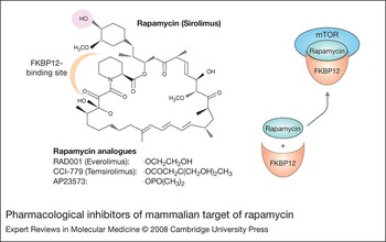
In addition to the prototype mTOR inhibitor rapamycin (sirolimus), three more mTOR inhibitors are available for clinical trials: CCI-779 (temsirolimus), RAD001 (everolimus) and AP23573 (Refs Reference Faivre, Kroemer and Raymond5, Reference Dancey150). All inhibitors are derivatives of rapamycin with which they share the same basic chemical structure; however, each has a different chemical group at the position of the C40 hydroxyl group of rapamycin. CCI-779 is essentially a prodrug because it is metabolised to rapamycin inside the organism, whereas RAD001 and AP23573 are metabolised differently (Fig. 3). To inhibit mTOR, all must initially form an intracellular complex with the immunophilin FKB12. This complex, in turn, binds the FRB domain of mTOR, inhibiting mTOR function. The inhibition of mTOR by rapamycin and its derivatives is regarded as quite specific, and rapamycin treatment of mice embryos induces the same developmental defects as knockout of the mTOR gene (Refs Reference Murakami151, Reference Gangloff152). Until recently, it was thought that rapamycin and its derivatives inhibit only the mTOR protein nucleated in mTORC1, which is mostly responsible for regulation of protein translation and autophagy. However, it has now been shown that prolonged treatment with rapamycin, in some types of cells, also can inhibit mTORC2, which is mainly responsible for activation of AKT (Ref. Reference Sarbassov153). This finding can be explained by the assumption that within the cell there exists an equilibrium between the pool of mTOR associated with Raptor (mTORC1) and that of mTOR associated with Rictor (mTORC2). Inhibition of mTORC2 by prolonged treatment with rapamycin is attributed to the exhaustion of mTOR available for binding to Rictor, because of the direct inhibition of mTOR bound to Raptor (Ref. Reference Sarbassov153).
Pharmacological inhibitors of mammalian target of rapamycin. The basic chemical structure of rapamycin is shared among all rapamycin derivatives (left). The shaded area corresponds to the position of different chemical groups in the derivatives shown below. This area is distinct from the rapamycin sites for binding to immunophilin FKBP12 and mammalian target of rapamycin (mTOR), so that the mechanism of mTOR inhibition is common for rapamycin and its analogues. Rapamycin, as well its analogues, associates initially with FKBP12, in order to inhibit mTOR (right). The combined surface of the rapamycin–FKBP12 complex binds, in turn, the FRB domain of mTOR in mTORC1 and inhibits its activity. It seems that FKBP12 presents rapamycin to mTOR in a conformation-favourable state for interaction with the FRB domain. Immunophilins, such as FKBP12, belong to a protein family with at least 12 members in humans, and function as accessory helper enzymes or chaperones, contributing to the stability of multiprotein complexes. A common feature of immunophilins is they can catalyse the interconversion of cis and trans amide bonds adjacent to proline residues in peptide substrates, the so-called PPIase (prolyl isomerase) activity altering the spatial rearrangement of protein domains. This activity is inhibited by binding of each immunophilin with the corresponding drug. However, it has been shown that inhibition of PPIase activity of FKBP12 by rapamycin binding is not essential for mTOR inhibition. Abbreviations: FKBP12, FK507 binding protein 12; FRB, FKBP-rapamycin binding; mTORC, mammalian target of rapamycin complex.

Unlike rapamycin, its derivatives do not induce significant immunosuppression when administered intermittently in the appropriate clinical setting (Refs Reference Faivre, Kroemer and Raymond5, Reference Dancey150). Rapamycin is produced in oral formulation for immunosuppression after kidney transplantation. CCI-779 (Wyeth Pharmaceuticals, Madison, NJ, USA) can be administered orally or intravenously. RAD001 (Novartis, Basel, Switzerland) is available in oral formulation and is in the advanced stage of development for use as an immunosuppressive in solid-organ transplantation. AP23573 (Ariad Pharmacheuticals, Cambridge, MA, USA) can be given orally or intravenously. Side effects common to all mTOR inhibitors include hypertension, oedema, hyperlipidaemia and gastrointestinal symptoms. Also, dose-limiting toxicities of rapamycin derivatives include reversible mucositis, asthaenia and thromphocytopaenia (Refs Reference Faivre, Kroemer and Raymond5, Reference Dancey150).
Clinical trials of mTOR inhibitors in lymphomas
Although rapamycin has been studied meticulously as an immunosuppressive drug, clinical experience using it as an antineoplastic agent is limited, especially for haematological malignancies (Ref. Reference Witzig and Kaufmann154). However, all rapamycin derivatives are currently used in Phase I/II clinical trials as antineoplastic agents and some Phase III clinical trials are in progress (Refs Reference Faivre, Kroemer and Raymond5, Reference Witzig and Kaufmann154, Reference Costa155).
One of the more promising results published to date regarding using mTOR inhibitors in lymphoma patients comes from a Phase II trial using CCI-779 in the treatment of MCL patients (Ref. Reference Witzig156). In this study, 34 patients with MCL, either refractory to previous therapy or relapsed, received CCI-779 as a single agent. Twelve patients showed partial response and one patient had a complete response, with an overall response rate of 38%. No deaths from the treatment were reported. The most frequent toxicity, which was also a cause for dose reduction, was thrombocytopaenia. Interestingly, reduction of the dose (from 250 mg to 25 mg intravenously once a week) seemed to decrease toxicities without affecting the response rate. Currently an international randomised Phase III trial comparing CCI-779 with control chemotherapy in MCL patients is ongoing.
In a Phase I/II trial of RAD001, which included four patients with MCL and six with CLL/SLL, three patients showed a 27–34% reduction of lymphadenopathy in radiological studies, and the drug was considered safe and worthy of further investigation (Ref. Reference Yee157). In another trial using AP23573 to treat patients with lymphoid malignancies one of nine CLL/SLL patients showed a partial response (Ref. Reference Costa155).
Additional clinical trials using rapamycin derivatives to treat Hodgkin lymphoma, PCM and other lymphomas, as monotherapy or in combination with other agents such as rituximab, are in progress, but the results are not yet published.
Research in progress and outstanding questions
Since preliminary clinical studies of mTOR inhibitors in cancer patients have shown promising results, further investigation will continue to accumulate considerable empirical knowledge about the spectrum of neoplastic diseases that will be successful targets for mTOR inhibitors. This will lead to better drug design and dosing, as well as the development of combinations of mTOR inhibitors with classical chemotherapeutical agents or new cell-signalling inhibitors that will be beneficial in various clinical settings.
However, many questions regarding the mTOR-signalling pathway that have clinical implications remain to be answered. What is the molecular signature that determines mTOR inhibition sensitivity in each clinical setting? Preliminary in vitro studies in PCM cells have shown that PTEN mutation and AKT overactivation are the most critical parameters of rapamycin sensitivity (Ref. Reference Shi141). Does this hold true for other lymphoid malignancies and, if not, what are the critical pathways that contribute to mTOR activation in each case? Also, why does mTOR inhibition induce only cell-cycle arrest in some lymphomas, whereas in others mTOR inhibition also induces apoptotic cell death? Regarding the resistance of some cancer cells to rapamycin, it seems that the internal wiring of the mTOR network may play an important role. For example, in vitro studies in PCM cells showed in a subset of cases that rapamycin treatment induced overactivation of AKT. This was due to decreased IRS serine phosphorylation as a result of cancellation of the inhibitory feedback between the downstream mTOR effector S6K1 and the IRS–PI3K link upstream of AKT (Ref. Reference Shi158). In which other neoplasms is this feedback connection important for rapamycin resistance? Will simultaneous inhibition at the level of AKT and mTOR by two independent inhibitors be able to overcome rapamycin resistance, as suggested in some studies (Refs Reference Shi158, Reference Sun159)? Some recent in vitro results suggest that prolonged rapamycin treatment – in addition to directly inhibiting mTORC1 – also has an indirect inhibitory effect on mTORC2 and therefore AKT activation through mTOR depletion (Refs Reference Sarbassov153, Reference Zeng160). Why is this true in some types of neoplasm and not in others, and is this a critical parameter of rapamycin resistance? The answers to these questions and many others of importance to our understanding of biology and patient care are likely to be forthcoming.
Acknowledgements and funding
We thank the referees for helpful suggestions to improve this manuscript.