Introduction
Abdominal angiostrongyliasis (AA) is a clinical entity characterized by a strong inflammatory reaction in the intestinal wall and by the onset of unspecific symptoms such as vomiting, anorexia, pain in the lower right quadrant, constipation and occasionally fever (Morera, 1985; Reference Morera1995). Diagnosis in the clinical laboratory continues to be one of the greatest challenges in the management of this helminthiasis. Since none of the stages of the parasite are shed in faeces, a coproparasitological diagnosis of abdominal angiostrongyliasis is impossible and the only way to confirm infection in human beings is through the finding of the parasite in histological preparations obtained after a surgical intervention.
Complementary diagnostic methods, such as serological tests to detect the presence of specific antibodies against A. costaricensis, have been implemented in the clinical laboratory. To date, a latex agglutination test (Morera, Reference Morera1987; Morera & Amador, Reference Morera and Amador1998 ) and an enzyme immunosorbent assay (ELISA) (Graeff-Teixeira et al., Reference Graeff-Teixeira, Agostini, Camillo-Coura and Ferreira-Da-Cruz1997; Geiger et al., Reference Geiger, Abrahams-Sandí, Soboslay, Hoffmann, Pfaff, Graeff-Teixeira and Schulz-Key2001) have been used in Costa Rica and Brazil, respectively, for the serological diagnosis of this disease. However, it has not been possible to standardize either of these two tests for their routine use in clinical laboratories. While for the latex test, also known as Morera's test, there is no published information on its sensitivity and specificity, for the ELISA test, cross-reaction with other helminthiasis and a low sensitivity have been the major problems. In order to minimize these cross-reactions as much as possible, a number of antigens have been tested, from somatic to purified fractions of different extracts, using techniques such as ELISA, Western blot and immunofluorescence (Graeff-Teixeira et al., Reference Graeff-Teixeira, Agostini, Camillo-Coura and Ferreira-Da-Cruz1997; Geiger et al., Reference Geiger, Abrahams-Sandí, Soboslay, Hoffmann, Pfaff, Graeff-Teixeira and Schulz-Key2001; Abrahams-Sandí et al., Reference Abrahams-Sandí, Hoffmann, Graeff-Teixeira, Schulz-Key and Geiger2004). To date, the immunoenzymatic test showing the highest specificity (91.1%) was developed by Geiger et al. (Reference Geiger, Abrahams-Sandí, Soboslay, Hoffmann, Pfaff, Graeff-Teixeira and Schulz-Key2001), using somatic extract of female adult forms as an antigen in order to determine specific IgG1. However, the sensitivity of this test reached only 76.2%.
Bender et al. (Reference Bender, Maurer, Fernandes da Silva, Ben, Barros, Aramburu da Silva and Graeff-Teixeira2003), in a study with human sera and using indirect immunofluorescence (IIF), described the high antigenicity and the low cross-reactivity found when employing A. costaricensis eggs as antigen. Some observations made in the experimental animal model had also shown the importance of eggs in the humoral response. In experimental infections with C57BL/6 and BALB/c mice strains, it was determined that an increase in the level of humoral response, mainly of IgG and IgG1, coincided with the maturity of the parasite and the start of oviposition by the parasite females (Geiger et al., Reference Geiger, Graeff-Teixeira, Soboslay and Schulz-Key1999, Abrahams-Sandí et al., Reference Abrahams-Sandí, Hoffmann, Graeff-Teixeira, Schulz-Key and Geiger2004.). Recently, working with antigens of different developmental stages, it was possible to demonstrate that the highest antibody levels in the animal model were detected using an egg extract as an antigen, with levels that remained high even through the post patent infection period (Abrahams et al., Reference Abrahams, Geiger, Fernández and Schulz-Key2005). Finally, in studies carried out with the BALB/c strain, most pathological changes observed occurred only after the production and release of eggs, which led to the conclusion that the local inflammatory response, the cellular infiltration, and the primary and secondary granulomatose reaction induced by the eggs and larvae are the main contributing factors to the pathogenesis of infection by A. costaricensis (Geiger et al., Reference Geiger, Graeff-Teixeira, Soboslay and Schulz-Key1999). Based on these previous studies and others we performed in animal models (unpublished data) we carried out this work with human sera to evaluate the diagnostic value of eggs through an ELISA.
Materials and methods
Antigen preparation
Whole egg and soluble egg-antigen (Egg-Ag) were collected and prepared as described previously by Abrahams et al. (Reference Abrahams, Geiger, Fernández and Schulz-Key2005) with some modifications. Briefly, fertile female worms were incubated in MEM (minimum essential Eagle medium) supplemented with antibiotics, at 37°C, 5% CO2 for 96 h. The culture medium was changed daily, pooled, and centrifuged for 20 min at 16,000 g at 4°C. The egg pellet was washed three times with phosphate-buffered saline (PBS) and stored at − 20°C. For the preparation of Egg-Ag, a suspension of eggs was sonicated four times on ice for 3 min (30% cycle, Cole Palmer Instruments 4710 series, Chicago, Illinois, USA) and centrifuged for 20 min at 16,000 g at 4°C. The protein concentration was determined by the Bradford assay using bovine serum albumin as standard. Finally, the antigen was stored at − 20°C pending further use.
Serum samples
Twenty-one serum samples of patients with A. costaricensis histopathological diagnosis (positive controls = C+) were kindly provided by Dr Pedro Morera (Pathology Service, Hospital San Juan de Dios, Costa Rica). Sera from patients with positive faecal examination for at least one of the following parasites were provided by Department of Parasitology, University of Costa Rica, and included as specificity controls (SC), Strongyloides stercoralis, Ascaris lumbricoides, Trichuris trichiura, Taenia sp., Enterobius vermicularis and hookworm (Necator americanus/Ancylostoma duodenale). Samples from healthy individuals living in an area with and without A. costaricensis active transmission and with negative faecal examination were also kindly provided by the Department of Parasitology, University of Costa Rica, and included in the testing as ‘negative controls from endemic zone’ (NCE) and ‘normal negative sera’ (NS).
Determination of parasite-specific IgG1 by enzyme-linked immunosorbent assay (ELISA)
The production of parasite-specific IgG1 against A. costaricensis Egg-Ag was determined by indirect ELISA, as described previously by Abrahams et al. (Reference Abrahams, Geiger, Fernández and Schulz-Key2005). In brief, high-binding ELISA plates (Costar) were coated with soluble egg antigen extract at 1 μg ml− 1 (50 μl/well) diluted in bicarbonate buffer (pH 9.6) at 4°C overnight. After washing three times in PBS/0.05% Tween-20, the plates were blocked with PBS/10% fetal calf serum (FCS) for 2 h at room temperature. Sera samples were diluted 1:100 in dilution buffer (PBS, 4% Tween-20, 10% FCS), 50 μl was added to each well and the plates were incubated for 90 min at 37°C. After being washed three times as described above, alkaline phosphatase-conjugate anti-human IgG1 diluted 1:500 was added (50 μl/well) and the plates incubated for 2 h at 37°C. Finally, after five washing steps, p-nitrophenyl phosphate at 1 mg ml− 1 (50 μl/well) was added and optical densities (OD) were recorded at 405 nm in a microplate autoreader (BioRad, Hercules, California, USA). The results were expressed as mean optical densities for duplicate determinations.
The OD cut-off value was determined from the sum of mean OD value plus two standard deviations of 29 SC (Strongyloides stercoralis, Ascaris lumbricoides, Trichuris trichiura, Taenia sp., Enterobius vermicularis, hookworms) and 8 NCE.
To calculate the specificity of the ELISA test 23 additional SC from patients with positive faecal examination for S. stercoralis, A. lumbricoides, hookworm and T. trichiura were used.
Results
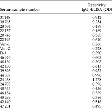
The OD cut-off value was established as 0.241, determined from the sum of mean OD value (0.0732) plus two standard deviations (SD = 0.084) of 29 SC and 8 NCE. An analysis of the C+ showed that 19 of the 21 sera had an absorbance over the OD cut-off value (table 1). On the other hand, only 3 of 23 additional SC had an absorbance over the cut-off. All of them were from patients with S. stercoralis faecal positive examination. According to these results, the sensitivity of the Egg-Antigen IgG1-ELISA was estimated to be 90.5% (19/21), while the specificity was 87.0% (20/23).
IgG1-ELISA results on sera from patients with confirmed abdominal angiostrongyliasis.
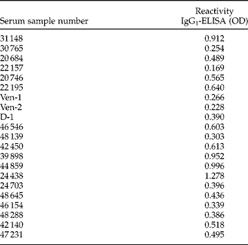
Cut-off point 0.241.
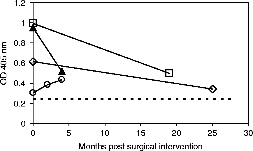
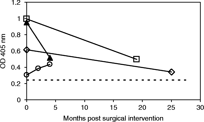
In order to visualize the kinetics of specific IgG1 in the human serum, the ELISA test using eggs as antigens was applied to paired sera of four patients with a confirmed AA diagnosis submitted to surgical treatment. A decline in the reactivity against the egg antigen was measured after the surgical procedure. Only in the sera of one of the patients tested was it possible to observe a slight increase in the immunoglobulin levels after surgery (fig. 1).
Decrease in the IgG1 reactivity against A. costaricensis eggs in serum samples from four patients with histopathological confirmation of the parasite. Samples were not available for the same time periods for all patients. (○) Patient 1: 0, 2 and 4 months post surgery (p.s.); (□) patient 2: 0 and 19 months p.s.; (▲) patient 3: 0 and 4 months p.s.; (⋄) patient 4: 0 and 25 months p.s. (- -) Cut-off point.
Discussion
In the present work, the results obtained confirmed the strong antigenicity of A. costaricensis eggs. With the indirect ELISA test developed, a sensitivity of 90.5% and a specificity of 87.0% were obtained, detecting a cross-reaction only in three of the sera with a previous diagnosis of S. stercoralis. This cross-reaction had already been reported in a previous work using crude antigen of adult forms (Graeff-Teixeira et al., Reference Graeff-Teixeira, Agostini, Camillo-Coura and Ferreira-Da-Cruz1997). The results of the present study suggest that common antigens in the eggs of both parasites could also be involved in such a reaction. If the parasitic cycle of S. stercoralis is reviewed, the presence of eggs and the eclosion of larvae in this helminthiasis, as well as in abdominal angiostrongyliasis, take place in the intestinal wall, and the histopathological reactions observed surrounding the above-mentioned parasitic forms, evidence their antigenicity.
Cross-reactivity has also been reported in the very few studies that have used eggs as antigen in the development of a serodiagnostic test. For example, in the ELISA developed for the diagnosis of ocular and visceral toxocariasis using Toxocara canis embryonated eggs (Glickman et al., Reference Glickman, Schantz, Dombroske and Cypess1978), it was necessary to preadsorb the serum samples with A. suum eggs in order to achieve an adequate sensitivity and specificity (Cypess et al., Reference Cypess, Karol, Zidian, Glickman and Gidin1977; Pollard et al., Reference Pollard, Jarret, Hagler, Allain and Schantz1979). In the case of schistosomiasis, the major problem of the test developed by Colmenares et al. (1993) using S. mansoni eggs as antigen was the low specificity, i.e. 85%, considered inadequate for application in communities where the prevalence of schistosomiasis is low and polyparasitism frequent. In order to improve the specificity of these immunoassays, Alarcón de Noya et al. (Reference Alarcón de Noya, Colmenares, Lanz, Caracciolo, Losada and Noya2000) proposed the elimination of carbohydrates responsible for the cross-reactivity by treating the antigen with sodium metaperiodate. This approach allowed improving the specificity up to 97%, without affecting the sensitivity of test, which remained at 99%.
The use of an extract of A. costaricensis eggs as an antigen for the detection of a specific isotype of immunoglobulin, i.e. IgG1, evaluated in the present study, resulted in the highest concurrent specificity and sensitivity values that have been reported to date for an immunodiagnostic test for abdominal angiostrongyliasis. Although cross-reactivity against S. stercoralis was determined, differential diagnosis could be achieved through a coproparasitologial analysis performed in parallel to the ELISA, because, in contrast with an abdominal angiostrongyliasis, in the case of a strongyloidiasis most patients excrete the larval forms in faeces.
In the present study, and by applying the indirect ELISA test using egg antigens, it was possible to evidence, through the analysis of paired samples, a decrease with time of the reactivity of IgG1 against eggs, after surgical treatment. The degree by which this reactivity decreased was variable, and it was possible to detect optical densities over the cut-off point in samples taken 19 and 25 months after surgery. A similar tendency has been reported by Geiger et al. (Reference Geiger, Abrahams-Sandí, Soboslay, Hoffmann, Pfaff, Graeff-Teixeira and Schulz-Key2001) when working with adult-form antigens. However, in contrast to our results, most of their sera presented IgG reactivity below the cut-off 6 months after surgery. In our study, a moderate but constant increase of IgG1 levels was observed in the sera samples of one A. costaricensis human case, even 4 months after surgery. This could be explained by the presence of some forms of the parasite, eggs or larvae, in anatomical sites different from the section of intestine that was removed during surgery, or by the diversity in the immune response usually found in natural infections. No additional samples were available in the laboratory for further follow-up of this case.
In conclusion, the results obtained in the present work suggest that it is feasible to apply this technique both in epidemiological studies and in clinical diagnosis of AA. The proper dilution of sera, as well as the definition of a cut-off value, that allow the differentiation between acute infections and past infections, could contribute to a better interpretation of the results. However, it is important to mention that many cases are asymptomatic and do not require surgical intervention. Therefore, as described for other helminthiases and other diagnostic methods, the interpretation of these techniques, mainly when used as a diagnostic tool in the clinical laboratory, should not be performed in an isolated way but rather complemented by clinical criteria.
Currently, studies to improve the efficiency of egg collection and to identify the most important antigenic peptides are being performed in our laboratory in order to improve the test methodology.
Acknowledgements
This study was supported by the ‘Vicerrectoría de Investigación’, University of Costa Rica, project number 803-A7-037. We would like to thank Martha Conejo for the provision of sera samples and Arnoldo Castro for his technical assistance.