INTRODUCTION
Polyclad research of the European Atlantic coasts has mainly focused on the coastlines of Scandinavia, northern France and the United Kingdom (Müller, Reference Müller1776; Montagu, Reference Montagu1815; Claparéde, Reference Claparéde1861; Giard, Reference Giard1888, Reference Giard1894; Bergendal, Reference Bergendal1890; Gamble, Reference Gamble1893, Reference Gamble1900; Hallez, Reference Hallez1893, Reference Hallez1894; Plehn, Reference Plehn1896; Bock, Reference Bock1913; Moore, Reference Moore1937; Hendelberg, Reference Hendelberg1974). By the 21st Century, specific records can be found within the literature for the Iberian Peninsula, but usually from faunistic studies of a particular area (e.g. Faubel & Warwick, Reference Faubel and Warwick2005) or due to the appearance of an invasive species (Faubel et al., Reference Faubel, Sluys and Reid2007).
The Iberian Peninsula forms part of the South European Atlantic Shelf within the Lusitanian ecoregion. The characteristics of this region (Spalding et al., Reference Spalding, Fox, Allen, Davidson, Ferdaña, Finlayson, Halpern, Jorge, Lombana, Lourie, Martin, Mcmanus, Molnar, Recchia and Robertson2007) suggest a great invertebrate biodiversity (Noreña et al., Reference Noreña, Marquina, Pérez and Almon2014). Nevertheless, no previous records of Polycladida are known for the Cantabrian Sea in the scientific literature (although photographs taken by marine photographers do exist). This fact reflects the knowledge gap about the biodiversity of this taxonomic group on the Northern Iberian coast. Here, we report the presence of five previously described species and the description of a new one: Notoplana vitrea (Lang, Reference Lang and Engelmann1884); Pleioplana atomata (Müller, Reference Müller1776); Imogine fafai sp. nov.; Discocelis tigrina (Blanchard, Reference Blanchard1847); Leptoplana tremellaris (Müller, Reference Müller1773); and Cycloporus papillosus (Sars, Reference Sars1878).
The Cantabrian Sea, characterized by strong waves and north-westerly winds, is a transitional sea between the cold waters of the North Atlantic Ocean and the temperate waters of the tropics. Cantabrian coastal waters are warm in summer (22–24°C) and cold in winter (10–12°C). The average salinity is 35‰, though lower salinities are possible due to high rainfall in the area. The tidal range, which is under the influence of the Gulf Stream, the Rennell Current and the North Atlantic Drift, is large.
Because of the Gulf Stream and the North Atlantic Drift, similar fauna would be expected on the American and European shores. However, despite a few exceptions (Thysanozoon brocchii (Risso, Reference Risso1818) and Pleioplana atomata (Müller, Reference Müller1776)), there are no common species for both Atlantic coasts. Moreover, the Rennell Current, a warm marine current considered a ‘side arm’ of the Gulf Stream and North Atlantic Drift, runs ‘counter clockwise’ along the coastal shore. The Rennell Current, in turn, facilitates the distribution of species from the Galician coast to the Atlantic coastline of France (Noreña et al., Reference Noreña, Damborenea, Faubel and Brusa2007; Ax, Reference Ax2008) (Figure 1).
Map of the Cantabrian Sea, with the Gulf Stream (GF), the North Atlantic Drift (NAD) and the Rennell Current (RC) indicated by white dots.

MATERIALS AND METHODS
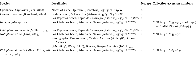
A survey of polyclad biodiversity was performed in the Cantabrian Sea (northern Iberian Peninsula, Figure 1) between September and November of 2012. Collection localities of specimens studied are listed in Table 1. Specimens were collected in the intertidal zone, on sand or on gravel under stones. In addition, the historical collection of the Iberian Fauna oceanographic campaign II was revised and two specimens of Cycloporus papilosus were histologically examined (Table 1).
Collection localities of the Cantabrian specimens studied in this paper.
*, both photographs are from the same exemplar.
Animals were anaesthetized in 7.5% magnesium chloride and fixed in Bouin's fluid, following the Newman & Cannon (Reference Newman and Cannon2003) method. Photographs were taken of both live and fixed specimens to retain maximal information regarding shape, colour and external morphology. The material was subsequently embedded in Paraplast® , serially sectioned at 7 µm and stained with AZAN (trichrome staining method). Reconstructions of internal anatomy were derived from serial sagittal sections. All measurements were determined from fixed material.
Type specimens and voucher exemplars were deposited into the invertebrate collection of the Museo Nacional de Ciencias Naturales of Madrid (MNCN, CSIC) in Spain.
A search for polyclad flatworm photographs on Iberian websites specializing in natural history has revealed several accounts of Notoplana vitrea. To increase the knowledge about the distribution of this species in the Iberian Peninsula and to make this information available to the scientific community, these records were collected and codified as follows: Asturnatura.com (AN) and Biodiversidadvirtual.org (BV), followed by the number assigned to each photograph on the website. The polyclad flatworm photographs included from these websites are up to date as of January 2014. Locality information is summarized in Table 1.
SYSTEMATICS
Suborder ACOTYLEA Lang, Reference Lang and Engelmann1884
Superfamily LEPTOPLANOIDEA Faubel, Reference Faubel1984
Family NOTOPLANIDAE Marcus & Marcus, Reference Marcus and Marcus1966
Genus Notoplana Laidlaw, Reference Laidlaw1903
Notoplana vitrea (Lang, Reference Lang and Engelmann1884) Bock, Reference Bock1913
(Figure 2)
LOCALITY
Los Chalanos Beach, Muros de Nalón (Asturias). Intertidal zone, among sand and gravel or under stones.
EXAMINED MATERIAL
One mature sagittally sectioned specimen in 18 slides MNCN 4.01/745–762.
DESCRIPTION
External characters
Elongated worms 21 mm long and 7 mm wide. Body leaf-shaped with light wavy margins, rounded anterior and slightly pointed posterior ends. Dorsal surface smooth. Coloration whitish, translucent, with characteristic white ratios-like prolongations at the anterior end (Figure 2C). Ventral side pale. Without tentacles. Tentacular eyes form small clusters of 10–15 eye spots; cerebral eyes scattered in two groups (Figure 2D, E). Characteristic ruffled pharynx near the anterior end (second quarter of the body), ventrally oriented; oral pore in the centre of the pharyngeal cavity. Main digestive trunk forward and backwards from the pharynx extended. Male and female genital pores clearly separated and located in the middle of the body
Notoplana vitrea Lang, Reference Lang and Engelmann1884: (A) sagittal reconstruction of the reproductive system; (B) histological section of the reproductive system; (C) dorsal view of a living specimen; (D) sagittal histological section of the cerebral eyes; (E) cerebral and tentacular eyes in a fixed specimen; (F) sagittal reconstruction of a whole specimen. Anterior to the left in A, B and F. b, brain; ce, cerebral eyes; de, ductus ejaculatorius; fp, female pore, i, intestine; ib, intestinal branch; Lv, Lang's vesicle; op, oral pore; ovd, oviduct; ph, pharynx; pp, penis papilla; pv, prostatic vesicle; s, stylet; sd, seminal duct; sv, seminal vesicle; v, vagina; vd, vas deferens; ve, vagina externa; vi, vagina interna.
.
Reproductive system
Male copulatory apparatus is located anterior to male pore and immediately posterior to the pharyngeal cavity. Orientation mainly dorso-ventrally, but also directed caudally. The male system consists of a well developed, muscular seminal vesicle, a true interpolated prostatic vesicle with a glandular epithelium and a penis papilla armed with a long and slender stylet (Figure 2A, B, F). The vasa deferentia, running from the lateral sides of the body, enter separately into the seminal vesicle. The seminal vesicle is elongated and opens through the sperm duct into the prostatic vesicle. The prostatic vesicle is interpolated, pear-shaped, and its glandular epithelium forms a few longitudinal chambers. The stylet is surrounded by a sheath.
The female pore is clearly separated from the male pore, and the epithelium between both openings is higher and more developed than the rest of the ventral surface (Figure 2A, B). The female system is characterized by a short female atrium, the muscular vagina externa and the Lang's vesicle. Abundant cement glands are located around the female pore and the distal part of the vagina. The ovaries are dorsal. The oviducts, running lateral to the body, join together in a short common duct that opens ventrally into the proximal end of the internal vagina. Few developed cement glands can be observed along the female system. Posterior to the common oviduct's entry appears the well developed Lang's vesicle, which extends posteriorly to the female pore.
Taxonomic remarks
Due to the presence of tentacular and cerebral eyes clusters, the ruffled pharynx, a muscular penis papilla, a long and slender stylet and the muscular vagina externa of the female apparatus, the studied species belongs to the genus Notoplana and specifically to Notoplana vitrea. A small difference can be found between our exemplar and the original description of Lang (Reference Lang and Engelmann1884) and Bock (Reference Bock1913), and that is the size of the Lang's vesicle, larger and wider in our exemplars.
DISTRIBUTION
Until now, Notoplana vitrea was considered to be a Mediterranean endemism (Marquina et al., Reference Marquina, Osca, Rodríguez, Fernández-Despiau and Noreña2014). This species is known for several localities within the Mediterranean Sea: Gulf of Naples (Lang, Reference Lang and Engelmann1884); Gulf of Trieste (Micoletzky, Reference Micoletzky1910); and Catalonia (Novell, Reference Novell2003).
The specimen described here and the photographs (see Table 1) from the Bay of Biscay constitute the only known records of this species for the Cantabrian Sea and the Atlantic Ocean.
Family PLEIOPLANIDAE Faubel, Reference Faubel1983
Genus Pleioplana Faubel, Reference Faubel1983
Pleioplana atomata (Müller, Reference Müller1776) Faubel, Reference Faubel1983
(Figure 3)
LOCALITY
Los Chalanos Beach, Muros de Nalón (Asturias). Intertidal zone among sand and gravel under stones.
MATERIAL EXAMINED
One mature sagittally sectioned specimen in 72 slides MNCN 4.01/763–834.
DESCRIPTION
Few morphological differences between our specimens and the description of Bock (Reference Bock1913) are detected. Therefore, an updated description is provided.
External characters
Body shape leaf-like, anterior end wide and posterior end pointed, 2 cm long and 0.7 cm wide. Body margin slightly undulated. Coloration yellowish brown or beige, translucent. Without tentacles (Figure 3A). Tentacular and cerebral eyes clearly separated. The tentacular eyes are formed of clusters of 9–10 eye spots each; cerebral eyes scattered in two groups (Figure 3B). Frontal or marginal eyes absent. The ruffled pharynx has deep folds and lies in the first third of the body (Figure 3C). Oral pore near the posterior end of the pharynx. The main digestive trunk is highly branched but without anastomosis. Female and male gonopores separated by a corrugated surface and located posterior to the middle of the body (Figure 3D, E).
Pleioplana atomata (Müller, Reference Müller1776) Faubel, Reference Faubel1983: (A) dorsal view of a living specimen; (B) cerebral and tentacular eyes in a fixed specimen; (C) dorsal view of a fixed specimen; (D) dorsal view of the copulatory complex in a fixed specimen; (E) sagittal reconstruction of the reproductive system; (F) histological section of the prostatic vesicle; (G) histological section of the prostatic vesicle and the penis papilla; (H) histological section of the vagina externa; (I) sagittal reconstruction of a whole specimen. Anterior to the right in E, F, G, H and I. b, brain; cs, corrugated surface; de, ductus ejaculatorius; fp, female pore; ovd, oviduct; ph, pharynx; pv, prostatic vesicle; rLv, residual Lang's vesicle; s; stylet; sv, seminal vesicle; ve, vagina externa; vi, vagina interna.
Reproductive system
Male reproductive system characterized by a large seminal vesicle, an interpolated, pear-shaped prostatic vesicle (Figure 3E, F) and a copulatory organ armed with a long, slender stylet (Figure 3E, G, I). Vasa deferentia, running from lateral sides of the body, collect the sperm from the ventral testes and open proximally into the seminal vesicle. Seminal vesicle elongated, muscular and located posterior to pharynx (Figure 3E, I). Seminal and prostatic vesicles connected by a long and winding ejaculatory duct that crosses the prostatic vesicle almost to its distal end. Prostatic vesicle with many elongated tubular chambers that give it, in transversal section, the appearance of a citrus fruit (Figure 3F) with the lumen of the ejaculatory duct in the centre. Long stylet directed backwards, housed in an entirely ciliated penis sheath.
Female reproductive apparatus well developed (Figure 3E, H, I). Female pore broad. Vagina externa (also called vagina bulbosa in this species) covered with multiple folds (Figure 3H) and muscle layers. Proximal part of vagina externa enlarged but smooth and without folds. Ovaries are dorsal. The vagina interna runs backwards and ventrally receives the common oviduct. A reduced Lang's vesicle is present following the entry of the oviduct.
Taxonomic remarks
Due to the presence of tentacular and cerebral eyes clusters, the absence of tentacles, a continuous ejaculatory duct with a large slender stylet and the presence of a muscular vagina bulbosa, the described species belongs to the genus Pleioplana, concretely to P. atomata. Only the poor developed Lang's vesicle differs from the description given by Bock (Reference Bock1913) and Faubel (Reference Faubel1983) for Pleioplana atomata.
DISTRIBUTION
Pleioplana atomata is well known for the coasts of Europe including Scotland (Fleming, Reference Fleming1823) and Plymouth (Gamble, Reference Gamble1893), United Kingdom; the Faroe Islands, Denmark (Steinböck, Reference Steinböck1931); Norway (Steinböck, Reference Steinböck, Römer and Schaudinn1932); and for North America, Massachusetts, Maine (Verrill, Reference Verrill1893) New Hampshire (Rawlinson et al., Reference Rawlinson, Bolaños, Liana and Litvaitis2008) and Alaska (Hyman, Reference Hyman1953), United States and Canada (Hyman, Reference Hyman1940).
This is the first record for the Cantabrian Sea and the North Atlantic shores of the Iberian Peninsula. It was previously recorded for the Mediterranean coast (Catalonia) of the Iberian Peninsula (Novell, Reference Novell2003).
Superfamily STYLOCHOIDEA Faubel, Reference Faubel1984
Family STYLOCHIDAE Stimpson, Reference Stimpson1857
Genus Imogine Marcus & Marcus, Reference Marcus and Marcus1968
Imogine fafai sp. nov.
(Figure 4)
TYPE LOCALITY
Los Chalanos Beach, Muros de Nalón (Asturias). Intertidal zone among sand and gravel under boulders.
TYPE
Holotype: one mature sagittally sectioned specimen in 73 slides MNCN 4.01/835–907.
ETYMOLOGY
The last author, Carolina Noreña, dedicates this species to Fernando Ángel Fernández-Álvarez (FAFA), who collected the specimens and thus began the study of Polycladida in Asturias.
DIAGNOSIS
Imogine with a wide, heart-shaped body. Margins with numerous deep folds. Gonopores close to each other but clearly separated. Medially located sperm duct opening; ductus ejaculatorius and penis papilla short.
DESCRIPTION
External characters
Oblate body shape with a firm consistency, 14 mm long and 10 mm wide. Anterior end of body wider than posterior end. Body margin highly undulated. In live specimens, colour bright orange on dorsal and ventral surfaces with small dark spots on the dorsal side. Conical tentacles present with numerous eyes; tentacular eyes on the inner tentacle's surface. Cerebral eyes in two not well separate clusters. Few frontal eyes; marginal eyes present in the first quarter anterior body edge. Pharynx short, located at the second third of the body. Oral pore in middle of the body, opening anterior to the centre of the pharyngeal cavity. The main digestive trunk extends forwards to the level of the brain and backwards without reaching the copulatory system. Gonopores located near the posterior end of the body. Male and female pores are very close to each other.
Reproductive system
Male reproductive system with an anchor-shaped seminal vesicle, a free prostatic vesicle and a small penis papilla housed in an atrium. Testes ventrally located and scattered on both sides of body. Vasa deferentia enlarged, form spermiducal bulbs at end of their trajectory. Seminal vesicle (400 µm long and 438 µm wide) shows the characteristic tripartite, anchor-shaped pattern of the genus. The sperm duct is well developed, large and often bent. The prostatic vesicle is of the ‘djiboutiensis’ type (after Faubel, Reference Faubel1983), large (approximately 675 µm long and 404 µm wide), with a thick muscular envelope, a glandular folded inner epithelium and extravesicular glands that pierce the wall and secrete into the lumen. The prostatic vesicle overlies the seminal vesicle and displaces it ventrally. Prostatic duct joins medially to the ductus ejaculatorius that opens at the tip of the small papilla, which is housed in a non-ciliated deep atrium.
Female system posterior to male copulatory organ. Male and female gonopores in close proximity but clearly separated. Ovaries dorsally arranged. Oviducts enter separately and ventrally at the proximal end of the vagina interna. Vagina runs upwards and forwards and turns downwards to female pore. Female atrium a straight, narrow and non-ciliated cavity. Cement glands open into vagina externa. Lang's vesicle absent.
Taxonomic remarks
Due to the presence of an anchor-shaped seminal vesicle enclosed in a strong muscular bulb, a polyglandular prostatic vesicle and an unarmed penis papilla, the new species belongs to the genus Imogine Marcus & Marcus, Reference Marcus and Marcus1968.
Within the genus Imogine, three groups can be distinguished (Marquina et al., Reference Marquina, Osca, Rodríguez, Fernández-Despiau and Noreña2014): (1) species with near but separated gonopore openings; (2) species with gonopores sharing a common atrium; and (3) species with gonopores clearly separated. Imogine fafai sp. nov. belongs to the first group with I. ceylanica Laidlaw, Reference Laidlaw1904, I. hamanensis Kato, Reference Kato1944, I. hyalina Bock, Reference Bock1913, Imogine izuensis Kato, Reference Kato1944, I. megalops (Schmarda, Reference Schmarda1859), I. meganae Jennings & Newman, Reference Jennings and Newman1996, I. minima Palombi, Reference Palombi1940, I. nebulosa (Girard, Reference Girard1853), I. rutilis Yeri & Kaburaki, Reference Yeri and Kaburaki1918 and I. stellae Marquina et al., Reference Marquina, Osca, Rodríguez, Fernández-Despiau and Noreña2014.
Imogine stellae, I. megalops, I. minima, I. nebulosa and I. rutilis show either a proximal or distal seminal-prostatic junction in the prostatic duct, which differs from the medially located junction of I. fafai. Imagine hyalinus, I. ceylanica, I. izuensis and I. meganae have a larger penis papilla, which is very short in I. fafai, as well as in I. hamanensis and I. rutilis.
The new species can be distinguished from I. rutilis by the arrangement of the cerebral eyes: in I. fafai, they are arranged as two elongated diffuse patches, whereas in I. rutilis, they are distributed in a single patch between both tentacles. Furthermore, the body margins are highly undulated in I. fafai but smooth in I. rutilis, and the frontal eyes are scarce and only in the distal most frontal body edge in I. fafai (Figure 4D, E), but numerous and distributed along the entire frontal region in I. rutilis (see Yeri & Kaburaki, Reference Yeri and Kaburaki1918).
Imogine fafai sp. nov.: (A) dorsal view of a living specimen; (B) dorsal and (C) ventral views of a fixed specimen; (D) dorsal and (E) ventral views of the anterior end of a fixed specimen focusing on the tentacles and the cerebral and marginal eyes; (F) sagittal reconstruction of a whole specimen; (G) sagittal reconstruction of the reproductive system. Anterior to the left in F and G. b, brain; ce, cerebral eyes; cg, cement glands; de, ductus ejaculatorius; fp, female pore; i, intestine; ma, male atrium; mp, male pore; op, oral pore; ovd, oviduct; pd, prostatic duct; ph, pharynx; pv, prostatic vesicle; sd, seminal duct; sdb, spermiducal bulb; sv, seminal vesicle; t, tentacle; te, tentacular eyes; ts, testes; v, vagina.
Imogine fafai differs from I. hamanensis in several characteristics. The pigmentation of I. hamanensis is pale with dorsal brown spots, whereas in I. fafai, it is bright orange on both the dorsal and ventral surfaces (Figure 4A). The highly undulated heart-shaped body of I. fafai (Figure 4B, C), in which the width, at times, appears to be greater than the total length of the animal (Figure 4C, D), differs from the body shape of I. hamanensis. Other morphological differences in I. fafai (although they may be due to the maturity of the animal) include a larger prostatic vesicle and the presence of spermiducal bulbs, which were not described in I. hamanenesis. And last but not least, the distribution is very different in both species: Imogine hamamensis was captured in Lake Hamana, Totomi, Japan (Pacific Ocean) and I. fafai in Muros de Nalón, Asturias, Spain (Cantabrian Sea, Atlantic Ocean).
Well-known polyclad species newly recorded for the study area
The following species were mainly identified on the basis of their characteristic external anatomy.
Order ACOTYLEA Lang, Reference Lang and Engelmann1884
Superfamily LEPTOPLANOIDEA Faubel, Reference Faubel1984
Family LEPTOPLANIDAE Stimpson, Reference Stimpson1857
Genus Leptoplana Ehrenberg, 1831
Leptoplana tremellaris (Müller, Reference Müller1773) Oersted, 1843
(Figure 5A)
Leptoplana tremellaris belongs to the family Leptoplanidae and is well known for the Atlantic coast of Europe. This species is characterized by an elongated, oval shaped body with wavy margins. Pigmentation brownish, translucent. Without tentacles. Tentacular eyes in two rounded clusters, and cerebral eyes in two patches, between or slightly anterior to the tentacular eye clusters. Pharynx ruffled, located in the first half of the body, often darker as the body pigmentation. Ovaries dorsal and testes ventral. Reproductive organs posterior to the pharynx. Male copulatory organ with interpolated prostatic vesicle. Penis papilla without stylet or cirrus. Female system with the typical truncated S-shape, with few muscles and abundant cement glands. A characteristic genital sucker is located between the male and female organs.
DISTRIBUTION
One specimen was found in Las Represas Beach, Tapia de Casariego (Asturias), intertidal region among sand and gravel under boulders. The identity of the specimens was confirmed based on the characteristic external morphology (Figure 5A) and the internal morphology, which was examined in histological sections. No remarkable morphological differences were found with the description by Faubel (Reference Faubel1983) or with later descriptions (Faubel & Warwick, Reference Faubel and Warwick2005).
Species identified mainly through external anatomy: (A) Leptoplana tremellaris, dorsal view of the fixed specimen; (B–E) Discocelis tigrina, (B) dorsal view of an alive specimen (C) detail of cerebral and tentacular eyes, (D) dorsal view of a cleared specimen (E) detail of the marginal eyes from the ventral side; (F–I) Cycloporus papillosus, (F) dorsal view of a fixed and cleared specimen (H) ventral view of a fixed and cleared specimen, (G) detail of the cerebral and tentacular eyes in dorsal and (I) ventral view.
Leptoplana tremellaris is a well-known species from the eastern coasts of the North Atlantic Ocean, particularly from Scotland (Fleming, Reference Fleming1823); Belgium (van Beneden, Reference van Beneden1860); Saint Malo, France (Keferstein, Reference Keferstein1868); the Irish Sea (Gamble, Reference Gamble1893); the Isles of Scilly (Faubel & Warwick, Reference Faubel and Warwick2005); and the North Sea (Müller, Reference Müller1773; Bock, Reference Bock1913). The Mediterranean distribution of this species is controversial; it is likely that all of the Mediterranean records correspond to L. mediterranea (Bock, Reference Bock1913) (see Gammoudi et al., Reference Gammoudi, Egger, Tekaya and Noreña2012 for a detailed discussion). Novell (Reference Novell2003) reported the presence of L. tremellaris in Catalonia, but according to Gammoudi et al. (Reference Gammoudi, Egger, Tekaya and Noreña2012), the morphology of this specimen is closer to L. mediterranea than L. tremellaris. Thus, the specimen reported in this paper is the first Iberian record for L. tremellaris and represents its southernmost locality.
Superfamily ILYPLANOIDEA Faubel, Reference Faubel1984
Family DISCOCELIDAE Laidlaw, Reference Laidlaw1903
Genus Discocelis Ehrenberg, 1836
Discocelis tigrina (Blanchard, Reference Blanchard1847) Lang, Reference Lang and Engelmann1884
(Figure 5B–E)
Discocelis tigrina is characterized by a wide oval or elongated body shape, a yellow-whitish coloration marked with brown spots and a characteristic reproductive complex. Male and female systems empty into a common genital atrium where muscular dorsal folds form the penis papilla. Seminal vesicle absent. The prostatic secretion is provided by numerous prostatic glands (prostatoid organs) that open into the papilla and atrium. The female apparatus is characterised by a winding duct and horseshoe-shaped Lang's vesicle.
DISTRIBUTION
One specimen found in the intertidal zone of Las Represas Beach, Tapia de Casariego and another in Rodiles Beach, Villaviciosa (Asturias). The specimens were immature, therefore they were determined through the external morphology, characterized by a dorsal surface covered with dots and dark spots on a pale background, definite cerebral and tentacular eyes clusters, marginal eyes along the anterior periphery and a ruffled pharynx (see Figure 5B–E).
Discocelis tigrina is known for the Mediterranean Sea (Coast of Sicily (Blanchard, Reference Blanchard1847), Gulf of Naples (Lang, Reference Lang and Engelmann1884), Catalan coasts (Novell, Reference Novell2003)), and for the North Atlantic Ocean (western Sahara and Mauritania (Palombi, Reference Palombi1939)). The records provided here are the first ones for the Cantabrian Sea and represent, to date, the northernmost Atlantic record for the species.
Suborder COTYLEA Lang, Reference Lang and Engelmann1884
Superfamily EURYLEPTOIDEA Faubel, Reference Faubel1984
Family EURYLEPTIDAE Lang, Reference Lang and Engelmann1884
Genus Cycloporus Lang, Reference Lang and Engelmann1884
Cycloporus papillosus (Sars, Reference Sars1878) Lang, Reference Lang and Engelmann1884
(Figure 5F–I)
Cycloporus papillosus is characterized by an elongated, but wide, or plump body, with slightly undulated margins and rounded anterior and posterior ends. Dorsal surface with characteristic numerous papillae. The coloration varies from orange to yellowish or translucent grey. With tentacular and cerebral eyes. Tentacles, marginal and short. The ventral side is smooth and pale. Sucker in the middle of the body. Tubular pharynx anterior and frontally oriented. Oral pore behind the brain. Male and female genital pores separated and behind the pharynx.
Male organ with a short armed penis papilla (stylet), a true prostatic vesicle with a smooth glandular epithelium and a well-developed muscularized seminal vesicle; vasa deferentia sometimes very dilated. Female apparatus with a short female atrium, a female duct (vagina) and characteristic uterine vesicles.
DISTRIBUTION
One individual from Oyambre Cape, San Vicente de la Barquera (Cantabria) at a depth of 135 m recollected during the ‘Fauna Ibérica’ Oceanographic campaign II of the Cantabrian Sea realized during the summer (June) of 1991. Photographs from the fixed material were taken (Figure 5F–I). The Cantabrian individual was formaldehyde fixed and the serial sections were slightly damaged. However, it was possible to recognize the main diagnostic characters of the species. For the determination, the external characters were decisive, as it were; the characteristic numerous papillae, the tentacular and cerebral eyes, the marginal tentacles, the presence of a ventral sucker and the tubular pharynx frontally oriented. Moreover, the specimen captured in Cantabria does not show differences with specimens previously found and described for Arosa Estuary, Galicia, (Noreña et al., Reference Noreña, Marquina, Pérez and Almon2014) or with specimens from other localities of the North Atlantic Ocean.
Cycloporus papillosus is considered a likely cosmopolitan species. It has been recorded in the North Atlantic Ocean (Bergen, Norway (Jensen, Reference Jensen1878), Plymouth, United Kingdom (Gamble, Reference Gamble1893), Porto Praia, Cape Verde (Laidlaw, Reference Laidlaw1906) and Ría de Arosa, Galicia, Spain (Noreña et al., Reference Noreña, Marquina, Pérez and Almon2014)). It is also found in the Mediterranean Sea (Rovigno, Croatia (Vàtova, Reference Vàtova1928)), and the eastern Pacific Ocean (Susaki near Simoda, Japan (Kato, Reference Kato1944)). This is the first record for the Cantabrian Sea.
DISCUSSION
Although polyclad flatworms can easily be found in the marine environments of the Cantabrian Sea, no taxonomic studies cataloguing polyclad biodiversity have been performed until now. A focal study of localities along the Cantabrian Sea has revealed a new species of the genus Imogine, a new record for the Iberian Peninsula and three new records for the Cantabrian Sea. Minor differences were noted between the original descriptions and the exemplars described here for Notoplana vitrea Lang, Reference Lang and Engelmann1884 and Pleioplana atomata (Müller, Reference Müller1776) Faubel, Reference Faubel1983. Thus, updated descriptions are provided for both taxa.
Until now, N. vitrea was considered endemic to the Mediterranean Sea. The new record and the photographs posted on natural history websites (Table 1) demonstrate the presence of this species in the Cantabrian Sea. The relatively short geographical distance between, and the similar environmental conditions of the Cantabrian and Mediterranean Seas facilitate the dispersion of this species along the Iberian Peninsula coastline. Similarly, Discocelis tigrina (Blanchard, Reference Blanchard1847) Lang, Reference Lang and Engelmann1884, has a known distribution that comprises the Mediterranean Sea (Lang, 1885, Prudhoe, Reference Prudhoe1985) and the western Sahara and Mauritania coasts (Palombi, Reference Palombi1939). This new record of D. tigrina for the Cantabrian Sea represents the northernmost locality in the Atlantic Ocean, even though new samples should be made in order to ascertain their real distribution.
Although an Iberian record of Leptoplana tremellaris (Müller, Reference Müller1773) Oersted, 1843 exists in the literature (Novell, Reference Novell2003), the morphology and distribution of these specimens point to a misidentification with a closely related species, L. mediterranea (see Gammoudi et al., Reference Gammoudi, Egger, Tekaya and Noreña2012 for a detailed discussion). Therefore, the specimen described here, in which the identity was confirmed by anatomical examination, is the first record of L. tremellaris for the Iberian Peninsula. The known distribution of this taxon occurs in Northern Europe (Bock, Reference Bock1913); however, this new Cantabrian Sea record extends the distribution range towards the south. Pleioplana atomata, which has been widely reported in both the north-western and north-eastern parts of the Atlantic Ocean and in the Mediterranean Sea, is reported, for the first time, in the Cantabrian Sea.
The cosmopolitan species Cycloporus papillosus is reported, for the first time, for the Cantabrian coast. The existence of cosmopolitan marine invertebrate species is a commonly described phenomenon in the literature. Recently, however, several of these so-called cosmopolitan species have proven to be cryptic species complexes (e.g. Casu & Curini-Galletti, Reference Casu and Curini-Galletti2006; Sanna et al., Reference Sanna, Lai, Francalacci, Curini-Galletti and Casu2009; Kawauchi & Giribet, Reference Kawauchi and Giribet2010; Curini-Galletti et al., Reference Curini-Galletti, Casu and Lai2011; Jörger et al., Reference Jörger, Norenburg, Wilson and Schrödl2012).
Two specimens were used to describe a new species, Imogine fafai sp. nov. To date, this species is only known from its type locality (Muros de Nalón, Asturias, Iberian Peninsula). Thus, it is not yet possible to hypothesize on its distribution.
Despite providing new data here, polyclad fauna for the Cantabrian Sea is still poorly known. Therefore, new sampling campaigns and studies are necessary for better assessing the actual level of biodiversity of this group of organisms, as occurs with other marine organisms (Fernández-Álvarez, in press). Recently, the importance of non-scientific natural history websites, containing photographs taken by amateur and/or professional naturalists, has increased. In fact, several of these websites provide the first evidence of a taxon for a specific region (e.g. the Iberian records of N. vitrea, see Table 1) or could constitute the basis for a scientific publication (e.g. García et al., Reference García, Menéndez Valderrey and Torralba-Burrial2013; Merino-Sáinz et al., Reference Merino-Sáinz, Fernández-Álvarez and Prieto2013, Annexe II). Searching and indexing the information of these websites provides valuable resources as it makes the work of amateur and/or professional naturalists available for scientific research and provides information on distribution and habitat, which can be integrated into the scientific literature.
ACKNOWLEDGEMENTS
We would like to thank Luis Ángel Díaz Álvarez (AN11586), César Fernández González (AN11823, BV291881) and Juancar Diéguez (BV282937) for taking photographs of Notoplana vitrea and making them freely available online. We are indebted to Melinda Modrell for carefully proofreading the English text and the valuable comments made on the manuscript. Thanks to two anonymous referees for their helpful comments and suggestions, which substantially improved the manuscript.
FINANCIAL SUPPORT
This study was supported by the Spanish Ministry of Economy (I + D project grants CGL 2010-15786/BOS and CGL2011-29916). We thank the support of the European Community–Research Infrastructure Action under the FP7 ‘Capacities’ Specific Programme (ASSEMBLE grant agreement no. 227799).