Extracorporeal membrane oxygenation (ECMO) has become an important mechanical support for the failing circulation in patients with congenital heart disease.Reference Alsoufi, Al-Radi and Gruenwald 1 – Reference Agarwal, Hardison and Saville 3 It allows hemodynamic stabilisation of a critically ill patient giving time to recover or to plan further treatment. In some instances, additional interventions are necessary before weaning from ECMO. Cardiac catheterisation may reveal new information not available from other diagnostic studies, leading to modification of the therapeutic plan.Reference Agarwal, Hardison and Saville 3 – Reference Callahan, Trucco, Wearden, Beerman, Arora and Kreutzer 5 It also gives an opportunity to address newly recognised or residual lesions.Reference Callahan, Trucco, Wearden, Beerman, Arora and Kreutzer 5 – Reference Panda, Alphonso, Govindasamy, Anderson, Stocker and Karl 7 However, cardiac catheterisation during ECMO support comes with several challenges, including but not limited to transporting the patient with the ECMO circuit to and from the cath-lab, obtaining vascular access in heparinised patients who are also often vasoconstricted secondary to inotropic and vasoactive medications, and catheter manipulation in the presence of large cannulas in the major vessels. Standard percutaneous vascular access techniques have been traditionally used in these patients or direct to the great vessels or cardiac chambers through an open chest.Reference Agarwal, Hardison and Saville 3 , Reference Callahan, Trucco, Wearden, Beerman, Arora and Kreutzer 5 , Reference Panda, Alphonso, Govindasamy, Anderson, Stocker and Karl 7 Complications of cardiac catheterisations while on ECMO are not uncommon and are often related to the access site.Reference Callahan, Trucco, Wearden, Beerman, Arora and Kreutzer 5 , Reference Kato, Lo Rito and Lee 8 We describe a technique for direct access into the ECMO circuit to perform emergent interventional cardiac catheterisations.
Case presentation
Technique of accessing the ECMO circuit
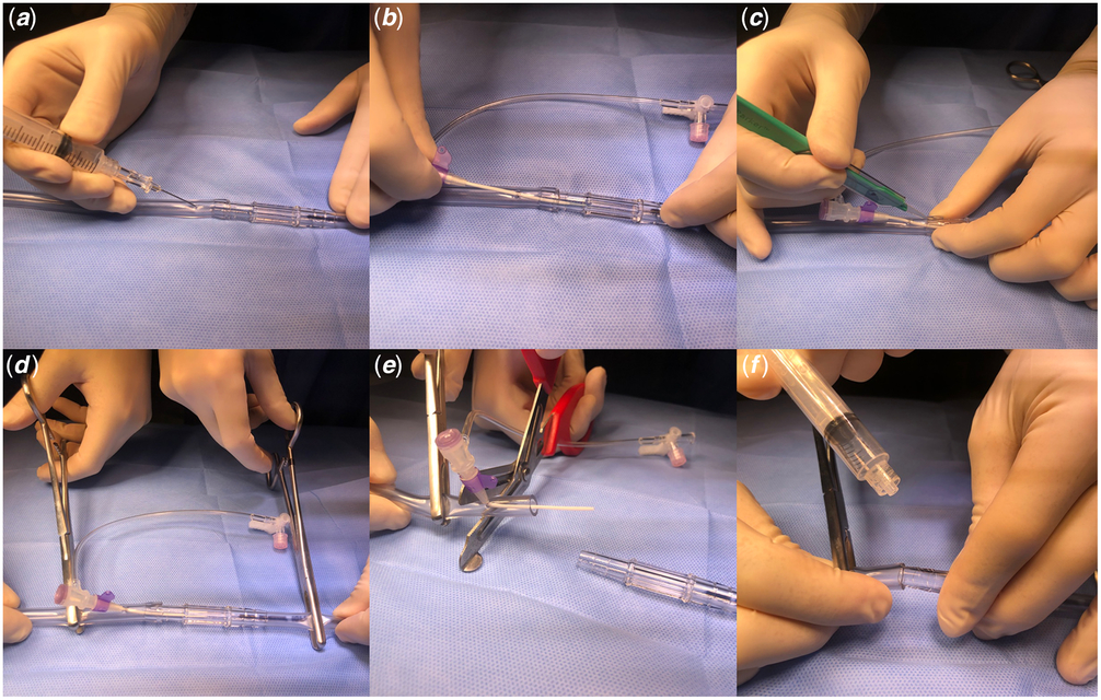
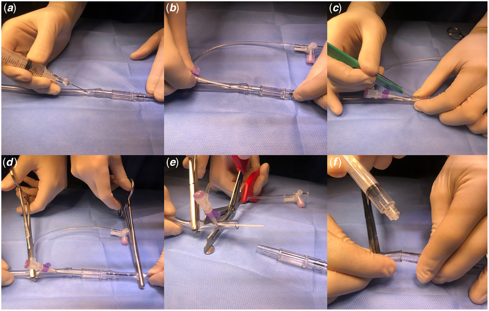
The patient is positioned head to toe inverted for easier access to the cannulas in the neck, which allows smoother manipulation of interventional equipment and improves communication between team members for overall efficiency of the procedure (Supplementary Figure S1). To facilitate screening in this inverted position, fluoroscopy settings need to be adjusted to maintain the usual orientation on the imaging screen. After sterile preparation of approximately 10 cm of the ECMO tubing and the adjacent 5 cm of the cannula, the circuit tubing is punctured with a butterfly needle or short argon needle, approximately 5 cm from the connection with the arterial cannula (Fig 1, Supplementary Video S1). Next, using a Seldinger method, a short 3.3- or 4-Fr sheath is introduced into the circuit, aiming in the direction of flow. Extreme care is taken to avoid introducing air into the circuit; however, the pressure in the arterial circuit minimises this likelihood. At this stage, if the arterial cannula is too deep to allow navigation of a wire to the desired head and neck vessel, light traction is applied to improve its position, taking care not to dislodge it. Once the procedure is finished, with the focus of the perfusion and cardiology teams, the sheath is partially withdrawn, and two clamps were placed on the arterial line, one on the arterial cannula just distal to the tip of the sheath, the second on the tubing proximal to the sheath’s entrance point. For a brief moment (5–10 seconds), the ECMO flow is ceased, and the tubing is cut proximal to the sheath entrance and reconnected to the arterial cannula.
Accessing the extracorporeal membrane oxygenation circuit for cardiac catheterisation. The tubing was punctured with a needle ( a ) just before connection with the arterial cannula and a short 3.3-Fr sheath was introduced ( b ). The tubing was cut longitudinally to enhance quick disconnection of the circuit ( c ). Two clamps were placed on the arterial line, one on the arterial cannula just distal to the tip of the sheath, the second on the tubing proximal to the sheath entrance point ( d ). The tubing was cut proximal to the sheath and reconnected to the arterial cannula ( e , f ).
Case one: balloon angioplasty of a systemic-to-pulmonary artery shunt
A 49-day-old infant (male, 3.6 kg) with a diagnosis of hypoplastic left heart syndrome underwent a balloon atrial septostomy, and aortic valvuloplasty followed with Norwood stage 1 with a 3.5-mm innominate artery to main pulmonary artery shunt. This shunt was surgically revised to address a distal stenosis, with the distal end enlarged to 4 mm. During recovery in the intensive care unit, the infant was found to be in atrial flutter and was electrically cardioverted. Shortly after cardioversion, the infant became bradycardic, hypoxemic, hypotensive and had a cardiac arrest. He was emergently placed onto venoarterial ECMO. An 8-Fr arterial cannula was placed in the right internal carotid artery, and a 12-Fr cannula was placed in the internal jugular vein. On transthoracic echocardiogram, flow through the shunt could not be demonstrated, and the patient was taken to the cath-lab for further evaluation.
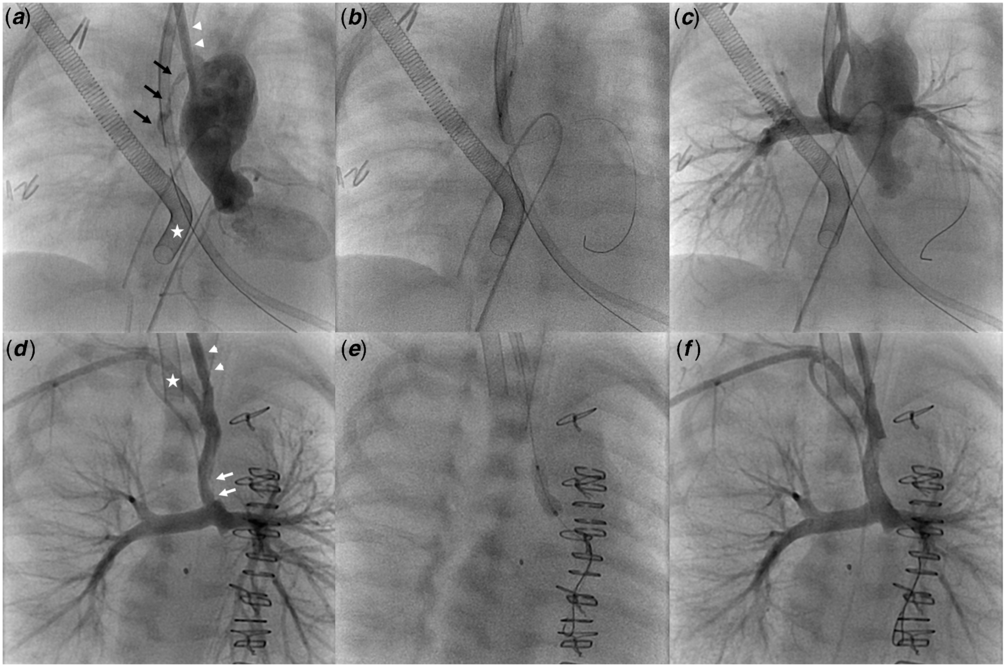
The arterial ECMO cannula was accessed as described above with a 3.3-Fr sheath (Mongoose; PediaCath, Chagrin Falls, OH, USA). An angiogram was performed through the sheath in the arterial cannula, demonstrating near-complete occlusion of the shunt with presumed thrombus (Fig 2a–c, Supplementary Video S2). With mild traction on the cannula as described above, a 0.014″ wire was inserted through the sheath into the shunt and placed in the distal left pulmonary artery. The shunt was dilated with a 3.5 mm × 12 mm Maverick balloon (Boston Scientific, Marlborough, MA, USA). Post-dilation angiography demonstrated resolution of the clot with mild proximal shunt stenosis. The balloon was inflated again at the proximal end of the shunt. A final contrast injection showed normal filling of the shunt without any evidence of thrombus or stenosis. Fluoroscopy and procedural times were 5 and 24 minutes, respectively. A total of 15 ml of contrast was administered. The radiation dose was 622 μGy m2 (16 mGy). The patient was transferred to the intensive care unit and was successfully weaned from ECMO 7 days later.
Balloon dilation ( a – c ) and stent implantation ( d – e ) to a systemic-to-pulmonary artery in patients on venoarterial (white star and arrowheads) extracorporeal membrane oxygenation. An angiography through a short sheath placed in the arterial cannula (arrowheads) demonstrated severe occlusion of the systemic shunt with a thrombus (black arrows) ( a ). The shunt was serially dilated with a coronary balloon ( b ). Post-dilation angiography demonstrated the shunt with normal filling and no evidence of thrombus or stenosis ( c ). An angiography showed a mural thrombus in the distal half of the shunt and a moderate narrowing at the anastomosis site (white arrows) ( d ). A 4 mm × 9 mm coronary stent was implanted ( e ). Post-stenting angiography demonstrated the shunt with normal filling and no evidence of thrombus or stenosis ( f ).

Case two: stent implantation in a systemic-to-pulmonary artery shunt
A 27-day-old infant (male, 3.3 kg) with a diagnosis of hypoplastic left heart syndrome and a restrictive atrial septum underwent a balloon atrial septostomy followed with Norwood stage 1 with a 3.5-mm innominate artery to main pulmonary artery shunt and subsequent atrial septectomy. After stable recovery in the intensive care unit, the patient decompensated unexpectedly due to presumed shunt obstruction. With oxygen saturations in the 20’s he was emergently cannulated onto venoarterial ECMO in a similar manner to the previous patient. The patient was brought to the cath-lab to asses and intervene on the shunt. The arterial ECMO cannula was accessed with a 4-Fr sheath (Glidesheath; Terumo, Somerset, NJ, USA). An angiogram through the sheath demonstrated an area of mural thrombus in the distal half of the shunt and a moderate narrowing at the anastomosis site with the pulmonary artery (Fig 2d–e, Supplementary Video S2). A 4 mm × 9 mm Integrity stent (Medtronic, Minneapolis, MN, USA) was introduced over a 0.014″ wire and deployed in the distal segment of the shunt. Final angiography showed a satisfactory result with uniform lumen throughout the shunt. Fluoroscopy and procedural times were 3 and 23 minutes, respectively. A total of 5 ml contrast was administered. The radiation dose was 81 μGy m2 (3 mGy). The patient was transferred to the intensive care unit and successfully weaned from ECMO 2 days later.
Discussion
In patients supported with ECMO, cardiac catheterisation may be conducted for diagnostic or therapeutic indications via the ECMO cannulas without placing percutaneous sheaths. New findings, not available with other diagnostic modalities, were discovered in up to 78% of patients, and most of them were subsequently addressed with interventional procedures.Reference Agarwal, Hardison and Saville 3 Most recent reports show the rate of transport-related and catheterisation-related complications ranging between 0% and 18% using traditional access approaches.Reference Callahan, Trucco, Wearden, Beerman, Arora and Kreutzer 5 , Reference Zahn, Dobrolet, Nykanen, Ojito, Hannan and Burke 6 , Reference Kato, Lo Rito and Lee 8 Callahan et al. reported 15% rate of complications with all but one being vascular-related.Reference Callahan, Trucco, Wearden, Beerman, Arora and Kreutzer 5 Our technique for cardiac catheterisation while on ECMO negates the need for obtaining vessel access in these fragile patients and adds to alternative routes for performing interventional cardiac catheterisations.Reference Davenport, Lam and Whalen-Glass 9 This could potentially lead to fewer complications and shorter procedural times, as found in the patient examples described.
In our experience, a successful implementation of this technique revolves around excellent communication and slick performance of sheath introduction and, at the end of the procedure, removing the punctured segment of the tubing. Changing the orientation of the patient on the table may give better access to the lines and enhance crucial steps of the procedure to be conducted, although this may be dependent on the local cath-lab environment and protocols. Excellent communication with the ECMO team, particularly when introducing the sheath or clamping the arterial line and removing the sheath, is of utmost importance. All the team members need to be bought into this concept. At the end of the procedure, one operator places the clamps, and another removes the sheath with the adjacent tubing. As with any manipulation and connections performed in an active ECMO circuit, extreme care is taken to remove all air before reconnecting the ECMO tubing to the cannula using a primed wet-to-wet connection. In both reported patients we were able to clamp, cut, disconnect and reconnect the ECMO circuit in under 30 seconds with minimal blood loss.
In both cases, we could easily puncture the tubing with a butterfly needle or short argon needle, and used short, low-profile 018″ introducer sheaths. In all cases we have not used more than a 4-Fr system and have not seen any change in ECMO parameters with these sheaths in the circuit. As with accessing any vessel, great care was taken to avoid introduction of air into the circuit. This never occurred in either of these cases or in our other practice where we enter venous and arterial circuits for diagnostic assessments on ECMO.
During catheterisation, we did not observe any significant interference of the access sheath positioned in the arterial cannula with the ECMO flow. Flow was not routinely decreased during angiography. In one patient we had to put backward traction on the cannula to improve orientation and simplify access to the Blalock-Taussig shunt. Otherwise, we did not encounter any major limitation to the movement of wires and catheters. The access is effectively like an innominate arterial sheath placement and gives excellent orientation for the entire aortic arch and systemic-to-pulmonary artery shunts when present. Likewise, a venous cannula from the internal jugular vein usually allows the catheterisation of whichever cardiac chambers are allowed by the patients’ anatomical disposition, as well as the pulmonary arteries when they are connected normally.
We speculate that this technique of accessing the ECMO circuit could have wider potential interventional applications. Entering the arterial limb of the circuit could give access for the treatment of arch obstruction, restenosis in a stented arterial duct, stenosis of a central shunt or shunt-supported pulmonary arteries. Accessing the venous line may allow us to address a restrictive atrial septum, right ventricular outflow tract obstruction or stenosed pulmonary arteries.
We acknowledge several limitations of this technique, including the inherent risks associated with directly accessing the circuit and the need for a brief cessation of ECMO flow when reconnecting. The latter could potentially lead to haemodynamic instability in patients purely dependent on ECMO flow, although that has not been seen with very brief clamp times recorded in our experience.
Conclusion
Direct Seldinger access to ECMO circuits is a potentially safe and effective alternative to percutaneous approach for performing an emergent interventional and diagnostic cardiac catheterisation.
Supplementary material
To view supplementary material for this article, please visit https://doi.org/10.1017/S1047951119001446
Acknowledgements
Dr Goreczny would like to thank the Polish–U.S. Fulbright Commission for supporting his research projects with a Senior Award Scholarship.
Financial Support
None.
Conflicts of Interest
None.
Ethical Standards
The authors assert that all procedures contributing to this work comply with the ethical standards of the relevant national guidelines on human medical regulations and with the Helsinki Declaration of 1975, as revised in 2008, and has been approved by the ethical committee of Colorado Children’s Hospital.