There is general agreement that a common arterial trunk, or persistent truncus arteriosus, is best defined as the presence of a solitary arterial trunk leaving the base of the heart through a common arterial valve, with the arterial trunk then supplying directly the aortic, pulmonary, and coronary arterial pathways.Reference Van Praagh and Van Praagh1–Reference Crupi, Macartney and Anderson3 Almost always, this pattern of branching of the arterial pathways is seen in the setting of usual atrial arrangement, and with concordant atrioventricular connections. The greatest anatomic variability is found in the pattern of the branching of the common trunk, and this has served as the basis for subclassification. The traditional system introduced in 1949 by Collett and EdwardsReference Collett and Edwards4 was based on the variability in the pulmonary arterial pathways. In their so-called “type I” variant, a confluent pulmonary arterial channel arose from the common trunk itself, and then branched to become the right and left pulmonary arteries. In their type II variant, these pulmonary arteries were described as arising separately from the posterior and leftward part of the common trunk, while in the much rarer type III variant, the pulmonary arteries arise from the right and left sides of the posterior aspect of the intrapericardial segment of the common trunk. There has been much discussion concerning their type IV variant, characterized by absence of the intrapericardial pulmonary arteriesReference Edwards and McGoon5. It is now considered that this pattern is better described in terms of a solitary arterial trunk, with clinical presentation more akin to that seen in patients with tetralogy of Fallot and pulmonary atresia.Reference Thiene and Anderson6 The other commonly used system for describing the anatomic variability in patients with common arterial trunk also took note of the different arrangements of the pulmonary arteries, but stressed also the importance, when present, of interruption of the aortic arch.Reference Calder, Van Praagh and Van Praagh2, Reference Crupi, Macartney and Anderson3 This system also emphasized the importance of distinguishing patients with a common trunk guarded by a common valve from those having an aortopulmonary window, since as we will discuss, some patients with the latter lesion, characterized by presence of separate aortic and pulmonary valves, can also have an effectively common intrapericardial arterial channel.
For better or worse, nonetheless, it is now accepted that the cardinal diagnostic feature is the presence of the common truncal valve guarding the entrance to the common intrapericardial arterial channel. The clinical features of patients united in this fashion are related to the amount of flow of blood to the lungs, and the degree of truncal insufficiency, if present. As we will show, cross-sectional echocardiography, when combined with Doppler interrogation and assessment using colour flow, has created the ability accurately to diagnose the common valve and common arterial trunk, without the need for other diagnostic modalities. As we will also explain, in the current surgical era, there is no role for palliative surgery. Elective repair is usually undertaken within the neonatal period. Subsequent to successful surgical repair, outcomes over the longer term depend on three separate factors, namely the adequate function of the conduit almost always inserted to restore flow to the pulmonary arteries as part of surgical reconstruction, competency of the truncal valve, which becomes the aortic valve subsequent to repair, and pulmonary vascular resistance.
Anatomy
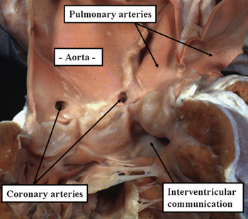
Common arterial trunk is, perhaps, the most obvious congenital cardiac malformation that benefits from direct description. When the entity is described in terms of persistent truncus arteriosus, or expansions of the term implicating persistence of an embryonic condition, it becomes necessary to provide a definition of the lesion as seen in the postnatal heart. As we will discuss in the subsequent section devoted to morphogenesis, the so-called truncus in the developing heart gives rise to the intrapericardial arterial pathways. And, as already discussed, the essential feature of common trunk seen as a congenital cardiac malformation in humans, rather than the presence of a common intrapericardial arterial channel, is the presence of a common ventriculo-arterial junction, guarded by a common truncal valve.Reference Van Praagh and Van Praagh1–Reference Crupi, Macartney and Anderson3 Such persistence of the common ventriculo-arterial junction requires failure of septation of the proximal outflow tract, or conus, during development, rather than the distal outflow tract, or truncus. Thus, those preferring embryological descriptions should be describing persistent conus arteriosus!! Description of the entity as a common arterial trunk negates the need to pass through such descriptive minefields, albeit that there remains the need to provide a definition for the entity seen in the presence of a common ventriculo-arterial valve. Thus, as stated in our introduction, the malformation now recognized as common arterial trunk is best defined on the basis that the trunk takes its origin from the ventricular mass through a common ventriculo-arterial junction, and then gives rise directly to the systemic, pulmonary and coronary circulationsReference Van Praagh and Van Praagh1–Reference Crupi, Macartney and Anderson3 (Fig. 1).
The specimen shows the features of common arterial trunk, with a solitary arterial vessel exiting from the ventricular mass through a common ventriculo-arterial junction guarded by a common arterial valve, and supplying directly the coronary, systemic and pulmonary circulations.

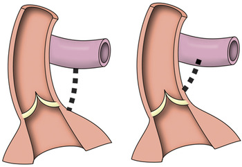
Common arterial trunk as thus described is but one example of a patent solitary arterial trunk exiting from the ventricular mass. Solitary aortic trunk, and solitary pulmonary trunk, are easy to differentiate in the postmortem room, since their diagnostic feature is the presence of an atretic second trunk arising directly from the ventricular mass, albeit that atretic trunks represented by no more than fibrous strands may be harder to distinguish in the clinical setting. The branching pattern of the solitary trunk, nonetheless, will almost always permit diagnosis. The fourth type of trunk is also easy to distinguish, but harder to describe. This is the entity in which there is complete absence of the intrapericardial pulmonary arteries. In this setting, it is impossible to know whether, had it been present, the pulmonary trunk would have arisen from the heart. In this instance, the solitary trunk would be an aorta. It is also possible, very rarely, for the atretic remnant of the pulmonary pathway to arise directly from the arterial trunk itself. This indicates that the trunk, initially, had been a common vessel.Reference Schofield and Anderson7 The only sensible way of describing the vessel arising from the heart in absence of the intrapericardial pulmonary arteries, therefore, is as a solitary arterial trunk (Fig. 2). Others may describe this as tetralogy of Fallot with pulmonary atresia and absent pulmonary arteries. Indeed, patients with this malformation, when encountered clinically, certainly have more in common with tetralogy of Fallot and pulmonary atresia than with patients having a common arterial trunk. This is because, in most patients with absence of the intrapericardial pulmonary arteries, the pulmonary arterial supply is derived, at least in part, from systemic-to-pulmonary collateral arteries.
The cartoon shows how, in absence of the intrapericardial arteries, it is impossible to know whether they would have taken origin from the heart, or from the arterial trunk itself (dotted lines). There is no way of knowing, therefore, whether the trunk was destined to become an aorta or a common trunk. Because of this uncertainty, the solitary vessel is best described as a solitary trunk. Patients with this arrangement, nonetheless, are best considered as a subset of those with tetralogy and pulmonary atresia.

Common arterial trunk as defined here, therefore, is but one form of single outlet from the heart. It must be anticipated to coexist with all possible segmental combinations. Again, as discussed in our introduction, most usually there will be usual atrial arrangement, with concordant atrioventricular connections. Examples can be found, nonetheless, in combination with discordant atrioventricular connections, or with absence of the right atrioventricular connection.Reference Malec, Mroczek, Pajak and Kordon8 Indeed, any possible combination must be anticipated. Furthermore, while the atrioventricular junctions themselves are usually separate, and guarded by mitral and tricuspid valves, a common trunk can also be found in association with an atrioventricular septal defect and a common atrioventricular valve.Reference Atik, Soares and Aiello9
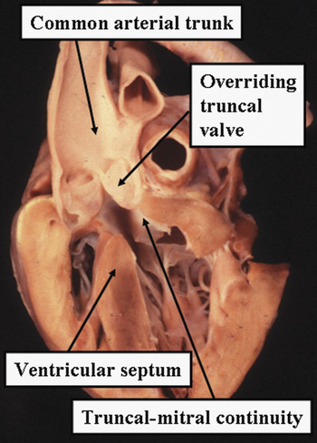
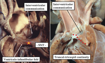
In the presence of the common trunk, almost always the truncal valve is supported above the cavities of both ventricles, the common truncal orifice overriding the ventricular septal crest, and typically with the leaflets of the truncal valve in fibrous continuity with those of the mitral valve in the roof of left ventricle (Fig. 3). In such a setting, there is a juxtaarterial interventricular communication, almost always large. The defect opens into the right ventricle between the limbs of the septomarginal trabeculation, or “Y” of the septal band, and is roofed by the leaflets of the truncal valve. When the trunk overrides in this fashion, a cone of space is subtended from the leaflets of the truncal valve to the ventricular septal crest. It is the right ventricular margin of this cone that is closed by the surgeon during repair. This plane, therefore, should be considered to represent the ventricular septal defect. In the majority of cases, fusion of the inferior limb of the septomarginal trabeculation with the ventriculo-infundibular fold along this right ventricular margin produces muscular discontinuity between the leaflets of the tricuspid and the truncal valves (Fig. 4 – left-hand panel). In the absence of such fusion, there is continuity between the leaflets of the tricuspid and truncal valves, making the defect perimembranous (Fig. 4 – right-hand panel). If present, as it is in the majority of patients, the muscular bar in the postero-inferior margin protects the atrioventricular conduction axis. In most instances, when the valvar leaflets are closed, there is a space between their ventricular aspect and the crest of the septum during ventricular diastole, permitting interventricular shunting. This space may sometimes be reduced, or the leaflets may even close directly on the septal crest during diastole. This latter arrangement has been suggested to represent an intact ventricular septum.Reference Carr, Bharati, Kusnoor and Lev10 This is misleading because, even in this arrangement, an interventricular communication is present when the truncal valve opens during ventricular systole. Furthermore, hearts can be found when the ventricular septum is truly intact, the common trunk then arising exclusively from the right ventricle. The ventricular septal defect can be restrictive when the common trunk takes an exclusive origin from one or other ventricle. Such a restrictive defect is more likely to produce problems when the trunk arises exclusively from the right ventricle, since it then represents the outflow tract for the morphologically left ventricle.
This long-axis section through a heart with common arterial trunk shows the typical pattern of the truncal valve overriding the crest of the muscular ventricular septum, with the valvar leaflets supported in both ventricles, but typically, as shown, in fibrous continuity with the leaflet of the mitral valve.

The two hearts illustrate the different morphology of the plane of space closed by the surgeon so as to reconnect the arterial trunk with the morphologically left ventricle. In the left-hand panel, the ventriculo-infundibular fold fuses with the postero-inferior limb of the septomargical trabeculation (SMT), producing a muscular bar (asterisk) which protects the atrioventricular conduction axis during surgical correction. In contrast, in the heart shown in the right-hand panel, the ventriculo-infundibular fold does not fuse with the postero-inferior limb of the septomargical trabeculation. Instead, the postero-inferior margin of the defect is made up of fibrous continuity between the leaflets of the tricuspid and truncal valves, making the defect perimembranous, and putting the atrioventricular conduction axis (red dotted line) at greater potential risk during surgical correction.

The truncal valve has 3 leaflets in approximately two-thirds of patients, with either 2 or 4 leaflets found in most other instances. The truncal valvar leaflets are almost always in fibrous continuity at some point with the antero-superior leaflet of the mitral valve (Fig. 3). There can be a completely muscular subtruncal infundibulum, particularly when the common trunk arises exclusively from the right ventricle. Insufficiency of the truncal valve is not uncommon, most usually due to thickened and dysplastic leaflets, or to prolapse of unsupported leaflets as a result of dilation of the ventriculo-arterial junction. Truncal valvar stenosis is relatively uncommon, and if present is usually the consequence of dysplastic or myxomatous valvar leaflets. These leaflets are different from those typically seen in valvar aortic or pulmonary stenosis. Dysplastic truncal valves look more like the immature arterial valvar leaflets seen in the fetus.
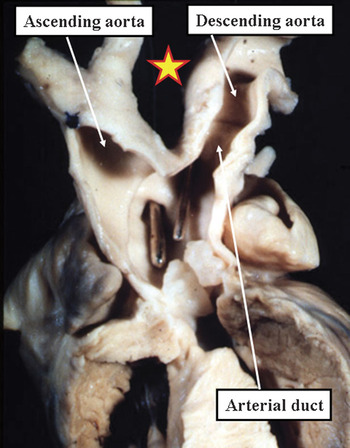
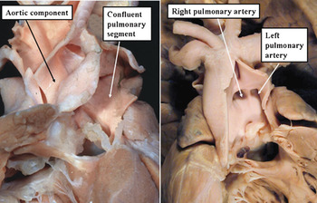
The greatest anatomic variability is found in the pattern of the branching of the common trunk. This feature, therefore, should play, and has played, a role in classification. As discussed already, attention has been focused on either the pulmonaryReference Collett and Edwards4 or aorticReference Van Praagh and Van Praagh1, Reference Calder, Van Praagh and Van Praagh2 pathways. The aortic arch is right-sided in up to one-third of patients. Hypoplasia of the aortic arch, with or without coarctation, is found infrequently, with complete interruption of the arch being the commonest malformation of the aortic pathway (Fig. 5). It was hearts with such interruption that were selected to make up one of the major sub-groups recognised in the popular alpha-numeric system used by Van Praagh and his colleaguesReference Van Praagh and Van Praagh1, Reference Calder, Van Praagh and Van Praagh2 for classification, providing the so-called Van Praagh Type 4. In the setting of such interruption, the persistently patent arterial duct feeds the descending thoracic aorta and part of the brachiocephalic circulation, with the precise proportion depending on the site of interruption. Retro-oesophageal origin of the right subclavian artery is also frequently seen. Apart from those hearts with severe coarctation or interruption, or in which the pulmonary arteries are discontinuous, and one is fed through a patent duct, this being Type 3 in the classification of Van Praagh, it is rare to find ductal patency coexisting with common arterial trunk, although it does exist. While the integrity of the aortic arch is, perhaps, the most significant clinical associated malformation, it was variation in the origin of the pulmonary arteries that formed the basis of the system proposed by Collett and EdwardsReference Collett and Edwards4 for classification. The pulmonary arteries typically arise from the left posterolateral aspect of the common trunk, taking origin a short distance above the truncal valve. Very rarely, they can take their origin directly from a truncal arterial valvar sinus. In the more usual situation, the presence of a short confluent pulmonary arterial segment produces the so-called type I variant (Fig. 6 – left-hand panel). Most of these cases only have confluence on the lateral aspect of the apparent pulmonary arterial portion of the trunk. The bifurcation frequently occurs very close to the entrance to the aortic pathway, even though when viewed externally there seems to be a pulmonary arterial confluence. There are obviously separate origins of the right and left pulmonary arteries from the posterior aspect of the trunk in the type II lesion categorized by Collett and EdwardsReference Collett and Edwards4 (Fig. 6 – right-hand panel). Because most examples of their first category also have essentially separate right and left pulmonary arteries, many have suggested that this pattern should be recognised as “type 1½”. The essence of the first pattern is anterior or lateral origin of the right and left pulmonary arteries from the trunk, while in the second type the origin of the pulmonary arteries tends to be more posterior. In the third type defined by Collett and Edwards,Reference Collett and Edwards4 which is rare, the pulmonary arteries arise from the right and left sides of the posterior aspect of the intrapericardial segment of the common trunk. In some instances, only one pulmonary artery arises from the common trunk, the other being supplied initially through a duct that becomes ligamentous, producing the clinical feature of apparent unilateral absence of one pulmonary artery. Almost always the artery presumed to be absent can be identified as a patent, albeit hypoplastic, channel within the hilum of the lung. In some circumstances, the pulmonary artery feeding the right lung is to the left at its origin from the common trunk when judged relative to the origin of the artery running to the left lung. The two arteries then spiral as they extend to the pulmonary hilums. This entity is called crossed pulmonary arteries.Reference Becker, Becker and Edwards11 In the original categorisation of Collett and Edwards,Reference Edwards and McGoon5 there was also a fourth pattern described, characterized by absence of the intrapericardial pulmonary arteries. As already discussed, this arrangement is better described as a solitary arterial trunk, patients with this variant presenting clinically as a subset of those with tetralogy of Fallot with pulmonary atresia.Reference Thiene and Anderson6 It is also possible for one pulmonary artery to arise directly from the ascending aorta, while the other takes its origin from the right ventricle. This entity should not be described as hemitruncus, since patients with this lesion have separate ventriculo-arterial junction, guarded by separate aortic and pulmonary arterial valves. They cannot, therefore, exhibit a common arterial trunk as currently defined. It is also important to distinguish patients having a common arterial trunk guarded by a common truncal valve from those with aortopulmonary windows. Indeed, such windows can be sufficiently large effectively to produce a common intrapericardial arterial trunk. The feature of patients with aortopulmonary windows, however, is the presence of separate ventriculo-arterial junctions for the aortic and pulmonary pathways, albeit that one of the junctions, on occasion, can be atretic. It is also the case that the most proximal parts of the aortic and pulmonary pathways are discrete and separate within the pericardial cavity, showing that the lesion truly is an aortopulmonary window rather than a completely common intrapericardial arterial trunk.Reference Anderson, Brown, Webb and Henderson12
In this specimen, the systemic pathways are interrupted between the left common carotid and left subclavian arteries. The arterial duct feeds the descending aorta and the left subclavian artery. The probes are placed in the pulmonary arteries, which arise from the back of the common trunk. Note that the trunk itself arises almost exclusively from the left ventricle.

The panels show the common variants distinguished in the categorization of Collett and Edwards. In the heart shown in the left-hand panel, a short confluent pulmonary arterial segment interposes between the common trunk and the origin of the right and left pulmonary arteries. This is the so-called “type I” variant. In the heart shown in the right-hand panel, the right and left pulmonary arteries take separate origin from the leftward and posterior aspect of the common trunk, producing the so-called “type II” variant.

Anomalies of the origin and distribution of the coronary arteries are frequent.Reference Suzuki, Ho, Anderson and Deanfield13 Unlike the situation when there are separate aortic and pulmonary valves, in which virtually without exception the coronary arteries arise from one or other of the aortic valvar sinuses adjacent to the pulmonary trunk, and usually both sinuses, there is no constant pattern of sinusal origin when there is a common ventriculo-arterial junction guarded by a common truncal valve. The coronary arteries can arise from any of the truncal valvar sinuses, albeit that almost always there are two coronary arteries, with the left artery giving rise to anterior interventricular and circumflex branches. The arteries often arise close to a zone of apposition between the valvar leaflets, and origin above the sinutubular junction is quite common. This can produce potential difficulties during surgical correction should the high origin be adjacent to the origin of the pulmonary arteries.
Knowledge of location of the atrioventricular conduction axis is important when planning surgical repair. As explained, in those hearts in which a muscular bar interposes between the attachments of the truncal and tricuspid valves in the postero-inferior margin of the septal defect, the atrioventricular conduction axis is distant from the rim of the defect (Fig. 4 – left-hand panel). In patients in whom the ventricular septal defect is perimembranous, in contrast, the conduction tissue passes directly along the left aspect of the fibrous posteroinferior rim of the defect (Fig. 4 – right-hand panel). In terms of associated malformations, a defect within the oval fossa has been noted in up to one-fifth of patients, persistence of the left superior caval vein draining to the coronary sinus in up to one-tenth, and an aberrant subclavian artery in between one-tenth and one-twentieth.Reference Calder, Van Praagh and Van Praagh2, Reference Crupi, Macartney and Anderson3 Partially anomalous pulmonary venous connection has also been reported.Reference Calder, Van Praagh and Van Praagh2
Morphogenesis
The morphology of the heart with the presence of a common arterial trunk supports very strongly the notion that, during development, there has been failure of septation of the ventricular outlets and the outflow segment of the heart tube. It has been suggested that the lesion results from failure of formation of the subpulmonary infundibulum, with the common arterial trunk in essence representing the aorta.Reference Van Praagh and Van Praagh1, Reference Calder, Van Praagh and Van Praagh2 No evidence from study of developing hearts has accrued over the last decades to support this latter notion. There is much evidence, nonetheless, to contradict the notion that the arterial trunk is essentially an aorta, with failure of formation of the pulmonary trunk. From the stance of morphology, hearts with common arterial trunk show no evidence of a blind-ending subpulmonary outflow tract, such as is seen in tetralogy of Fallot with pulmonary atresia. It is the latter lesion that unequivocally represents underdevelopment of the subpulmonary outflow tract. Additionally, if the arterial root truly represented the aorta, then the coronary arteries might be anticipated to arise in patterns comparable to those seen in the normal heart. This is rarely the case, since the origins and course of the coronary arteries are frequently bizarre.Reference Suzuki, Ho, Anderson and Deanfield13 Alternative explanations for abnormal coronary arterial origins include interposition of some rudimentary pulmonary valvar tissue between the aortic leaflets. This would explain the usual finding of asymmetry of valvar leaflets when four or five are present, unexpected if this were a true truncal valve. In the developing heart, the valvar leaflets are sculpted from four cushions, with the two central ones dividing to produce a total of six arterial leaflets and sinuses in the definitive heart. One might expect, therefore, to find four leaflets in the truncal valve. In fact, three leaflets is most common, followed by four, then two, then five. It is extremely rare to find six leaflets.
From the stance of cardiac development, studies on both normal and abnormal hearts show that the initially common ventricular outflow tract is septated by endocardial cushions, or ridges.Reference Van Mierop, Patterson and Schnarr14–Reference Bartelings and Gittenberger-de Groot16 Failure of fusion of these cushions during embryological development was shown to result in common arterial trunk as long ago as 1978.Reference Van Mierop, Patterson and Schnarr14 Much work over the past 15 years has provided further evidence of the importance of the outflow cushions in dividing the initially common ventricular outflow tract. Ongoing studiesReference Hutson and Kirby17 have shown that cells from the neural crest migrate through these cushions to reach the heart. Perturbation of such migration can produce, amongst other lesions, a common arterial trunk. The genetic basis for such malformations is supported by observations made in the homozygous mutant Splotch mouse.Reference Conway, Henderson, Kirby, Anderson and Copp18 Common arterial trunk has been produced when there is deficiency of sox4, a gene which also normally populates the endocardial cushions of the developing outflow tracts.Reference Schilham, Oosterwegel and Moerer19 Significantly, some of the afflicted embryos in these experiments had doubly committed ventricular septal defects rather than common arterial trunk. The morphology of the outflow tracts is almost identical in such hearts with the situation seen in common arterial trunk, apart from the finding of separate aortic and pulmonary valvar orifices in the hearts with the doubly committed ventricular septal defects.
Clinical Features
Patients with common arterial trunk, postnatally, often exhibit mild to moderate cyanosis. There is generally complete mixing proximal to the truncal valve, with some streaming of blood from the right ventricle to the pulmonary arteries. The clinical features are related to the amount of flow of blood to the lungs and, if present, the degree of truncal valvar insufficiency. Immediately after birth, the systemic vascular resistance increases with removal of the low resistant umbilical placental circulation, as well as other hormonal and vascular mediators associated with the transition form the fetal to the newborn circulation. Pulmonary vascular resistance decreases immediately with ventilation of the lungs, and gradually with maturation of the small pulmonary vessels. Initial arterial oxygen saturations may range from 75 to 80%, increasing to 90% as pulmonary vascular resistance diminishes over time. Alternatively, if there is stenosis within the pulmonary arterial segments, there may be limited flow of blood, and cyanosis may be the more pronounced clinical presentation. In presence of moderate-to-severe truncal insufficiency, there are signs and symptoms of congestive cardiac failure, such as increased respiratory rate and hepatomegaly. In consequence, poor feeding will develop earlier than in patients without truncal valvar insufficiency. Patients often exhibit normal to bounding pulses associated with a wide pulse pressure as the flow of blood to the lungs increases compared to systemic flow. The widened pulse pressure may also be exacerbated by truncal valvar insufficiency. A hyperdynamic precordial impulse may be exhibited due to biventricular hypertrophy. There may be an associated ejection click, a long systolic ejection murmur, or an early diastolic murmur, as well as a single second heart sound associated with truncal valvar abnormalities. Electrocardiographic findings are nonspecific, but may show signs of biventricular hypertrophy over time. Chest radiography will demonstrate cardiomegaly, with developing increased pulmonary vasculature. A right aortic arch will be noted in about one-third of patients.
Imaging
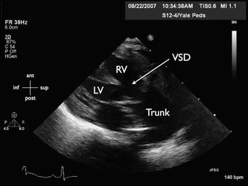
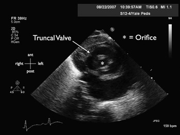
Cross-sectional echocardiography, when combined with Doppler interrogation and analysis of colour flow, has made it possible accurately to diagnose common arterial trunk without the need for other diagnostic modalities. Using bedside echocardiographic imaging, the parasternal long-axis view allows for defining the often large subarterial ventricular septal defect, as well as the extent of sharing of the common ventriculo-arterial junction between the ventricles (Fig. 7). It is unusual to have associated ventricular septal defects, but the entire septum should be evaluated for their presence. The truncal valve, which is almost always abnormal in patients with a common arterial trunk, can be evaluated from a number of echocardiographic views. To assess the structure of the truncal valve, the parasternal short-axis view is the most useful. In addition to evaluating the morphology and number of truncal leaflets, this view allows for the assessment of additional valve abnormalities such as inadequate coaptation of the leaflets, yielding valvar insufficiency (Fig. 8). When assessing for truncal valvar stenosis, the four-chamber apical and subcostal views are typically the most enlightening. Using colour flow and Doppler interrogation, it is possible to study the amount of insufficiency or stenosis of the valve. As described above, most commonly, the truncal valve will be seen to have two, three or four separate leaflets, though truncal valves with as few as one or as many as six leaflets have been described. Irrespective of the number of leaflets within the valve, and their function, parts of the leaflets adjacent to the mitral valve are usually in fibrous continuity with its anterior-superior leaflet. This feature is well-demonstrated from the parasternal long-axis view.
This image is obtained from cross-sectional echocardiographic imaging at the bedside of a neonate with common arterial trunk. The image is obtained from the long-axis view and reveals the large subarterial ventricular septal defect (VSD), as well as the extent of sharing of the common ventriculo-arterial junction between the right ventricle (RV) and left ventricle (LV). The common arterial trunk (Trunk) is shown heading superiorly and posteriorly.

This image is obtained during diastole from the parasternal short-axis view from the same neonate as in the previous figure. The truncal valve is large and, in this instance, possesses four leaflets. The edges of the leaflets are thickened and do not coapt well, yielding a diastolic orifice (*) through which there is truncal insufficiency.

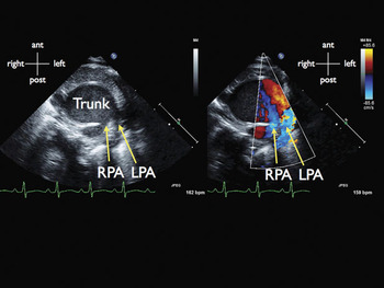
The parasternal short-axis view allows for excellent evaluation of the origin of the pulmonary arteries, especially with the application of colour flow Doppler mapping (Fig. 9). Their origin, size, and patency should be evaluated thoroughly. From this view, the origin and course of the coronary arteries should also be established. It is also important to look from the suprasternal notch for the sidedness and integrity of the aortic arch. Interruption of the aortic arch, typically between the left common carotid and subclavian arteries, is an important association with common arterial trunk, particularly when microdeletions of chromosome 22q11 are present. The chromosomal composition of these patients should be assessed with fluorescent in situ hybridization. When there is interruption of the aortic arch, the distal aorta will be supplied by an arterial duct. When the aortic arch is normal, however, an arterial duct is rare. Nonetheless, the echocardiographic examination should dedicate care to the evaluation of its presence in all cases of common arterial trunk. It is also important to evaluate for the presence of other defects that may be associated with the common arterial trunk. Included in these would be an atrial septal defect, and a left superior caval vein draining to the coronary sinus.
The echocardiographic images in this panel are obtained from the parasternal short-axis view and demonstrate the origin of the pulmonary arteries from the common arterial trunk (Trunk). The image on the left demonstrates the posterior origin of both the right pulmonary artery (RPA) and the left pulmonary artery (LPA) from the large trunk. Colour flow Doppler is applied in the image on the right of the panel and demonstrates the pattern of flow from the trunk into the right and left pulmonary arteries.

Cardiac catheterization and angiography is usually not necessary in the patient with a common arterial trunk, but can be useful to define stenosis in the pulmonary arteries, anomalies of the aortic arch, or to evaluate pulmonary vascular resistance in older children prior to surgical repair. Magnetic resonance imaging is excellent for demonstrating the anatomy and origin of the pulmonary arteries, as well as the morphology of the aortic arch. The technique also demonstrates flow through the pathways, and does not have the limitation of rapid dissipation of contrast material in cases with torrential flow to the lungs. Magnetic resonance imaging may be particularly useful in assessing right ventricular size and volume, as well as the integrity of the pulmonary arteries in the follow-up through adolescence and into adult life of those who have undergone repair of the common arterial trunk.
Management
Medical management of congestive cardiac failure is used to optimize the cardiac and nutritional state prior to surgical correction. In the presence of 22q11 deletion, issues should be addressed such as hypocalcaemia and immune deficiency. In the current surgical era, there is no place for palliative surgery for patients with common arterial trunk. The large left-to-right shunt with pulmonary volume overload, truncal valvar insufficiency, and associated ventricular dysfunction lead to haemodynamic deterioration, so if the patient is stable, and is appropriately medicated against congestive cardiac failure, elective repair is preferred within the neonatal period.Reference Bove, Lupinetti and Pridjian20 It was shown early in the era of primary surgical correction that repair undertaken at 6 to 12 months of age was associated with double the mortality occurring when performing surgery at 6 weeks to 6 months of age.Reference Ebert, Turley, Stanger, Hoffman, Heymann and Rudolph21
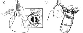
The first successful definitive surgical repair of common arterial trunk was reported in 1968.Reference McGoon, Rastelli and Ongley22 The repair involved separating the pulmonary arteries from the common arterial trunk, then repairing the defect created in the trunk, closing the ventricular septal defect with a patch, and establishing continuity between the right ventricle and the pulmonary arteries (Fig. 10). Debate continues as to the optimal technique for reconstructing the path to the pulmonary arteries. Most insert a conduit, but there is no agreement as to whether the conduit should contain a valve. It is hypothesized that a valveless conduit may avoid the obstruction seen with degeneration of valved conduits. Choices of valved conduits include homografts of pulmonary or aortic origin, as well as heterografts, such as the Contegra, Hancock and Shelhigh models.Reference Almeida, Wyse, de Leval, Elliott and Stark23–Reference de Leval, McGoon, Wallace, Danielson and Mair26 Some reports have suggested that homografts are superior to heterografts.Reference Almeida, Wyse, de Leval, Elliott and Stark23, Reference Reddy, Rajasinghe, McElhinney and Hanley24 Recent experience reported from the University of Michigan covering the period from July 2000 to December 2007 exemplifies this dilemma concerning the choice of conduit. Of 33 patients undergoing surgery, all have surgical correction between 7 and 90 days of life, 13 received pulmonary homografts, 6 aortic homografts, 3 had placement of Shelhigh heterografts, 8 had Contegra, and 3 Hancock prostheses. All but one of the patients was discharged alive from hospital. Of the group, 6 patients thus far have required replacement of their conduits without problem, endorsing the well recognized low risk for such replacement.
The illustrations show the steps of surgical repair. Panel (a) shows the separation of the pulmonary arteries from the common arterial trunk, with repair of the aortic defect, while panel (b) shows the establishment of continuity between the right ventricle and the pulmonary arteries.

Severe truncal insufficiency increases the risk of operative correction and reoperation.Reference Henaine, Azarnoush and Belli27, Reference Thompson, McElhinney, Reddy, Petrossian, Silverman and Hanley28 In the cohort of patients undergoing surgery at the University of Michigan, 6 had moderate-to-severe truncal valvar insufficiency. Of these, 5 required repair of the truncal valve, repeated in 2 patients, while the valve was replaced in the other patient. Interruption of the aortic arch was found in 8 patients. This association did not result in increased mortality in this cohort, but the combination of common trunk and interrupted aortic arch still emerges as a confounding feature when analysis is made of more widespread contemporary experiences.Reference Konstantinov, Karamlou and Blackstone29
Outcomes
After successful surgical repair, long term outcomes depend on three separate factors, namely adequate function of the conduit placed between the right ventricle and the pulmonary arteries, the competency of the truncal valve placed in aortic position subsequent to repair, and pulmonary vascular resistance. The factors significantly associated with poorer survival over time have been reported as weight at the initial operation of less than 2.5 kilograms, and the need for truncal valvar replacement. Survival after follow-up of 24 years has been reported at 86% when operative repair was performed prior to the age of 2 years.Reference Allen, Gutgesell, Clark and Driscoll30 As indicated, although the association with interruption of the aortic arch did not prove to be a risk factor for the patients treated at the University of Michigan, experiences reported recently from the Congenital Heart Surgeon’s Society reveal increased morbidity, need for reintervention, and mortality compared to the surgical repair of either common arterial trunk or interrupted aortic arch when these lesions are treated in isolation.Reference Konstantinov, Karamlou and Blackstone29
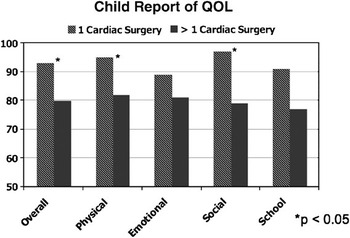
Replacement of the conduit adds unavoidable morbidity, albeit with low mortality. Reported freedom from replacement at 5 years ranges from 28% to 84%. A survey of both the patients receiving conduits and their parents demonstrated decreases in the markers for mental, physical, and overall quality of life. It also showed a significantly decreased overall quality of life for children requiring more than one cardiac operation compared to those requiring only a solitary surgical intervention (Fig. 11). There is also a trend for poorer markers of quality of life reported by the parents of those receiving conduits when compared to similar patients with transposition, and even compared to patients with functionally univentricular hearts.Reference Mussatto and Tweddell31
The table shows a significant decrease in physical, social and overall quality of life for children with greater than one cardiac operation compared to those undergoing a solitary surgical procedure.