Introduction
Eurypterids (Arthropoda, Chelicerata) are extinct aquatic chelicerates that achieved widespread geographic distribution between their origination in the Middle Ordovician and extinction at the end of the Permian (Tetlie, Reference Tetlie2007; Lamsdell et al., Reference Lamsdell, Briggs, Liu, Witzke and Mckay2015). Within the order are significant differences in the extent to which some taxa were dispersed, and this could be a reflection of relative swimming abilities, morphospace occupation, and the eurypterid marine–freshwater environmental transition that took place in the mid-Paleozoic (Lamsdell and Braddy, Reference Lamsdell and Braddy2010; Lamsdell and Selden, Reference Lamsdell and Selden2017). For example, in the transitional Silurian–Devonian, walking-legged stylonurines were confined to the paleocontinents that make up Laurussia (Tetlie, Reference Tetlie2007); most stylonurids known from this time occur in the USA and Scotland (eastern Laurentia), with only a few known from mainland Europe. A few Silurian–Devonian-age swimming eurypterine taxa are known from eastern Europe, the Baltics, Ukraine, and Russia (Plotnick, Reference Plotnick1983, Reference Plotnick1999; Tetlie, Reference Tetlie2007, and references therein), but stylonurines in this region are rare, having only been found (but not named) in Belarus (Plax et al., Reference Plax, Kruchek and Barbikov2009; Obukhovskaya et al., Reference Obukhovskaya, Kruchek, Pushkin, Nekryata, Plax, Sachenko, Obukhovskaya and Antipenko2010; Plax and Barbikov, Reference Plax and Barbikov2012, Reference Plax and Barbikov2013).
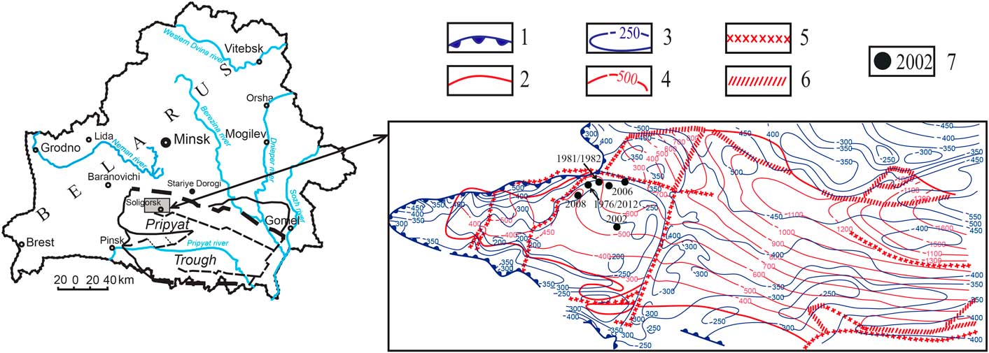
Over the last 40 years, several stylonurine specimens have been obtained during mining excavations in the Belarusian Starobin potassium-salt evaporite basin, all from within the Osovets Beds of the Upper Devonian Soligorsk Formation (Plax et al., Reference Plax, Kruchek and Barbikov2009; Fig. 1). These specimens are exceptionally preserved as carbonized, nearly fully articulated bodies (to 34 cm in length) and isolated appendages. Aside from eurypterids, the only other organismal remains known from these beds are rare terrestrial plants and local miospores. Although the occurrence and general morphology of the eurypterids in these potassium-salt deposits has been documented previously (Plax et al., Reference Plax, Kruchek and Barbikov2009; Obukhovskaya et al., Reference Obukhovskaya, Kruchek, Pushkin, Nekryata, Plax, Sachenko, Obukhovskaya and Antipenko2010; Plax and Barbikov, Reference Plax and Barbikov2012, Reference Plax and Barbikov2013), they have not yet been described within a detailed morphological or systematic context.
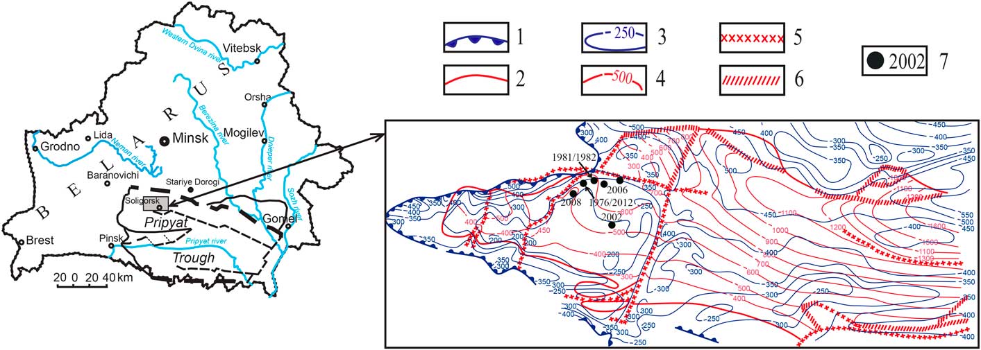
Map of Belarus and the area of study, and structural geology map of the Upper Devonian (middle–upper Famennian) evaporite deposits bounding the Soligorsk Formation. 1=distribution boundary of the halite deposits; 2=distribution boundary of the potassium salt (sylvinite) beds; 3=isohypses of the roof of halite deposits; 4=isohypses of the bottom of sylvinite deposits; 5=known rifts in salt deposits; 6=potential rifts in salt deposits; 7=sites of eurypterid discoveries (Soligorskopterus tchepeliensis n. gen. n. sp.) with years of discovery: 1976/2012, specimen lost/BKM 1052 (holotype); 1981/1982, BSUM P-276/P-170; 2002, specimen lost; 2006 (2 dots), BKM 942; 2008, BKM 943. See Table 1 for the exact latitude/longitudes of individual eurypterid occurrences. Modified from Plax et al. (Reference Plax, Kruchek and Barbikov2009).
In this paper, we present a comprehensive morphological description of these stylonurine eurypterids and describe their systematic and phylogenetic affiliation. All specimens are assigned to Soligorskopterus tchepeliensis new genus new species, the first eurypterid taxon formally described from Belarus. We consider the paleoecology and unusual preservation of S. tchepeliensis n. gen. n. sp. in this evaporitic lithofacies, and the paleogeography of stylonurids in the Late Devonian. The exceptional preservation of eurypterid material from this region provides a unique perspective on global eurypterid distributions at a critical time in their history.
Geological setting and stratigraphy
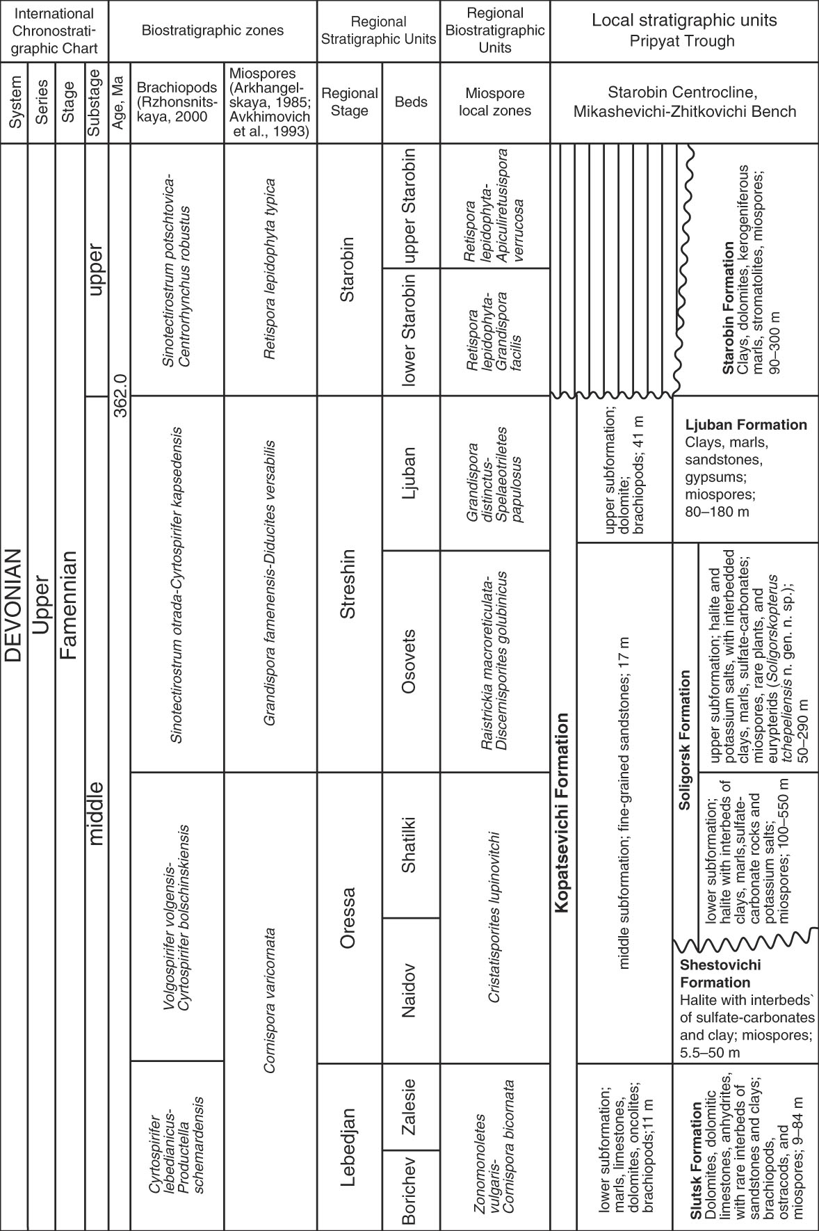
All specimens described herein were found in the Upper Devonian Soligorsk Formation (middle Famennian; Streshin regional stage; Osovets Beds) in the northwestern edge of the Pripyat Basin in the Pripyat Trough (or Rift) of Belarus (Kruchek et al., Reference Kruchek, Makhnach, Golubtsov and Obukhovskaya2001; Obukhovskaya et al., Reference Obukhovskaya, Kruchek, Pushkin, Nekryata, Plax, Sachenko, Obukhovskaya and Antipenko2010) (Fig. 2). The Pripyat Basin was formed during tectonic rifting that began before the middle Frasnian (ca. 369 Ma) and extended to the Famennian (367−364 Ma) (Garetsky et al., Reference Garetsky, Konishchev and Aizberg2001). As tectonic activity decreased in the Late Devonian, the area and water depth of the sedimentation basin was concomitantly reduced. An arid climate and repeated oceanic influxes into the continental margin at this time resulted in thick evaporite deposits, primarily composed of halite (NaCl) or potassium salt minerals (carnallite: KMgCl3·6H2O), that were later altered by subsurface brines to sylvite (KCl) or a mixture of halite and sylvite known as sylvinite (Konishchev et al., Reference Konishchev, Garetsky and Aizberg2001). These thick evaporite units are interbedded with thinner layers of carbonates, clays, and marls, as well as significant amounts of plant remains and miospores. The siliciclastic deposits and plant remains suggest that the evaporative and restricted environment in the basin was occasionally interrupted by periods of humidity and freshwater input from temporary creeks or small rivers in the basin that carried terrigenous sediments (Vysotsky et al., Reference Vysotsky, Garetsky and Kislik1988; Plax et al., Reference Plax, Kruchek and Barbikov2009).
Stratigraphy and biostratigraphic zonation of the Upper Devonian (middle–upper Famennian) Pripyat Trough of Belarus. Formation thicknesses are indicated in meters. See Table 1 for the subsurface depths at which individual specimens of Soligorskopterus tchepeliensis n. gen. n. sp. were discovered in the Soligorsk Formation. Adapted from Obukhovskaya et al. (Reference Obukhovskaya, Kruchek, Pushkin, Nekryata, Plax, Sachenko, Obukhovskaya and Antipenko2010).
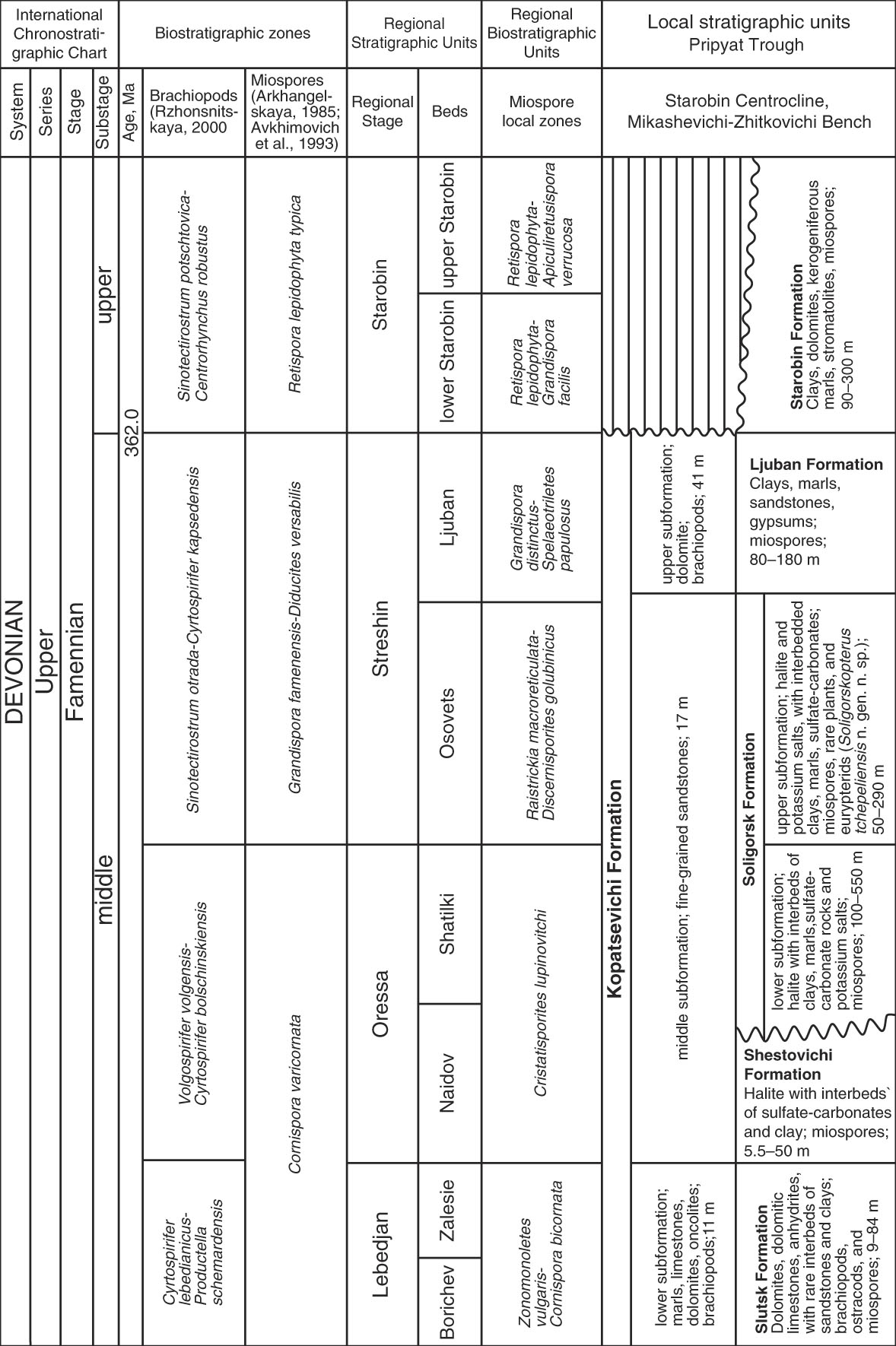
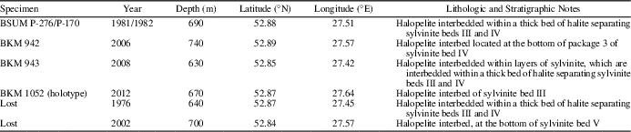
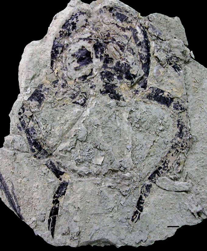
The subsurface Soligorsk Formation in the Storobin region is one of several evaporitic units formed during an oceanic influx and comprises five thick halite and sylvinite deposits, numbered I–V. Multiple specimens of a single eurypterid species, Soligorskopterus tchepeliensis n. gen. n. sp., were found during mining activity in the upper Soligorsk Formation, at 630–700 m depth, at several localities within an approximately three kilometer wide region (Table 1). Soligorskopterus tchepeliensis n. gen. n. sp. occurs as isolated, dark grey-black carbonized remains within thin, light gray to greenish halopelite (halite-bearing clay) beds interbedded within layers of potassium-salt minerals (carnallite, sylvite). The halopelite interbeds vary in thickness from a few millimeters to 10–15 cm and occur within thicker, multiyear, rhythmic halite and sylvinite beds (Garetsky et al., Reference Garetsky, Vysotsky and Kislik1984; Vysotsky et al., Reference Vysotsky, Garetsky and Kislik1988). In at least one collected specimen (BKM 1052; Fig. 3), a very thin halopelite bed containing a eurypterid lies directly adjacent to a thicker sylvite layer.
Stratigraphic and locality information for all specimens of Soligorskopterus tchepeliensis n. gen. n. sp. examined in this study.

Eurypterid specimens are oriented parallel to the bedding plane, and specimen completeness ranges from almost fully articulated exoskeletons to isolated tagma. The generally excellent preservation of the eurypterids was previously taken as evidence that they were not transported far prior to burial and thus represent an autochthonous assemblage (Plax et al., Reference Plax, Kruchek and Barbikov2009). Rare, carbonized, and unidentified terrestrial plant remains and miospores (Discernisporites Neves, Reference Neves1958, Retispora Staplin, Reference Staplin1960) have been found with the eurypterids at some localities in the formation and are the only other organic material to occur in this unit.
Materials and methods
Specimens were photographed using a Canon 600 D camera with a 24–105 mm objective lens. Interpretive drawings were prepared in CorelDRAW X3 (Corel Corporation, USA).
Eurypterid terminology largely follows Tollerton (Reference Tollerton1989) for morphology of the carapace, metastoma, lateral eyes, prosomal appendages, genital appendage, opisthosomal differentiation, telson, and patterns of ornamentation. The terminology of the ventral plate morphologies follows Tetlie et al. (Reference Tetlie2008). Terminology for prosomal structures and cuticular sculpture, and the labeling of the appendages, follows Selden (Reference Selden1981) with additions from Lamsdell et al. (Reference Lamsdell, Briggs, Liu, Witzke and Mckay2015). Minor modifications to the terminology used in these papers follows Lamsdell (Reference Lamsdell2011).
Repositories and institutional abbreviations
Types, figured, and other specimens examined in this study are deposited in the following institutions in Belarus: BKM, Trudovoy Slavy Museum, Soligorsk; BSUM, Geological Museum of Belarusian State University, Minsk.
Systematic paleontology
Arthropoda Von Siebold in Von Siebold and Stannius, Reference von Siebold and Stannius1848
Chelicerata Heymons, Reference Heymons1901
Eurypterida Burmeister, Reference Burmeister1843
Stylonurina Diener, Reference Diener1924
Stylonuroidea Diener, Reference Diener1924
Stylonuridae Diener, Reference Diener1924
Soligorskopterus new genus
urn:lsid:zoobank.org:act:46990205-E94F-4ACA-87D6-545DAEEB5FC5
Type species
Soligorskopterus tchepeliensis n. gen. n. sp., by monotypy.
Diagnosis
Stylonurid of medium size, 12–34 cm in length. Prosoma is horseshoe-shaped with a relatively narrow marginal rim. Compound lateral eyes are crescentic, located in the anterior half of the prosoma. Opisthosoma is wide, the metasoma bearing slight epimera, and tapering into a styliform telson. Prosomal appendages are robust, gradually increasing in length anteriorly to posteriorly. Prosomal appendages II−IV are spiniferous Ctenopterus-type; prosomal appendages V and VI are nonspiniferous Parastylonurus-type. The metastoma is petaloid in shape, markedly narrower than long with an angular anterior notch and a flattened posterior margin. Type A genital appendage is long, reaching beyond the fifth opisthosomal segment, and is composed of at least two segments with a median ridge. Type B genital appendage is short and narrow. The pretelson is elongated and bears a pair of ridges ventrally comprised of large overlapping scales. Cuticular ornament is in the form of small, contiguous or separate, semilunate, linguoid-shaped and acicular scales, which in some places transform into ridges and crests.
Etymology
The genus name is derived from the words Soligorsk (the town in Belarus closest to where the specimens were found) and pterus, meaning wing.
Remarks
Soligorskopterus n. gen. is differentiated from all other stylonurid taxa in possessing a prosomal shield and opisthosoma morphologically similar to Parastylonurus Kjellesvig-Waering, Reference Kjellesvig-Waering1966, but prosomal appendages reminiscent of Pagea Waterston, Reference Waterston1962 and Laurieipterus Kjellesvig-Waering, Reference Kjellesvig-Waering1966.
Soligorskopterus tchepeliensis new species
urn:lsid:zoobank.org:act:A0A05255-FE4B-42F0-8CDF-26F1FBFE8AE7
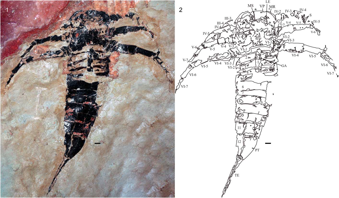
Soligorskopterus tchepeliensis n. gen. n. sp., holotype, BKM 1052, specimen preserving the ventral surface: (1) specimen photograph (note the pink-red sylvinite directly adjacent to and under the specimen); (2) interpretive drawing. GA, genital appendage; LE, lateral eyes; M, metastoma; MR, marginal rim; MS, median suture; PT, pretelson; S, spines; TE, telson; VP, ventral plate; 1–12, sternites. Prosomal appendages are labeled with Roman numerals (I−VI), and individual podomeres are labeled with Arabic numerals (1−9), ordered proximally to distally. Scale bar=10 mm.

Soligorskopterus tchepeliensis n. gen. n. sp., paratype BKM 943; specimen preserving the ventral surface: (1) specimen photograph; (2) interpretive drawing. Abbreviations as in Figure 3. Scale bar=10 mm.

Soligorskopterus tchepeliensis n. gen. n. sp., paratype BSUM P-276/P-170; prosoma with appendage pair VI viewed from the ventral surface. Scale bar=10 mm.

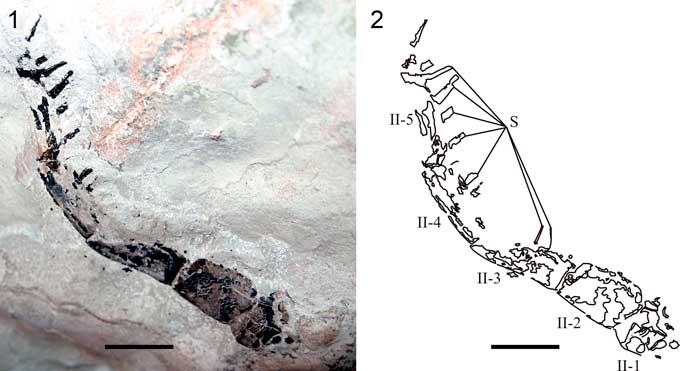
Soligorskopterus tchepeliensis n. gen. n. sp., paratype BKM 942; prosomal appendage II: (1) specimen photograph; (2) interpretive drawing. Abbreviations as in Figure 3. Scale bar=10 mm.

Type specimens
Holotype, BKM 1052, Upper Devonian (middle Famennian), upper Soligorsk Formation, Pripyat Basin in the Pripyat Trough of Belarus. Paratypes, BKM 942, 943; BSUM P-276/P-170.
Diagnosis
As for the genus.
Occurrence
Upper Devonian (middle Famenian, Streshin regional stage), upper Soligorsk Formation (Osovets beds), near Soligorsk, Belarus.
Description
The holotype (BKM 1052; Fig. 3) is the largest specimen of Soligorskopterus tchepeliensis n. gen. n. sp. recovered, with a length of 34 cm, and represents a mostly complete individual preserved in ventral aspect. The prosoma (71 mm in length; 80 mm in width; length/width ratio 0.89) is horseshoe-shaped with a comparatively narrow (3 mm) marginal rim. The lateral eyes are crescentic, located in the anterior half of the prosoma. The prosomal ventral plates are of Eurypterus-type, bearing a single median suture. Transverse sutures are evident on the ventral plates, expanding forward anteriorly. The prosomal appendages are incomplete, with appendages III–VI evident on the left and appendages IV–VI evident on the right. On appendage III, only the third and fourth podomeres are fragmentarily preserved, largely in the form of an imprint. The appendages of limb pair IV are also preserved fragmentarily, being known from the second, third, fourth, and fifth podomeres. Podomeres four and five bear multiple rows of fixed spines. The cuticular ornament of these podomeres consists of weakly expressed small scales. Appendages V and VI are better preserved than the previous appendage pairs. Appendage V is known from the coxa to the seventh podomere and is nonspiniferous. Appendage VI, also known from the coxa to the seventh podomere, also lacks spines. The fourth podomere of appendage VI is the longest, with subsequent podomeres reducing in length consecutively. Both appendages exhibit cuticular ornamentation of a row of small, closely situated semilunate scales along the anterior third of each podomere. The lateral margins of the podomeres are also ornamented with a series of thickened scales. The metastoma is petaloid in shape with an angular anterior notch and a flattened posterior margin.
The opisthosoma is generally broad but does not exceed the maximum width of the prosomal carapace. The opisthosoma consists of 12 segments, divided between the six-segmented mesosoma anteriorly and the six-segmented metasoma posteriorly. The mesosoma is of generally constant width, with the metasoma narrowing from the seventh opisthosomal segment onward. Because the specimen is preserved in ventral aspect, the dorsal tergites of the mesosoma are not seen; however, the ventral structures of the genital appendage and opisthosomal opercula are evident. The genital operculum comprises a clear median and posterior opercular plate, with evidence of a narrow anterior opercular plate provided through imprints within the sediment. The median opercular plate is 9 mm long, with a posterior width of 70 mm, whereas the posterior opercular plate has a length of 12 mm and a posterior width of 76 mm. A large Type A genital appendage is present (8 mm wide, at least 38 mm long), extending to at least the fourth opisthosomal segment. A deltoid plate is apparent to the right of the insertion of the genital appendage into the median opercular plate. The genital appendage itself has a carina running along its length and is composed of at least two segments, with the division occurring slightly posterior to the posterior margin of the third opisthosomal segment. The first postgenital operculum (corresponding to the third opisthosomal segment) is 13 mm in length with a posterior width of 75 mm. The operculum of the fourth segment has a length of 13 mm with a posterior width of 74 mm, that of the fifth segment has a length of 14 mm and a posterior width of 74 mm, and that of the sixth segment has a length of 15 mm and a posterior width of 71 mm.
The metasoma is represented by the sternites, which are fused laterally to the tergites to form ankylosed segments. The seventh segment has a length of 11 mm and a posterior width of 69 mm; the eighth segment has a length of 15 mm and a posterior width of 68 mm; the ninth segment has a length of 16 mm and a posterior width of 61 mm; the tenth segment has a length of 17 mm and a posterior width of 54 mm; the eleventh segment has a length of 21 mm and a posterior width of 45 mm. The pretelson is elongated, having a length of 40 mm and a posterior width of 20 mm, resulting in a length/width ratio of 2.0. The posterolateral margins of each metasomal segment are produced into short epimera. The epimera of the pretelson are reduced to appear almost absent. The cuticular sculpture of the metasomal sternites consists of small, closely situated semilunate scales. These scales exponentially increase in size (from 0.4–0.5 mm to 5 mm) on sternites 10–12, with some becoming acicular and forming a pair of crests running in parallel flanking the center of the sternites.
The telson is styliform and 73 mm in length. Where the cuticle has worn away impressions of the sediment reveal the presence of a moderate dorsal keel. The ventral surface of the telson is shown to bear a continuation of the enlarged scale ridge that runs down the center of the telson, with the scales progressively decreasing in size toward its tip.
The first paratype (BKM 943; Fig. 4) is an almost intact specimen in ventral view preserving the opisthosoma and portions of the prosomal appendages and metastoma. The total length of the specimen is 25 cm. The prosoma is fragmentary, preserving some evidence of a narrow marginal rim and the ventral plate and its transverse suture. The prosomal appendages are incomplete, with only the proximal podomeres of appendages IV–VI preserved. Only the third and fourth podomeres of appendage IV are preserved, but they are fragmentary and do not preserve the armature. Appendages V and VI are both known from the coxa to the fifth podomere. Both appendages show the characteristic lateral border of thickened scales. The metastoma is petaloid, with an anterior notch and a truncated, flattened posterior. The lateral margins of the metastoma are remarkably straight, resulting in an almost rectangular appearance. A slight median ridge runs down the center of the metastoma. Small semilunate scales are present on the metastoma and the coxae.
The opisthosoma is relatively broad and tapers regularly throughout the metasoma. The mesosoma is generally well preserved. It is somewhat wider than the metasoma. The genital operculum is clearly composed of an anterior opercular plate, a median opercular plate (corresponding to the first opisthosomal segment), and a posterior opercular plate (corresponding to the second opisthosomal segment). The median opercular plate is 8 mm in length and 73 mm in width at the base, whereas the posterior opercular plate is 9 mm in length and 75 mm in width at the base. A clear suture is present between them. Both opercular plates have an ornament of small semilunate scales, whereas the posterior opercular plate also has acicular scales. A type B genital appendage is present, oval and elongated in shape, with a length of 13 mm and a width of 3 mm. The terminal part of the genital appendage is missing, and so it is impossible to ascertain how many segments it comprises. There are no deltoid plates evident alongside the insertion of the genital appendage, despite the region being well preserved.
The remaining opercula are generally uniform in morphology. The operculum of the third segment is 10 mm in length with a posterior width of 77 mm; that of the fourth segment is 11 mm in length with a posterior width of 77 mm; the fifth segment is 12 mm in length with a posterior width of 71 mm; and the sixth operculum is 12 mm in length and 64 mm in width at the base. The cuticular sculpture of the opercula comprises small semilunate and acicular scales. A median furrow is present on all the postgenital opercula, shallowing out just before the posterior margin of the sixth operculum. The opercula of segments 4–6 appear medially fused, however those of the third opisthosomal segment exhibit a median notch where they join, which indicates they might have been separate or only partially fused.
The metasoma is preserved in slightly rougher condition than the mesosoma. The metasomal segments narrow regularly posteriorly: the seventh segment is 14 mm in length, 62 mm in width across its posterior; the eighth segment is 14 mm in length, 58 mm in width; the ninth segment is 15 mm in length, 55 mm in width; the tenth segment is 16 mm in length, 50 mm in width; the eleventh segment is 18 mm in length, 40 mm in width; and the pretelson is 32 mm in length, 18 mm in width. The cuticular ornament of the metasomal sternites comprises small semilunate and acicular scales, which enlarge and form paired ridges on segments 10–12. Small epimera are present at the posterolateral margins of the metasomal segments. The telson is poorly preserved and present as only a few small fragments of cuticle posterior to the pretelson.
The second paratype (BSUM P-276/P-170; Fig. 5) is represented by a fragmentary prosoma comprising the outline of the prosomal carapace with the prosomal appendage coxa, metastoma, and a complete paired appendage VI. The prosoma is horseshoe-shaped with a length of 75 mm and a width of 83 mm, resulting in a length/width ratio of 0.90. The narrow 2–3 mm wide marginal rim extends around the anterior and lateral edge of the carapace and tapers to its posterior. The metastoma is petaloid in shape, narrow, with relatively straight sides and a shallow anterior notch. The cuticle at its posterior is absent but imprints in the sediment suggest it had a truncated, flattened posterior margin. Paired appendage VI is preserved in its entirety, revealing a narrow coxa with no anteriorly expanded ear. The appendages are long and nonspiniferous, with the fourth podomere being the longest and all subsequent podomeres shortening consecutively. No modified podomere 7a is present. Distal podomeres are not expanded into a swimming paddle. The anterodistal margin of podomere VI-8 is drawn out into a long projection, extending for three-fifths the length of podomere VI-9. The ornamentation on the appendages comprises small semilunate scales, with a thickened border of scales along the lateral podomere margins. Dense scale ornament is also visible on the coxa.
The final paratype (BKM 942; Fig. 6) is a partially exfoliated but fairly complete prosomal appendage II. The appendage tapers in width from 10 mm proximally to 3 mm distally; its total length is 75 mm and it is comprised of six podomeres, including the coxa. The coxa and second podomere are devoid of armature, whereas podomeres II-3–II-5 bear numerous short (5–8 mm) and thin (0.9–1.1 mm) spines, at least five per podomere. These spines bear slight striations running along their length. The surface of the first two podomeres of the appendage has sculpture in the form of dense semilunate and acicular scales.
Etymology
The species name is derived from the village of Tchepeli, near where one of the eurypterid specimens was found.
Remarks
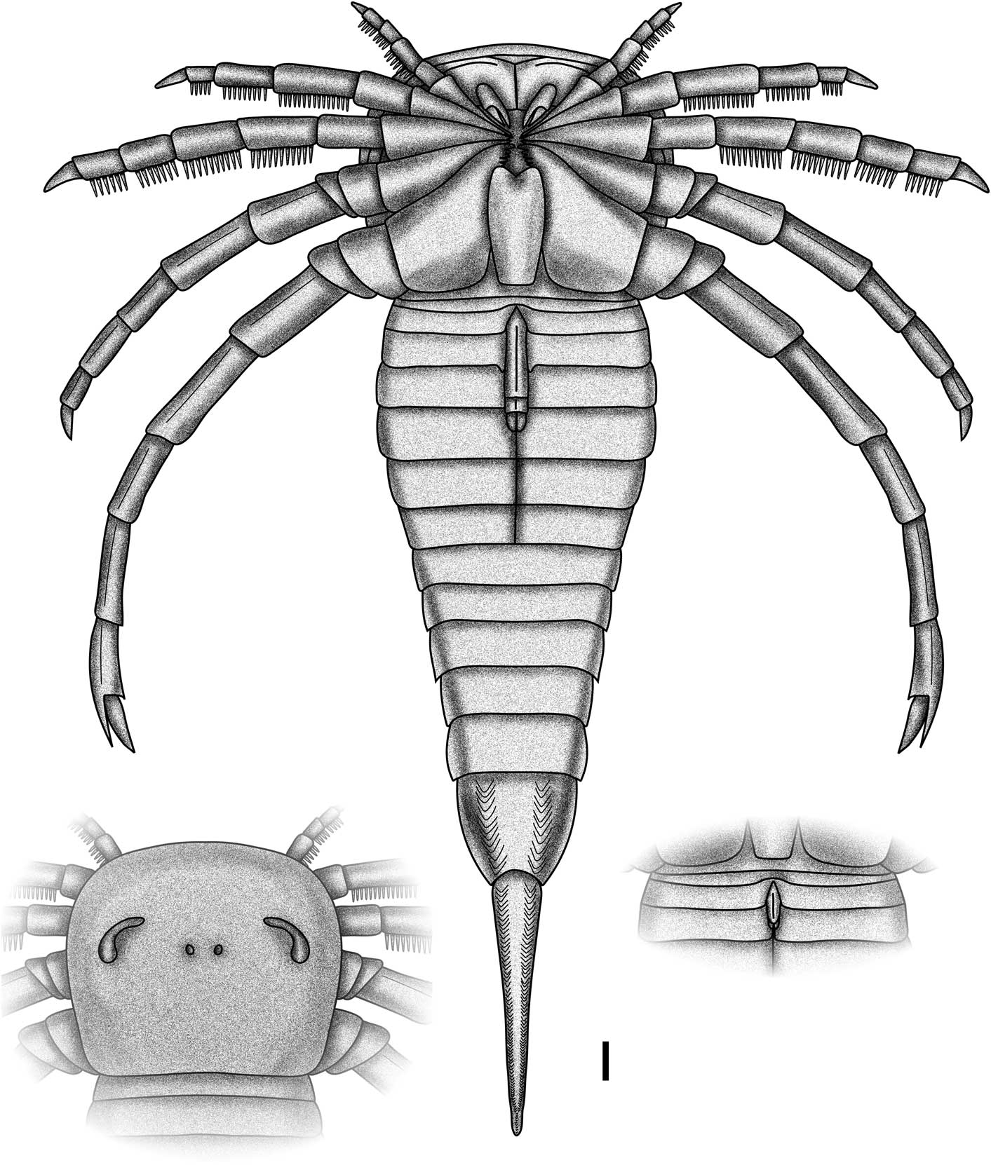
The new eurypterid (Fig. 7) exhibits a number of characteristics that place it firmly within the stylonurines. The most obvious of these is the retention of prosomal appendage VI as a walking limb lacking a modified podomere 7a, however the assignment is also supported by the lack of an expanded ear on coxa VI, the transverse suture on the prosomal ventral plates, and the possession of a visible anterior opercular plate (Lamsdell et al., Reference Lamsdell, Braddy, Loeffler and Dineley2010b). Within the Stylonurina, Soligorskopterus n. gen. exhibits closest affinities to the Stylonuroidea, lacking as it does the spiniferous appendage V characteristic of Kokomopteroidea (Tetlie, Reference Tetlie2008; Lamsdell et al., Reference Lamsdell, Braddy, Loeffler and Dineley2010b) and the cleft posterior metastoma and sweep-feeding modifications seen in Mycteropoidea (Lamsdell et al., Reference Lamsdell, Braddy and Tetlie2009; Lamsdell, Reference Lamsdell2013). The multiple spines per podomere on appendages II–IV also preclude the new species from assignment to Rhenopteroidea (Lamsdell et al., Reference Lamsdell, Braddy and Tetlie2010a). Multiple spines per podomere are, however, known from some members of the Stylonuroidea. An assignment to Stylonuroidea is further supported by the flattened posterior of the metastoma in Soligorskopterus n. gen.
Reconstruction of Soligorskopterus tchepeliensis n. gen. n. sp. Complete ventral reconstruction showing type A genital appendage, flanked by dorsal view of the prosoma and ventral view of the type B genital appendage. Scale bar=10 mm.

Within Stylonuroidea, Soligorskopterus n. gen. bears closest similarity to Parastylonurus (Waterston, Reference Waterston1979), sharing a number of characteristics that are apparently plesiomorphic for the clade. The prosomal carapace in Parastylonurus and Soligorskopterus n. gen. has a more rounded outline, being horseshoe-shaped rather than quadrate as in species of Pagea (Waterston, Reference Waterston1962), Stylonurus Page, Reference Page1856 (Woodward, Reference Woodward1872), and Stylonurella Kjellesvig-Waering, Reference Kjellesvig-Waering1966 (Waterston, Reference Waterston1979), spatulate as in Laurieipterus (Waterston, Reference Waterston1962), or triangular as in Ctenopterus Clarke and Ruedemann, Reference Clarke and Ruedemann1912. The prosomal ventral plates are of the Eurypterus-type as in Parastylonurus and Stylonurella rather than possessing an epistoma as in Laurieipterus. The metastoma is generally narrow and straight-sided as in Parastylonurus and Pagea, quite unlike the broad metastoma of Laurieipterus. As in Pagea and Parastylonurus, Soligorskopterus n. gen. has a slight median ridge on the metastoma; however, the anterior notch is more reminiscent of Parastylonurus rather than the flattened anterior margin present in Pagea.
Soligorskopterus n. gen. differs from Parastylonurus primarily in the armature of prosomal appendages II–IV, which bear multiple spines per podomere and are of the Ctenopterus-type. This appendage morphology is shared with Laurieipterus, Pagea, and Ctenopterus, the appendages of Parastylonurus and Stylonurella only having a single pair of fully-developed spines per podomere. The spines of Soligorskopterus n. gen. are all equal in length along each podomere and so more closely resemble the armature of Lauriepterus and Ctenopterus than that of Pagea. The spines bear longitudinal striations, as do those of Laurieipterus and Stylonurella. The structure of prosomal appendages V and VI in Soligorskopterus n. gen. closely resemble those of Parastylonurus, with a row of enlarged scales present on the anterior third of each podomere and with each podomere possessing a margin of thickened overlapping scales. This type of appendage ornament is shared with Stylonurus, Laurieipterus, and Stylonurella. However, Parastylonurus and Soligorskopterus n. gen. are distinct in having the anterodistal margin of podomere VI-8 developed into a long spur. Soligorskopterus n. gen. differs from Parastylonurus in lacking the crenulate distal podomere margins.
The available material of the genital appendages in Soligorskopterus n. gen. reveals them to be almost identical to those in Parastylonurus, with the type A appendage being large, composed of at least two segments, and possessing a median ridge running along its length. The type B appendage is short, narrow, and also has a median ridge. The only other genital appendage known from Stylonuroidea is the type B genital appendage of Stylonurella, which agrees in overall morphology with those of Parastylonurus and Soligorskopterus n. gen. The postgenital opercula of both Soligorskopterus n. gen. and Parastylonurus also have small median keels running along their lengths. The overall shape of the opisthosoma is broad as in Parastylonurus and Stylonurus, although never expanding beyond the width of the prosomal carapace. The metasomal segments of Soligorskopterus n. gen. are extended into epimera, although they are short projections as in Pagea and not elongated like those of Parastylonurus. The epimera do, however, exhibit the same terraced ornamentation as observed in Parastylonurus. An elongate pretelson is common between Soligorskopterus n. gen., Parastylonurus, and Pagea, with Ctenopterus and Stylonurus having the regular, short pretelson morph. The general cuticular ornamentation, consisting of a mixture of semilunate and acicular scales, is also consistent between Soligorskopterus n. gen. and Parastylonurus. The scale ridges on the pretelson of Soligorskopterus n. gen. are not known from any other stylonuroid eurypterid and appear to be an autapomorphy for the genus; however, they could be an indication of trilobation as seen in Pagea.
Soligorskopterus n. gen. appears to be an intermediate between the traditionally considered parastylonurid (Parastylonurus and Stylonurella) and stylonurid (Stylonurus, Pagea, Ctenopterus, and Laurieipterus) groups. Parastylonurids are paraphyletic, with Stylonurella resolving closer to the stylonurids and Parastylonurus forming the sister taxon to all other stylonuroids (Lamsdell et al., Reference Lamsdell, Braddy and Tetlie2010a, Reference Lamsdell, Braddy, Loeffler and Dineley2010b). Soligorskopterus n. gen. likely resolves intermediately between Stylonurella and the Stylonurus/Pagea + Laurieipterus/Ctenopterus clade due to the armature of appendages II–IV comprising multiple spines per podomere (as in the Stylonuridae) and the morphology of appendage VI and the horseshoe-shaped carapace (which are parastylonurid in form).
Paleoecology of Soligorskopterus n. gen
The evaporitic nature of the eurypterid-bearing halopelite and the close proximity of the eurypterid-bearing beds to thick evaporite layers suggests deposition in a hypersaline environment. Moreover, potassium salts (e.g., carnallite, sylvite) are some of the last evaporite minerals to precipitate out of solution in marine settings (Warren, Reference Warren1997), and therefore the occurrence of one specimen (BKM 1052; Fig. 3) on a thin lamina of halopelite that is immediately adjacent to a sylvite layer would suggest deposition in or near a supersaline seawater brine. Yet despite the occurrence of Soligorskopterus tchepeliensis n. gen. n. sp. in this extreme environment, it is unlikely that this setting is representative of its preferred life habitat. Eurypterids, including stylonurids, in the Late Devonian were almost entirely constrained to freshwater habitats (Lamsdell and Braddy, Reference Lamsdell and Braddy2010). In addition, a previous study of eurypterids preserved in evaporitic settings has demonstrated that the onset of hypersaline conditions (and any subsequent formation of associated intrasedimentary evaporites) occurred after burial of the eurypterid specimens, and thus are not reflective of life habitat (Vrazo et al., Reference Vrazo, Brett and Ciurca2016).
Eurypterids have been suggested to congregate in sheltered regions to molt (Braddy, Reference Braddy2001; Vrazo and Braddy, Reference Vrazo and Braddy2011), and it is possible that the specimens of Soligorskopterus n. gen. are shed exuvia of individuals molting within a nearshore lagoon or tidal flat prior to the onset of hypersaline conditions. However, the majority of specimens are well articulated and do not exhibit any of the characteristic patterns of disarticulation or contortion characteristic of exuvia (Tetlie et al., Reference Tetlie, Brandt and Briggs2008; McCoy and Brandt, Reference McCoy and Brandt2009), and thus it seems more likely that the specimens are allochthonous carcasses transported into the environment of preservation following death. The co-occurrence of terrestrial plant material with the eurypterids indicates some terrestrial influence on the paleoenvironment and it is probable that the carcasses of Soligorskopterus n. gen. were transported into the Soligorsk environment via freshwater streams. Additionally, the general lack of disarticulation suggests that the individuals were only recently dead when transport occurred. Modern horseshoe crab mortalities have been shown to float intact for up to two days after death before settling on the sediment surface (Babcock et al., Reference Babcock, Merriam and West2000), facilitating the transport potential of recent carcasses (see also Allison, Reference Allison1986). Alternatively, living Soligorskopterus n. gen. might have been swept into the hypersaline lagoon during storm events before expiring in the toxic environment—a similar scenario to that suggested for occurrences of the horseshoe crab Mesolimulus Størmer, Reference Størmer1952 in the Upper Jurassic Solnhofen Limestone in Germany (Lomax and Racay, Reference Lomax and Racay2012). However, a large influx of meteoric water would likely have lowered the salinity of the lagoon at the same time that the eurypterids were swept in, and hence it seems more likely that the specimens were transported into the lagoon soon after death. Once in this setting, burial of the arthropod carcasses in fine-grained clays, and the onset of hypersaline conditions in the water column or subsurface, would have been inimical to growth of decay bacteria and conducive to long-term preservation (Seki and Taga, Reference Seki and Taga1963; McMahon et al., Reference McMahon, Anderson, Saupe and Briggs2016). The occurrence of Soligorskopterus n. gen. in what appear to be semiregular intervals in the Soligorsk Formation could also reflect a broader sequence stratigraphic control on preservation (see Vrazo et al., Reference Vrazo, Brett and Ciurca2017), and it is clear that further sedimentological and stratigraphic work is needed to fully understand the environmental factors that facilitated eurypterid preservation in this evaporitic lithofacies.
There is no direct evidence for the diet of Soligorskopterus n. gen.; however, analysis of the visual system of Rhenopterus diensti Størmer, Reference Størmer1936, a stylonurine eurypterid from the Early Devonian of Germany, indicates that at least some stylonurines had a visual acuity comparable to that of modern horseshoe crabs (Poschmann et al., Reference Poschmann, Schoenemann and McCoy2016). It is unlikely that Soligorskopterus n. gen. would have been an active predator like certain swimming eurypterids (Anderson et al., Reference Anderson, McCoy, McNamara and Briggs2014; McCoy et al., Reference McCoy, Lamsdell, Poschmann, Anderson and Briggs2015). Soligorskopterus n. gen. most likely had a diet like that of modern horseshoe crabs, consisting of small invertebrates (ostracods, gastropods, bivalves, polychaetes, etc.; Botton and Haskin, Reference Botton and Haskin1984). The morphology of appendages II–IV, with their rows of multiple spines per podomere, indicates that Soligorskopterus n. gen. was a dragnet-style sweep-feeder (Lamsdell et al., Reference Lamsdell, Braddy, Loeffler and Dineley2010b) and would have raked the spines of these appendages through the sediment to entangle and excavate partially buried prey.
Eurypterid paleogeography and patterns of extinction
Stylonuroidea was until now known with confidence from the lower Silurian (Llandovery) to the Lower Devonian (Pragian), with Ctenopterus (?) beecheri (Hall, Reference Hall1884), a species of questionable taxonomy, potentially extending the range of the group into the Famennian (Lamsdell and Braddy, Reference Lamsdell and Braddy2010). Soligorskopterus tchepeliensis n. gen. n. sp. is the first confirmed occurrence of Stylonuroidea in the Famennian and as such extends the age range of the clade some 50 million years.
The extension of Stylonuroidea into the Famennian means that four clades (Mycteropoidea, Kokomopteroidea, Stylonuroidea, and Adelophthalmoidea) persisted beyond the major drop in eurypterid diversity in the Early Devonian and into the Late Devonian biotic crisis (Lamsdell and Selden, Reference Lamsdell and Selden2017). However, each of these clades are represented by only a few species (in the case of adelopthalmoids and mycteropoids) or a single species (for kokomopteroids and stylonuroids), and the discovery of Soligorskopterus n. gen. does not suggest that Late Devonian eurypterid diversity was significantly higher than previously suggested. It is noteworthy that three of these clades are members of Stylonurina, which are consistently lower in diversity than the swimming Eurypterina and tend to have a more cryptic fossil record (Lamsdell et al., Reference Lamsdell, Braddy, Loeffler and Dineley2010b). The fact that the majority of Late Devonian eurypterids are stylonurines—including the new species described here—supports existing hypotheses regarding the global collapse of eurypterid diversity during the Devonian (Lamsdell and Selden, Reference Lamsdell and Selden2017). The nektonic and nektobenthic Eurypterina were particularly hard hit by the biotic crisis, dwindling exponentially in diversity from the latest Silurian through to the Middle Devonian. The survivors were relegated to the margins of eurypterid niche space and invaded continental freshwater ecosystems (Lamsdell and Braddy, Reference Lamsdell and Braddy2010; Lamsdell and Selden, Reference Lamsdell and Selden2017), although the exact timing of this transition is still uncertain (Vrazo et al., Reference Vrazo, Brett and Ciurca2017).
The morphological disparity of eurypterids has also been demonstrated to decrease over the course of the Devonian, with an associated reduction of morphospace occupation (Lamsdell and Selden, Reference Lamsdell and Selden2017). The discovery of Soligorskopterus n. gen. does not alter these patterns in disparity and morphospace occupation. Stylonuroid morphospace almost wholly overlaps with the kokomopteroid and mycteropoid morphospace, and as such does not expand the occupied morphospace of Famennian eurypterids. Soligorskopterus n. gen. does, however, expand the geographic occurrence of eurypterids during the Famennian. Previous records of Famennian eurypterids are known from western and central Laurussia and largely comprise stylonurines from the continental United States (Hall, Reference Hall1884; Ehlers, Reference Ehlers1935; Tetlie, Reference Tetlie2008), with a single stylonurine known from southwestern England (Lamsdell et al., Reference Lamsdell, Braddy and Tetlie2009) and another from Belgium (Fraipoint, Reference Fraipoint1889). The eurypterine genus Adelophthalmus Jordan in Jordan and von Meyer, Reference Jordan and von Meyer1854 is also known from the Famennian of the USA (Hall and Clarke, Reference Hall and Clarke1888) and Belgium (Fraipoint, Reference Fraipoint1889). Soligorskopterus n. gen. therefore extends the occurrence of Famennian eurypterids into eastern Laurussia. Despite Famennian eurypterids only being known from Laurussia, however, it is likely that they had a global distribution. In the Frasnian, eurypterids are known from both Laurussia (Ruedemann, Reference Ruedemann1921) and Gondwana (Tetlie et al., Reference Tetlie, Braddy, Butler and Briggs2004), and the same is true for the Tournasian (Waterston, Reference Waterston1985; Tetlie and Poschmann, Reference Tetlie and Poschmann2008).
Whatever the cause of the eurypterid shift from marine to freshwater habitats during the Devonian, it seems likely to have been the result of widespread, global pressure rather than an inherited trait through the chance survival of a few eurypterid groups within a single biogeographic region. As supported by phylogenetic information (Lamsdell and Selden, Reference Lamsdell and Selden2017), the concerted, independent habitat shift among eurypterids during this time period represents a case of macroevolutionary convergence driven by a selective trend (Lamsdell et al., Reference Lamsdell, Congreve, Hopkins, Krug and Patzkowsky2017; Congreve et al., Reference Congreve, Falk and Lamsdell2018). Such data are integral to expanding our understanding of biotic responses to environmental processes in the Late Devonian; further studies into other contemporaneous clades will undoubtedly refine our understanding of the events underpinning the biotic crisis that characterizes the end of the period (Stigall, Reference Stigall2010, Reference Stigall2012).
Acknowledgments
We are grateful to the miners of the Republican Unitary Enterprise of Production Amalgamation ‘Belaruskali’ who observed and saved the eurypterid specimens described in this study. B. Granovskiy is thanked for Russian translations. G. Andrews (West Virginia University) is thanked for converting Corel Draw files to PDF format. We thank J. Dunlop (Museum für Naturkunde, Berlin) and an anonymous referee for comments that improved the quality of the manuscript.