Isolated perimembranous ventricular septal defect is one of the most common congenital heart defect.Reference Hoffman 1 Although transcatheter device closure of the perimembranous ventricular septal defect is emerging as an effective and safe alternative to surgery, it is complicated by significant conduction disturbances. After initial disappointing reports of very high incidence of conduction disturbances following transcatheter perimembranous ventricular septal defect closure in young children with asymmetric device, recently there has been a renewed enthusiasm for transcatheter closure of perimembranous ventricular septal defect. Large-scale studies published in the later half of the last decade revealed very low incidence of complete atrioventricular block; however, their inclusion criteria precisely enrolled either children or adolescents. Although the incidence of complete atrioventricular block in these studies was as high as 1.82%, nearly 75% patients responded to steroids within 3–4 weeks, and permanent pacemaker implantation was required in only 0.25 to 0.35% of patients.Reference Yang, Li and Yang 2 – Reference Yang, Kong and Sheng 4 Empirical therapy with corticosteroid or non-steroidal anti-inflammatory drugs is reported to be effective when conduction disturbances occur in the early post-procedure period, and occasionally partially effective in late-presentation complete atrioventricular block;Reference Yang, Li and Yang 2 , Reference Zhou, Pan and Guan 3 , Reference Masura, Gao and Gavora 5 – Reference Yip and Zimmerman 8 however, the efficacy of prophylactic oral steroids is yet to be determined.
Materials and methods
Patient population and study design
This study was conducted as a prospective, single-centre observational study. All the patients were screened with transthoracic echocardiography in the long-axis parasternal view and apical five-chamber view to evaluate the rim under the aortic valve. Between 2007 and 2012, 297 patients with perimembranous ventricular septal defect, who accepted transcatheter closure using different devices, were considered for the study. The ethics committee of our institute approved the study, and informed written consent was obtained from each patient’s parents before the procedure.
Inclusion criteria for the study were as follows: children aged ⩾3 and ⩽18 years, maximum diameter of the defect ⩽12 mm by transthoracic echocardiography, and indication of transcatheter closure including ventricular septal defect-associated symptoms, haemodynamic changes – cardiomegaly and left atrium–left ventricle volume overload verified by echocardiography – or history of infective endocarditis.
Exclusion criteria for the study were as follows: body weight <10 kg, sub-aortic rim as shown by echocardiography in the five-chamber apical view <2 mm, aortic regurgitation or aortic valve prolapse, mean pulmonary artery pressure ⩾70 mmHg, and patient with contraindications to anti-platelet therapy. Patients with residual post-operative ventricular septal defect were excluded from the study.
Occlusion devices and occluder selection
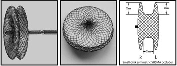
The devices used in this study were either symmetric double-disc perimembranous ventricular septal defect occluders (Block Aid-SHSMA, Shanghai Shape Memory Alloy Ltd, Shanghai and Cardi-o-fix, Starway Pvt Ltd, Beijing, China) or Amplatzer Duct Occluder I (AGA Medical Corporation, MN, USA). The perimembranous ventricular septal defect occluder is a self-expandable, double-disc (2-mm retention skirt) implant device (Fig 1). These devices have a woven mesh of 72% Nitinol (0.004–0.005-inch) wires with shape memory and a polyester mesh inside. The two discs are linked together by a short connecting waist corresponding to the size of the ventricular septal defect, and the waist thickness measures 2.5 mm. The inter-disc distance – waist thickness – is relatively smaller (1.5 mm) in the Amplatzer perimembranous device. Device sizes are available from 4 to 20 mm in 1-mm increment. The Amplatzer Duct Occluder is a cone-shaped device, 7 mm in length, made of a 0.004′′ nitinol wire mesh with a 2-mm retention skirt extending radially around the distal part of the device. It is available in sizes ranging from 6–4 to 14–12 mm at increments of 2 mm. The ventricular septal defect diameter was measured by transthoracic echocardiography and/or left ventricle angiography, and the selected occluder device was over-sized by 1–2 mm.
Symmetric disc devices used in the majority of patients in our study.

Procedure
The procedure has been reported previously in detail.Reference Gu, You and Zhao 9 In brief, the procedure was performed under general anaesthesia with transthoracic echocardiography and fluoroscopic guidance. After establishing femoral vascular accesses, heparin (100 units/kg) was infused intravenously. Left ventricle angiography was performed in two views – that is, the left anterior oblique 40/Cranial 25 and the left anterior oblique 60/Cranial 20 – to precisely define the perimembranous ventricular septal defect and the aortic rim. A device, 2 mm larger than the size of the ventricular septal defect, was selected for occlusion. Antegrade device closure was attempted using wire-maintaining technique in all patients. Left ventricle angiography was performed in all patients before and after delivery of the device to ensure the closure of the perimembranous ventricular septal defect.
All the patients received prophylactic steroid therapy consisting of 2 weeks of oral prednisolone (1 mg/kg/day) initiated immediately after the procedure independent of electrocardiography rhythm. In the event of bradyarrhythmia, the dose was escalated to 2 mg/kg/day for minimum 4 weeks, including a tapering dose over 2 weeks after reversal to sinus rhythm. After the 1st day of ICU care, cardiac rhythm was monitored daily by electrocardiography in the inpatient setting. Excluding patients with major complications, all the patients were discharged after 5 days of uneventful course and were advised to continue oral aspirin (5 mg/kg/day) for 6 months and tapering dose of oral steroids for 1–2 weeks depending on the evidence of new conduction disturbances. Patients were regularly followed-up at 1, 6, and 12 months and then annually for clinical, electrocardiography, and echocardiographic evaluation. Patients with post-procedure heart blocks underwent Holter monitoring after a minimum of 1 year interval.
Statistical analysis
Data were expressed as frequency or percentage for nominal variables, as the median (range) for categorical variables, and as mean±SD or median (range) for continuous variables.
Result
Demographics and clinical data
Between May, 2007 and August, 2012, 297 (127 females) eligible patients underwent an attempt at transcatheter device closure. The mean age and weight of the study patients were 9±3.12 years (3–17 years) and 21±8.27 (10–63 kg), respectively. Clinical symptoms related to ventricular septal defect were present in133 (46%) patients; two patients had a history of bacterial endocarditis. Pre-procedure resting electrocardiography revealed findings of either prominent left ventricle or biventricular forces in 19% of the patients; four patients had incomplete right bundle branch block before device closure.
The mean size of the perimembranous ventricular septal defect as measured by transthoracic echocardiography was 5±1.4 mm (2.5–10.2 mm). Trans-oesophageal echocardiography was used in only four cases with sub-optimal transthoracic window. Additional lesions of CHD were present in 12 (3%) patients, and included secundum atrial septal defect in five, patent ductus arteriosus in three, valvular pulmonary stenosis in two, bicuspid aortic valve in one, and coarctation of aorta in one. Mild tricuspid regurgitation was reported in 53/297 patients, whereas 19/297 patients had moderate tricuspid regurgitation; three patients had trivial aortic regurgitation without aortic valve prolapse. The mean defect size on the left ventricle angiography was 5.2±1.1 mm. In 107 (37%) patients, tricuspid septal leaflet aneurysm was restricting the right ventricle exit of defect. Perimembranous ventricular septal defects with multiple right ventricle exits were seen in 27 patients. Haemodynamic evaluation revealed a mean pulmonary artery pressure of 20±13.5 (10–62 mmHg).
Procedural data
Procedure was successful in 290/297 patients (97.6%). Seven patients were subjected to surgical repair after failed attempt of transcatheter closure. Three patients developed transient complete atrioventricular block during catheter manipulation and further procedure was abandoned. In four patients, the device had to be retrieved because of moderate residual shunt across the malaligned defect in two patients, significant device-related aortic regurgitation in one, and inability to securely place a device in one patient. Among all, nine patients had transient bradycardia with hypotension during railroad manoeuvre; however, haemodynamic stability recovered spontaneously in few minutes in all patients.
Transcatheter device closure was performed using the wire-maintaining technique, and the distal/left ventricle skirt of the device was pulled from the left ventricular apex to align at the defect in almost all patients. In one patient, the delivery system could not be positioned at the left ventricular apex and the opened left ventricle skirt was gradually withdrawn across the aortic valve; however, this patient required surgical repair of the aortic valve for tear-related significant aortic regurgitation on the 3rd day, despite complete device closure of the perimembranous ventricular septal defect.
In 257 patients (SHSMA-179, Cardio-o-fix-85), a symmetric double-disc device was used, whereas in 33 patients the Amplatzer Duct Occluder-I was used. The most frequently used symmetric device size was 6 mm (5–12) and duct occluder size was 8/6 mm (6/4–12/10). The most common sheath size used for device delivery was 8 Fr (6–10 Fr). Additional interventional procedures performed at the same setting included closure of the secundum atrial septal defect in three patients, patent ductus arteriosus occlusion in one, and balloon pulmonary valvuloplasty in two patients. The mean fluoroscopy and procedure times were 12.98±8.64 (3–70 minutes) and 32.4±10.25 (21–67.5 minutes), respectively. In 11 patients, the initial device was changed with a second device of larger size. In 14 patients, the delivery system required to be upsized by 1–2 F size, either because of sheath kinking or for upsizing of the device (n=11). Mean hospital length of stay was 5.2 days. Pre-discharge echocardiography revealed mild residual shunt in 32/290 patients, mild tricuspid regurgitation in 39/290, and moderate tricuspid regurgitation in 16/290 patients. At discharge, none of the patients had device malposition or more than mild residual shunt or aortic regurgitation.
Complications (Fig 2)
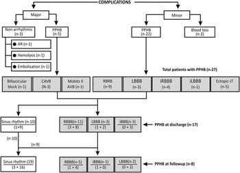
Flowchart of the mid-term outcome of complications after transcatheter device closure of perimembranous ventricular septal defect.

In total, 39 (13.4%) patients developed procedure-related complications, which included non-arrhythmic complications including major and minor in 12 (4%) and rhythm disturbances in 27 (9.3%) patients. The rhythm disturbances included five (1.7%) patients with major and 22 (7.5%) with minor post-procedure heart block. On the other hand, at discharge, only 17/27 (5.8%) patients had rhythm disturbances and these were all minor post-procedure heart blocks.
Major arrhythmias (n=5)
In total, five (1.7%) patients developed serious atrioventricular conduction disturbances within the 1st week after the procedure – two patients on the 2nd, and one each on the 3rd, 4th, and 6th day after the procedure, respectively. These included 3/290 (1%) children with complete atrioventricular block, one with symptomatic Mobitz’ type II atrioventricular block, and one with complete left bundle branch block with first-degree heart block; two patients with complete atrioventricular block had right bundle branch block preceding the complete atrioventricular block, and in one patient complete left bundle branch block immediately after the device closure progressed to the complete atrioventricular block on day 5. In these five patients with serious atrioventricular blocks, the steroid dose was immediately escalated to high dose (2 mg/kg/day). In all, three symptomatic patients with complete atrioventricular block required temporary pacemaker for 3, 3, and 4 days, respectively. Although complete atrioventricular block reverted to sinus rhythm within 72 hours – within 2 days in two patients and 3 days in one patient – of high-dose oral steroid treatment, temporary pacing support was discontinued after additional 24 hours observation. In addition, two patients with major arrhythmia – second-degree atrioventricular block and bi-fascicular block, left bundle branch block with first-degree atrioventricular block, respectively – were asymptomatic and responded to high-dose steroid treatment within 24 hours. All these patients were observed in the hospital for additional 7 days after rhythm recovery, and oral steroid therapy was continued for a total of 4 weeks including 2 weeks with tapering dose. At discharge, the bi-fascicular block patient had complete recovery, whereas significant atrioventricular blocks in four patients reverted to minor conduction disturbances – right bundle branch block in three patients and left bundle branch block in one patient.
Minor arrhythmias (n=22)
Minor post-procedure heart blocks were reported in 22/290 (7.5%) patients, with the right bundle branch block (nine) being the most common new rhythm disturbance. The other post-procedure heart blocks reported included incomplete right bundle branch block in four patients, left bundle branch block in three, incomplete left bundle branch block in one, and ectopic junctional rhythm in five patients. At discharge, junctional rhythm reverted to sinus rhythm in all five children, whereas new onset bundle branch block were persistent in 13 patients – right bundle branch block in eight, incomplete right bundle branch block in three, and left bundle branch block in two patients.
Follow-up
None of the study patients had new or worsening aortic regurgitation or tricuspid regurgitation. None of the study patients had left ventricular outflow tract obstruction during the study period. After 56 months of device closure, one patient succumbed to death at a district hospital due to viral hepatitis leading to hepatic encephalopathy and hepatorenal syndrome. At his last follow-up visit 6 months before the fatal event, he had normal electrocardiography and echocardiographic evaluation.
None of the patients with post-procedure heart block or junctional rhythm had new or worsening of conduction disturbances on Holter analysis at 1-year follow-up. At mean follow-up of 18.23±13.15 (6–70) months, nine out of 17 cases with post-procedure heart block reverted to normal sinus rhythm with normal conduction, whereas eight patients had persistent post-procedure heart block without any clinical or echocardiographic consequences. Further analysis of post-procedure heart block in the major and minor arryhthmia group revealed that, among five patients with serious bradyarrhythmias, only one patient had persistent right bundle branch block and one had incomplete left bundle branch block. Similarly, only six of 22 patients with minor arrhythmias had persistent bundle branch block – right bundle branch block in four and left bundle branch block in two patients – at the end of the study.
Discussion
In the majority of patients with perimembranous ventricular septal defect, the bundle and its bifurcation emerge into the sub-endocardium at the postero-inferior quadrant of defect, and therefore is always “at risk” for mechanical or inflammatory injury by any occlusion device.Reference Anderson, Ho and Becker 10 In their review of histopathological examination of septal defect after surgery, R. R. Latham and R. H. Anderson had postulated that haemorrhagic exudate can apparently tract in a retrograde direction along the conduction tissue as high as atrioventricular node node.Reference Latham and Anderson 11 They also observed that the right bundle is sensitive to oedema and pericellular exudate. Owing to this inherent high-risk anatomical substrate of perimembranous ventricular septal defect,Reference Holzer and de Giovanni 12 – Reference Predescu and Chaturvedi 15 conduction disturbances is the most important concern after transcatheter closure. In fact, the early enthusiasm of transcatheter device closure of perimembranous ventricular septal defect with asymmetric device was very soon counterbalanced by the unexpectedly higher incidence of conduction disturbances. The early studies have reported 5–7% incidence of complete atrioventricular block and 2–5% incidence of pacemaker implantation.Reference Holzer and de Giovanni 12 , Reference Carminati and Butera 13 This is of paramount importance when combined incidence of mortality and pacemaker implantation after surgical closure is <1% in the contemporary era.Reference Andersen, Leval and Tsang 16 , Reference Bol-Raap, Weerheim, Kappetein, Witsenburg and Bogers 17 In contrast to earlier studies, recent large-scale studies with intermediate term results have shown encouraging results of transcatheter device closure, with very low incidence of complete atrioventricular block, as low as <1%.Reference Yang, Li and Yang 2 – Reference Yang, Kong and Sheng 4 Observational analysis of these studies with extremes of incidence of complete atrioventricular block after transcatheter device closure have identified certain high-risk individuals as well as suitable candidates for the transcatheter device closure. From 2000 to 2006, observational analysis of published data of nearly 650 patients with similar inclusion criteria enrolled in eight different studiesReference Carminati and Butera 13 has revealed alarmingly high incidence of complete atrioventricular block; two-third of these patients who developed complete atrioventricular block were either <2 years old or had <8 kg weight. In fact, similar experience is reported with hybrid intervention as well.Reference Chen, Cao and Zhang 18 In a multi-centre prospective study by Holzer et al, patients with weight <10 kg had statistically significant higher incidence of device- or procedure-related complications.Reference Holzer, de Giovanni, Walsh, Tometki, Goh and Hakim 19 Perimembranous ventricular septal defect with inlet extension or Down syndrome also had higher incidence of complete atrioventricular block. Surprisingly, these groups of patients did not respond to steroid treatment satisfactorily, either because of profound mechanical trauma or inflammatory response. Delayed but persistent recovery of sinus rhythm in nearly 15% of the patients with permanent pacemaker implantation after device closure probably suggest early “inappropriate” permanent pacemaker implantation. Another unexplained observation was relatively the higher incidence (15–20%) of late complete atrioventricular block in this group of patients.
In our prospective study, high-risk candidates were not included and only grown-up children with body weight more than 10 kg were enrolled into the study. In addition, any patient with perimembranous ventricular septal defect extending into the inlet septum was excluded from this study. Only three (1%) of our study patients suffered transient complete atrioventricular block, and no patient developed late complete atrioventricular block at mean follow-up of 18.23±13.15 months. None of the study patients required permanent pacemaker implantation. All these patients developed symptomatic complete atrioventricular block during the 1st week of observation in the ward and recovered to sinus rhythm after high-dose oral steroid treatment and temporary pacemaker. Another important variable in our study was the device design. The symmetric double-disc perimembranous device with longer waist (2.5 mm with SASMA and Cardio-o-fix device) was the most commonly used device, whereas asymmetric double-disc device with a short waist (1.5 mm with Amplatzer) was not used in any patient. Duct occluder was used in 33 (11.3%) patients recruited in the later part of the study, and only one patient had transient minor post-procedure heart block that recovered to sinus rhythm before discharge. Yong Wen Qin et alReference Qin, Chen and Zhao 20 had quite encouraging results in 412 patients, enrolled in five different centres in China; six patients (four children, two adults) developed complete atrioventricular block within the 1st week, and the rhythm recovered within 3 weeks of dexamethasone and temporary pacemaker. Similar results of very low incidence of complete atrioventricular block (0.3%) requiring pacemaker implantation were reported by Zhou et al,Reference Zhou, Pan and Guan 3 in their single-centre study of 348 patients with perimembranous ventricular septal defect. The safety and efficacy of transcatheter device closure of perimembranous ventricular septal defect with very low incidence of serious complete atrioventricular block was further endorsed by Jian Yang et alReference Yang, Li and Yang 2 in their largest study with 848 patients (0.23% incidence of pacemaker implantation). Consistent with our study, two important and relevant observations in these studies with remarkably low incidence of conduction disturbances in large cohort of patients (~2500) at intermediate- to long-term follow-up were the age of the study patients and device design.Reference Yang, Li and Yang 2 – Reference Yang, Kong and Sheng 4 , Reference Qin, Chen and Zhao 20 , Reference Masura, Gao and Gavora 5 The younger the children are, the larger the ratio of device area/ventricular septum area. Accordingly, for the same size of device, the odds of affecting the conduction tissue are higher. Second, the longer-waist device is less likely to clamp or strain the defect edge, and thereby can cause mechanical trauma to the nearby conduction tissue;Reference Zhou, Pan and Guan 3 , Reference Pan and Zhao 6 however, Zhou et alReference Zhou, Shen and Zhou 21 reported 2.9% incidence of early complete atrioventricular block with asymmetric device in their study of 210 patients, but after high-dose steroid therapy none of the patients required permanent pacemaker implantation.
Steroid and conduction disturbances
The varied incidence and time course of atrioventricular block after device closure of the perimembranous ventricular septal defect are further complicated by the ill-defined natural history and treatment strategy for serious atrioventricular blocks. Approximately 90% of complete atrioventricular blocks appear within the first few weeks of procedure, and are defined as early complete atrioventricular blocks. Nonetheless, <10% of complete atrioventricular blocks have been reported after 3 months of procedure, with 2–3 cases documented as late as 3 years. The most common mechanism of early complete atrioventricular block is the oedema and secondary inflammation extending into the conduction tissue.Reference Fischer, Apostolopoulou, Rammos, Schneider, Bjornstad and Kramer 22 , Reference William, Zimmerman and Hijazi 7 In data published up to 2012, ~70 cases of early complete atrioventricular block are reported. In more than half (~60%) of these patients, complete atrioventricular block reverted to sinus rhythm within few weeks of steroid therapy and temporary pacemaker.Reference Yang, Li and Yang 2 , Reference Zhou, Pan and Guan 3 , Reference Pan and Zhao 6 , Reference Li, Li and Wang 23 Although the efficacy of steroids for the treatment of early complete atrioventricular block is evident in multiple case reports (58), the recovery of sinus rhythm can get delayed, and may lead to the situation of early “inappropriate” permanent pacemaker implantation. Of 29 cases of permanent pacemaker implantation described in different case series, seven patients (25%) did not require pacemaker activation after 2 months of device implantation. Walsh et alReference Walsh, Bialkowski, Szkutnik, Pawelec-Wojtalik, Bobkowski and Walsh 24 reported similar three cases of early complete atrioventricular block after device closure and they did not responded to initial high-dose steroid therapy; however, all three patients had sinus rhythm after few months of permanent pacemaker implantation. Although not supported by statistical or comparative data, we observed that short course of prophylactic steroid therapy in patients undergoing perimembranous ventricular septal defect device closure not only reduces the incidence of serious atrioventricular block but also accelerates the recovery of early complete atrioventricular block. All five patients in our study with high-grade atrioventricular block had sinus rhythm recovery with 3 days of high-dose steroid treatment. Similarly, 9/22 (45%) patients with minor arrhythmia also responded to conventional prophylactic steroid treatment and had reverted to normal electrocardiography. Certainly, the prophylactic oral steroid regimen could be one of the reasons for the very low incidence of serious bradyarrhythmia in our study, despite the relatively younger age of our study patients (mean age 8.6±3 years and mean weight 21±8.2 kg) compared with the recent studies with similar results where >75% study patients were either adolescents or adultsReference Yang, Li and Yang 2 – Reference Masura, Gao and Gavora 5 , Reference Qin, Chen and Zhao 20 (Table 1). Selective analysis of patients with clinically silent minor conduction disturbances revealed that out of 22/290 (7.5%) patients with minor post-procedure heart block after device closure, only 6/290 (2%) had bundle branch block at the end of the study. Similarly, many clinical studies have documented that majority of post-procedure heart blocks revert to normal, and when it is persistent they have a benign clinical course.Reference Yang, Li and Yang 2 – Reference Masura, Gao and Gavora 5 , Reference Qin, Chen and Zhao 20
Recent Studies showing the incidence of PPHB and steroid response.

ADO=amplatz duct occlude; AVB=atrioventricular block; AVSDO=asymmetrical ventricular septal defect occlude; CAVB=complete atrioventricular block; JR=Junctional rhythm; LAHB=left anterior hemi block; LBBB=left bundle branch block; MDVO=modified double-disk occlude; PPHB=post-procedure heart block; RBBB=Right bundle branch block; SHSMA=Shanghai Shape Memory Alloy; SVSDO=symmetrical ventricular septal defect occlude
Limitations
Our prospective study was an observational, single-centre study with strict inclusion criteria limited to grown-up children only. In addition to the prophylactic steroid treatment, the age of the patients and the device design were the two important variables that could have contributed to the higher safety and efficacy observed in this study.
Conclusion
Transcatheter device closure of perimembranous ventricular septal defect in children with prophylactic oral steroid resulted in excellent closure rate and acceptably low incidence of conduction disturbances at mid-term follow-up; however, long-term follow-up is essential.
Acknowledgements
The authors highly acknowledge the contribution by Dr Ashok Shah and Dr Jignesh Shah for their help, support, and critical review of the manuscript.
Financial Support
This research received no specific grant from any funding agency, commercial, or not-for-profit sectors.
Conflicts of Interest
None.
Ethical Standards
Institutional ethics committee approval was obtained for the study.