I. INTRODUCTION
One-dimensional (1D) nanostructures such as wires, rods and tubes often exhibit enhancement in electrical and thermal transport properties as compared with zero-dimensional (0D) nanostructures.Reference Xia and Yang1, Reference Xia, Yang, Sun, Wu, Mayers, Gates, Yin, Kim and Yan2 The 1D structures are also promising for fabrication of electronic, optoelectronic and electrochemical devices. Carbon nanotubes are one of the well-studied 1D nanostructures exhibiting unique mechanical and electrical properties.Reference Baughman, Zakhidov and de Heer3, Reference Yao, Kane and Dekker4 The usefulness of inorganic 1D nanostructures for applications in solar cells, light emitting diodes and transistors has been investigated for more than a decade.Reference Sun and Sirringhaus5–Reference Gonzalez-Valls and Lira-Cantu7 Unusual electronic properties in 1D nanostructure arise primarily from spatial confinement of electrons, phonons and electric field near the surface. A variety of methods have been developed for the shape- and size-controlled synthesis of 1D nanostructures of various materials, including templated growth,Reference Matsui, Kyotanni and Tomita8, Reference Shi, Pei, Xu and Li9 vapor-liquid-solid growth,Reference Wooten, Werder, Williams, Casson and Hollingsworth10–Reference Steinhagen, Akhavan, Goodfellow, Panthani, Harris, Holmberg and Korgel12 solvothermal route,Reference Wang and Li13, Reference Whittaker, Jaye, Fu, Fischer and Banerjee14 and hot-injection synthesis.Reference Kwon and Hyeon15, Reference Manna, Scher and Alivisatos16
For harnessing the unique properties of 1D nanostructure for practical applications, they need to be assembled over large areas. Assembled nanorods often provide enhanced properties as compared with those that are randomly oriented. For example, assembled semiconductor nanorods offer directed electron and hole transport leading to more efficient charge separation. Likewise, orientation-dependent surface plasmon resonance is observed in unidirectional assembled gold nanorods. It should be noted that controlled assembly of 1D nanostructures is distinct from their 0D counterpart as shape anisotropy can result in multiple orientation configurations. Large area assembly of nanorods requires appropriate control of the interparticle interactions that can be influenced by the solution chemistry or applied external field. A better understanding of nanorod self-assembly mechanism has enabled the development of strategies that can transform individual nanorods into well-defined two-dimensional (2D) or three-dimensional (3D) structures. Over the past decade, a number of assembly methods have been developed, e.g., with application of an applied electric field, drying kinetics, dipole-dipole interaction, nanorod surface chemistry, hydrogen bonding, using selective binding ligands, etc. Recently, the self-assembly and properties of various anisotropic nanostructures have been reviewed.Reference Baranov, Manna and Kanaras17–Reference Liu, Zhao and Kumacheva20 The primary objective of this review is to provide the readers a concise illustrative report on various nanorod self-assembly methods that have thus far been successfully demonstrated.
II. DIFFERENT SELF-ASSEMBLY ROUTES
A. Electric field-assisted assembly
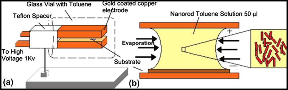
Assembly of 1D inorganic material utilizing external forces has been demonstrated in recent years. 1D wurtzite structures such as cadmium selenide (CdSe), cadmium sulfide (CdS) and zinc oxide (ZnO) generally possess a permanent dipole moment along their axes. The permanent dipole moment of wurtzite CdSe nanorods has been determined from transient electric birefringence measurements, and it has been postulated that dipole moment originates from noncentrosymmetric crystallographic lattice.Reference Li and Alivisatos21 The dipole moment of the material can be manipulated for the assembly of 1D nanostructures into 2D or 3D superstructures with application of an external electric field. Materials with low polarizability can also be induced to assemble using an electric field when the energy input exceeds the random thermal energy (kT). Ryan et al.Reference Ryan, Mastroianni, Stancil, Liu and Alivisatos22 reported placing CdS nanorods of dimension 30 nm (l) and 5 nm (d) suspended in toluene solution between parallel electrodes, with the solution then allowed to dry under an applied DC electric field of 1 V/µm. Packing of nanorods into a 2D structure on a substrate was achieved during the evaporation process. The induced dipole moment of CdS nanorods, estimated to be ∼220 D, was effectively utilized to vertically orient the nanorods along the applied electric field. Subsequent drying of the solvent brought the nanorods in close proximity to form a 2D array. Figures 1(a) and 1(b) show schematic representations of the experimental set up used for the electric field-assisted assembly process. 2D assembled domains obtained with this process consisted of ∼4 × 10³ nanorods. With higher concentration of nanorods, a 3D assembly hexagonal AB stacking was observed. A similar electric field-assisted assembly process was carried out on CdSe nanorods, in which poly(methyl methacrylate) (PMMA) polymer was used to freeze the nanorod assembly for preventing the destabilization of nanorods assembly when the electric field is turned off.Reference Gupta, Zhang, Emrick and Russell23 In a typical experiment, a droplet of chloroform solution of CdSe nanorods with PMMA was placed on a silicon oxide-coated silicon wafer. The droplet was allowed to evaporate with an applied electric field of 107 V/m. The CdSe nanorods used for this assembly experiment had a higher dipole moment value of ∼1450 D.
Schematic of electrode assembly for nanorod alignment. (a) 3D drawing. Each electrode measures 35 × 15 × 5 mm and is separated at a distance of 1.2 mm. The electrode assembly in a horizontal orientation is placed in a glass vial with 1 mL of toluene to create a saturated atmosphere. (b) 2D close up of trapped meniscus. The substrate (carbon-coated electron microscopy grid or silicon nitride membrane window) is placed between the electrodes, and 50 µL of nanorod toluene solution is deposited forming a meniscus between the upper and lower electrode. Reprinted with permission from Ryan et al.,Reference Ryan, Mastroianni, Stancil, Liu and Alivisatos22 copyright 2006 American Chemical Society.

B. Templated assembly
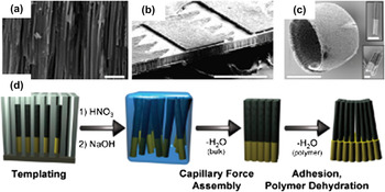
Templated assembly is another approach by which nanorods are assembled at predetermined locations to form secondary superstructures. Depending on the nature of the template, nanorod concentration and aspect ratio, different superstructures of assembled nanorods can be obtained. Saeedi et al.Reference Saeedi, Marcheselli, Shum and Parviz24 reported the assembly of nanorods on a prefabricated template. E-beam lithography was used to pattern a spin-coated poly(methyl methacrylate) (PMMA) resist. Deep reactive ion etching (DRIE) process was carried out after depositing the metal films (Cr/Au). Y- or U-shaped receptors were fabricated to trap the nanorods. For assembly of nanorods, the template was placed inside a centrifuge tube and a specific volume of the nanorod solution dropped inside the centrifuge tube. The tube was then centrifuged at different speeds. The centrifugal force pushed the nanorods and trapped them inside the receptor sites on the template. Nearly 92% of receptor sites were occupied with nanorods, out of which 38% were filled with single nanorods and remaining contained more than one nanorod. This assembly method is advantageous as it is not being limited to the nature of the material. Mirkin’s group reported the assembly of multiple component rod-like building blocks consisting of gold nanorods and polymer.Reference Park, Lim, Chung and Mirkin25–Reference Ciszek, Huang, Tsonchev, Wang, Shull, Ratner, Schatz and Mirkin27 They used templates for the synthesis of nanorods, which also played a significant role in the assembly process. Spheres, tubular and hemisphere-shaped mesoscopic superstructures were obtained by assembly. Gold nanorods were prepared by electrodepositing gold into anodized alumina template and then pyrrole was polymerized electrochemically. Nanorods were released from the template after etching with sodium hydroxide (NaOH). Self-organization of nanorods-polymer composite into various mesoscopic architecture including bundles, tubes and sheets consisting of hundreds of thousands of rods was demonstrated. Figures 2(a)–2(d) show schematic representations of nanorods assembly into superstructure. The shape and size of the final architecture could be controlled by adjusting the gold to polymer ratio. The authors proposed that interaction between polymer ends of the composite bring the nanorods closer to form secondary superstructures.
Superstructure shape control. (a) SEM image of the pores of a bare AAO membrane used as a template for nanorod generation. (b) The AAO template is patterned with photoresist (darker elevated pattern). The areas covered by photoresist are not exposed to the plating/polymerization solution and rods are electrochemically deposited exclusively in the uncovered areas (rods are the light areas at the bottom of the template). (c) After the template is removed the rods assemble into a superstructure (in this case a hemisphere) whose shape is determined by the photoresist pattern in conjunction with the template. The insets show typical two-part AuPpy rods, which comprise the curved superstructures. (d) A scheme showing each step of the assembly process. Scale bars in panels a, b, and c are 2, 300, and 100 μm, respectively. Inset scale bars are 1 μm. Reprinted with permission from Ciszek et al.,Reference Ciszek, Huang, Tsonchev, Wang, Shull, Ratner, Schatz and Mirkin27 copyright 2010 American Chemical Society.

C. Gravity driven assembly
Spontaneous assembly of anisotropic nanostructure to form columnar array with the aid of the gravitational force has been reported. Smith et al.Reference Smith, Kirby and Keating28 reported an interesting finding that controlled sedimentation of nanotubes with large density differences at the ends of nanotubes result in vertical assembly. For demonstration, partially gold-filled silica nanotubes were prepared by electrodepositing 2 μm of Au and Ag into alumina membranes, followed by coating of 30-nm silica, and finally completed with selective etching of Ag. The partially filled nanotubes were dispersed in water and a 100-μL aliquot was placed in a 0.64 cm2 glass coverslip to sediment on the surface. In this process, the nanotubes initially sedimented parallel to substrate surface, and subsequently with high particle surface coverage, vertically standing nanotubes were observed as dots in optical microscope images. The authors established that density difference between ends of nanotube was crucial to obtaining vertically standing arrays, otherwise only a smectic assembly was achieved. The maximum number of vertically standing tubes (nearly 90%) was obtained after 12.5 min of sedimentation. The vertically standing nanotube sedimentation percentage differed depending primarily on the concentration of nanotubes dispersion.
D. Surfactant and chemically assisted assembly
The use of surfactants that bind to specific surfaces of the nanorods is an effective approach for self-assembly. For example, assembly of gold nanorods into 2D structures by manipulating the pH, which effects the electrostatic interaction between the cationic surfactant bilayer around the nanorods and the negatively charged self-assembled monolayers, has been demonstrated using different dicarboxylic acid molecules.Reference Orendorf, Hankins and Murphy29 Cetyl trimethyl ammonium bromide (CTAB) binds to gold nanorods on their long side rendering them positively charged and carboxylate ends of adipic acid are negatively charged. Interaction between these two opposite charged molecules is triggered at high pH leading to side-by-side assembly of the nanorods. For confirming the assembly mechanism, the process was carried out using other carboxylic acids. Assembly using hexanoic acid (same carbon chain length as adipic acid) and dodecanedioic acid (twice the chain length of adipic acid) gave only isolated and disordered aggregates. Nonassembly in the case of hexanoic acid can be explained as it is a monocarboxylic acid and does not have the ability to bridge the nanorods. In the case of dodecanedioic acid, it was suggested that the acid binds to the nanorods surface in a looped conformation, which makes it chemically equivalent to hexanoic acid. The preferential attachment of CTAB molecule to the surface of gold nanorods plays a major role in the side-by-side assembly. Selective coating of nanorods surface with photochemically active molecules can also be utilized for the organization of nanorods, as demonstrated for gold nanorods.Reference Fava, Winnik and Kumacheva30 The side faces of the gold nanorods with face-centered cubic structure are terminated with (110) planes, whereas the tips display either the (111) or (100) planes. Exploiting the preferentially binding of CTAB on the (110) planes of Au, Fava et al.Reference Fava, Winnik and Kumacheva30 coated gold nanorods with CTAB on the side faces and poly(N-isopropylacrylamide) (PNIPAm) on the tips. The nanorods were then photoilluminated at the longitudinal plasmon band wave length. The nonradiative energy released from light absorption was sufficient to heat the PNIPAm polymer molecules above the lower critical solution temperature (LCST). This induced transformation of the polymer from the coil to globular form, thereby triggering end-to-end nanorod assembly. The proposed binding mechanism was confirmed by the red shift in the longitudinal absorption band. The hydrogen bonding ability of carboxylic acid groups can also be utilized for the end-to-end assembly of gold nanorods.Reference Thomas, Barazzouk, Ipe, Joseph and Kamat31 This mechanism was proposed in the assembly process using mercaptocarboxylic acid- and CTAB-protected gold nanorods.Reference Ni, Mosquera, Perez-Juste and Liz-Marzan32 As described earlier, CTAB preferentially binds to the side faces of the gold nanorods, making the tips available for the thiol group of mercaptocarboxylic acids to bind. Hydrogen bonding between the unbound carboxylic acid groups enables the nanorods to assemble in an end-to-end fashion, which was confirmed in transmission electron microscope (TEM) images as isolated linear arrays of gold nanorods. The hydrogen bond-assisted assembly was extended using 4-mercaptophenol, cysteine and glutathione molecules and the role of H-bonding in the end-to-end assembly was evidenced by measuring the extinction spectra at different pH. The use of chemicals that induce assembly of nanorods by selectively binding to specific faces on the surface was explored in yet another study of gold nanorods. A series of dithiol molecules were used for linking of the nanorods, including 1,3-propanedithiol, 1,5-pentanedithiol, 1, 6-hexanedithiol, 1,8-octanedithiol and 1,9-nonanadithiol.Reference Joseph, Ipe, Pramod and Thomas33 For the assembly process, gold nanorods of 2.9 aspect ratio were suspended in a mixture of water and acetonitrile and microliter quantity of dithiols were added to the mixture. The dithiol molecule-induced nanorod assembly was monitored by absorption spectroscopy, which showed a decrease in the intensity of the longitudinal surface plasmon band at 700 nm and the formation of interplasmon coupling band in the near infrared region.
The addition of dimercaptosuccinic acid (DMSA) to CTAB-coated gold nanorods solution was reported to induce assembly of the nanorods to form 1D, 2D, and 3D superstructures.Reference Sreeprasad, Samal and Pradeep34 The concentration of DMSA played a major role in the formation of the superstructures. At the lowest concentration (0.5 mM) of DMSA, nanorods were assembled side-by-side to form a tape-like 1D structure. With increasing the DMSA concentration to 0.8 mM, a perpendicular sheet-like structure was obtained, and increasing the concentration even further resulted in the formation of 2D sheets and 3D superstructure of parallel sheets. The mechanism for the formation DMSA-assisted superstructure was proposed as charge neutralization. When DMSA was added to the CTAB bilayer-coated gold nanorod solution, the negatively charge carboxylate ions neutralize the CTAB bilayer by formation of a CTA+-DMSA complex, thereby bringing the nanorods together to form a superstructure. Intermolecular disulfide bond formation in DMSA helps to further stabilize the superstructure assembly. The involvement of disulfide bond in the formation of nanorods assembly was tested using mercaptosuccinic acid, which yielded similar ordered superstructures as DMSA, but the extent of order was not as good.
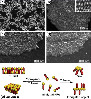
The end-to-end assembly of gold nanorods was further demonstrated using sodium citrate, which selectively binds to the tips of nanorods. In this study, nanorods with two different aspect ratios (3 and 16) were used for assembly.Reference Kawamura, Yang and Nogami35 Four different possible mechanisms were proposed for the observed assembly of gold nanorods, including electric charge neutralization of CTAB coated gold nanorods, interdigitation of carboxylic groups of CTAB caused by low coverage of the molecules on the nanorod surface, electrostatic interaction between cationic CTAB head group and citrate anions, and intermolecular hydrogen bonding of citrate carboxylate groups. The authors conceptualized that the end-to-end assembly in the case of gold nanorods with aspect ratio of 3 occurred through electric charge neutralization, and in nanorods with aspect ratio of 16 through interdigitation of carboxylic groups of CTAB. The selective functionalization of gold nanorods using a coordination complex-containing ligand bearing a thiol group can also induce end-to-end assembly. This was demonstrated recently using iron(II) coordinated with a thiol containing ligand 4′-(4-mercaptophenyl)-2,2′:6′,2′′-terpyridine (tpySH).Reference Selvakannan, Dumas, Dumur, Pechoux, Beaunier, Etcheberry, Secheresse, Remita and Mayer36 Zhao et al.Reference Zhao, Liu, Greener, Nie and Kumacheva37 reported an interesting approach to assemble Au-tipped CdSe heterostructure nanorods in which hydrogen bonding between carboxylic acid groups of 11-mercaptoundecanoic acid (MUA) attached to the gold tips was utilized to stabilize the self-assembled structure. In the Au-CdSe heterostructure nanorods, the CdSe surface was covered with octadecylphosphinic acid (ODPA) and hexylphosphonic acid (HPA) and the Au tips were capped with didodecyldimethylammonium bromide (DDAB) and dodecylamine (DDA). To achieve side-by-side assembly utilizing the anisotropic attraction force between the nanorods and solvents, ligands on the Au surface were exchanged with 11-mercaptoundecanoic acid (MUA) by adding a solution of MUA in dimethylformamide to the toluene solution of nanorods. The authors reported that the nanorods self-assembled for minimizing interaction between the nonpolar ligands and the polar solvent. SEM and dark-field TEM images of the Au-CdSe nanorod assembly are shown in Figs. 3(a)–3(d). In a subsequent publication, the authors reported the assembly of single tip Au-CdSe nanorods using the same method.Reference Zhao, Vickery, Guerin, Park, Winnik and Kumacheva38 A schematic representation of the assembly process is given in Fig. 3(e). Another novel method for assembly of binary nanostructures has recently been reported.Reference Nagaoka, Wang, Lynch, LaMontagne and Cao39 The assembly was carried out by adding toluene solution of triphenylphosphine (TPP) to a mixture of gold nanoparticles and CdSe/CdS nanorods. TEM images of the sample showed eight to ten close-packed nanoparticles aligned linearly between two nanorods, with a distinct spacing of 2.0 nm between the nanoparticles and 1.9 nm between the nanoparticles and nanorods. It is believed that the presence of triphenylphosphine additive is crucial for the formation of binary assembly, which binds to the both the gold nanoparticles and CdSe/CdS nanorods. Micrometer-range self-bundled arrays of CdS nanorods were prepared by a solution-based method, in which trioctylphosphine (TOP) and tetradecylphosphonic acids (TDPA) were used as cosurfactants.Reference Kang, Lai, Peng, Shyue and Chou40 The intermolecular force among the hydrocarbon chains of surfactant molecules was suggested as a reason for the assembly of nanorods, sufficient to overcome the interfacial force between the adsorbed head groups of surfactant and the surface of the nanorods. π-π stacking interaction between cyclic hydrocarbon surfactants of gold nanorods can also assemble them in a well-organized side-by-side manner.Reference Xue, Birel, Gao, Zhang, Dai, Urbas and Li41 To achieve such an assembly, perylene thiol (PDI-SH) monolayer protected gold nanorods were synthesized by mixing CTAB coated gold nanorods with perylene thiol.
(a) Dark-field TEM image of Au-tipped CdSe NRs in toluene, taken 1 h after NR preparation. (b) Dark field TEM image (top view) of the close-packed NR array, inset shows the magnified image and the scale bar represents 50 nm (c, d) Side view SEM images of the NR arrays. TEM and SEM images in (b–d) were taken 1 day after the addition of dimethylformamide solution of MUA. Reprinted with permission from Zhao et al.,Reference Zhao, Liu, Greener, Nie and Kumacheva37 copyright 2009 American Chemical Society. (e) Solution-based self-assembly of single-tip Au-CdSe NRs. Reprinted with permission from Zhao et al.,Reference Zhao, Vickery, Guerin, Park, Winnik and Kumacheva38 copyright 2011 John Wiley & Sons.

E. Nanorod assembly by Langmuir-Blodgett method
Langmuir-Blodgett (LB) method is perhaps the most promising scheme for the preparation of monolayer thick and highly ordered thin films. In the LB method, a molecular layer film spread at the water/air interface is transferred onto a solid substrate. This method has been utilized for the 2D monolayer assembly of nanorods.Reference Kim, Kwan, Akana and Yang42 Additionally, pressure-induced liquid crystalline phase transition (isotropic-nematic-smectic) can be achieved. To demonstrate these methods, BaCrO4 nanorods were prepared by reacting barium bis(2-ethylhexyl) sulfosuccinate [Ba(AOT)2] reverse micelle with Na2CrO4 containing NaAOT. The nanorods suspension was spread drop wise on the water surface of a Langmuir-Blodgett trough and consequently the surface was compressed to increase the surface pressure. When the surface pressure was raised up to ∼30 mN/m, nematic arrangement of nanorods was observed which then transformed to smectic arrangement at ∼35 mN/m surface pressure. The liquid crystalline 1D smectic phase was further transformed to 3D nematic on increasing the surface pressure to ∼38 mN/m.
F. Solution phase assembly of nanorods
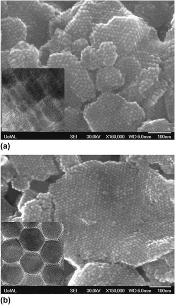
A simple solution phase nanorod assembly method utilizing the depleted attraction force was demonstrated recently.Reference Barano, Fiore, Huis, Giannini, Falqui, Lafont, Zandbergen, Zanella, Cingolani and Manna43 Depletion force can be defined as an attractive force between large spheres results from competition between large solute and small solvent particle entropies.Reference Melby, Prevost, Egolf and Urbach44 This type of attractive interaction force is comparable to other attractive forces, such as van der Waals and hydrogen bonding.Reference Oosawa and Asakua45 The depleted attraction force between nanorods is sufficient to induce the oriented binding of nanorods to form well-defined assembly. Assembly of trioctylphosphine oxide (TOPO) coated CdSe/CdS nanorods was induced by addition of oleic acid (OA) and poly(ethyleneglycol) methacrylate (PEG-MA) additives.Reference Barano, Fiore, Huis, Giannini, Falqui, Lafont, Zandbergen, Zanella, Cingolani and Manna43 Assembled nanorods were precipitated from the nanorod dispersion after injecting additives (OA and PEG-MA) above the threshold concentration. Nanorod assembly was observed as isolated 2D sheets in the size range of 50–200 nm in cryo-TEM images. By adjusting the concentration of rods and additives, interaction time and stirring conditions, the lateral size of 2D sheets could be increased to 1–2 μm. It has been noted that surfactants that are commonly used for the synthesis of nanocrystals, such as oleylamine, hexadecylamine, oleic, and stearic acids, can induce self-assembly in nanorods dispersion through depleted attraction force.Reference Barano, Fiore, Huis, Giannini, Falqui, Lafont, Zandbergen, Zanella, Cingolani and Manna43 We recently demonstrated a simple solution-based approach for the vertical assembly of nanorods utilizing the dipole-dipole interaction between the nanorods and the solvent.Reference Ramasamy, Zhang, Bennett and Gupta46 To demonstrate the assembly, wurtzite phase Cu2CdxZn1-xSnS4 nanorods were synthesized by reacting a mixture of thiol molecules with metal salts of copper, zinc/cadmium and tin at 150–155 °C, followed by nucleation and growth at a higher temperature. The synthesis process yielded wurtzite phase nanorods with uniform shape and size. For the vertical assembly, the samples were first dispersed in hexane by sonication and ethanol was then added to the dispersion. Assembly of the nanorods was accomplished by controlled centrifugation and air-drying. The assembly of the nanorods was observed in domains with maximum size of around 600–700 nm. High-resolution field emission scanning electron microscope (HR-FESEM) images of nanorod assembly in Fig. 4 indicate vertical assembly of nanorods. TEM images in inset to Figs. 4(a) and 4(b) clearly show smectic phase ordering of nanorods with uniform distances and well-defined lattice fringes. The self-assembly process can be explained based on dipole-dipole interaction of the nanorods and solvents. The absence of a center of symmetry in a hexagonal unit cell creates partial charge polarization in the nanorods, which induces a permanent dipole moment along the c-axis. Indeed, we observed that nanorods in nonpolar solvents, such as hexane or toluene, are randomly oriented as the dipole-dipole interactions and Columbic repulsions are in competition with each other. When a polar solvent is introduced into the randomly oriented nanorods, dipole-dipole interactions dominate, which cause the nanorods to assemble into a 3D structure. The dipole-dipole interaction energy provided by the polar solvent is sufficient to overcome the Brownian motion and the thermal energy barrier. The advantage of this assembly process is that it does not require an external electric field, functionalization of nanorods, or a substrate template. Moreover, the process is simple, reproducible and scalable to large quantities of the sample, and the assembly can be carried out on any substrate. However, the size and shape uniformity of the nanorods is vital for this assembly process.
(a and b) Top view SEM images of vertical nanorod assembly by solution method. Side and top view TEM images of nanorods assembly [inset to (a and b)]. Reprinted with permission from Ramasamy et al.,Reference Ramasamy, Zhang, Bennett and Gupta46 copyright 2012 Royal Society of Chemistry.

G. Nanorods to superparticles through self-assembly
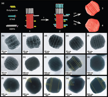
Superparticles are well-defined nano/mesostructures that form through ordered arrangement of the primary nanocrystals.Reference Zhung, Wu, Yang and Cao47 The superparticles exhibit collective properties of the building blocks due to coupling effect. Due to their potential as a new class of building blocks for nanofabrication, a considerable amount of work has been focused on the synthesis of superparticles using various methods. The synthesis of superparticles using various nanocrystals have recently been reviewed in detail, including theoretical considerations for the nucleation and growth of superstructures.Reference Wang, LaMontagne, Lynch, Zhuang and Cao48 Superstructure formation using colloidal nanorods was initially reported by Zhuang et al.Reference Zhung, Shaller, Lynch, Wu, Chen, Li and Cao49 In this work, CdSe/CdS nanorods in chloroform was mixed with an aqueous solution of dodecyl trimethylammonium bromide and the chloroform was removed by bubbling argon. The resultant mixture was injected into a vessel containing ethylene glycol to which dithiol (Tween-20-SH) was added. The mixture was heated to 80 °C and maintained at that temperature for 1 h. The isolated superparticles were disk-shaped with ∼56 nm height. The authors postulated that the formation of superparticles resulted from combined interparticle interaction between the CdSe/CdS nanorods and repulsive solvophobic interaction between the superparticles and ethylene glycol. Recently, the same group reported a detailed investigation of the growth of superstructures from the nanorods along with small angle x-ray scattering studies.Reference Wang, Zhuang, Lynch, Chen, Wang, Wang, LaMontagne, Wu, Wang and Cao50 They observed double-domed cylindrical structures when the number (N) of constituent nanorods was less than ∼80,000, and irregular multidomain or double-domed cylinders when N was more than this number. Figure 5 shows the synthesis scheme of superparticles and TEM images of the double-domed cylinder and irregular-multidomain particles. Further, when CdSe/CdS nanorods were incubated with octylamine for 6 days, single-domain elongated needle-like superparticles were obtained instead of cylindrical superstructures.
(a) Scheme for the synthesis of superparticles from CdSe-CdS nanorods: (i) nanorod-micelle formation, (ii) superparticle formation. Numbered images: (1) Proposed model for a CdSe-CdS nanorod functionalized with ODPA and octylamine; (2) proposed model for a nanorod micelle prepared using DTAB; (3) proposed model for a double-domed cylinder; (4) proposed model for an irregular-multidomain particle. (b–p) TEM images of superparticles made from nanorods of l = 28.0 ± 1.5 nm and d = 6.7 ± 0.3 nm. Reprinted with permission from Wang et al.,Reference Wang, Zhuang, Lynch, Chen, Wang, Wang, LaMontagne, Wu, Wang and Cao50 copyright 2012 American Association for the Advancement of Science.

H. Nanorods assembly using biomolecules
The use of biomolecules to influence nanorod assembly is being actively explored to produce well-organized assemblies of nanomaterials. Following the ability of gold nanospheres to self-assemble on immobilizing oligonucleotides and hybridization to form duplexes, the process has been extended to gold nanorods to investigate the assemblage of anisotropic nanomaterials.Reference Dujardin, Hsin, Wang and Mann51 For this work, gold nanorods were prepared by converting gold anodic material into nanoparticles in a coelectrolytic surfactant system. The nanoparticles were subject to treatment with two oligonucleotide systems—a two strand complementary system each consisting of thiolated oligonucleotides immobilized on to nanoparticle suspensions individually and then allowed to hybridize by mixing, and a three strand system where two short noncomplementary thiolated oligonucleotides were immobilized on the nanoparticles and treated with a nonthiolated third strand, which was complementary to both the immobilized strands. TEM images from both the two and three strand hybridized systems showed bundles of nanorods aligned in parallel stacks. The 3D nanorod bundles were observed to have alternate and superimposed stacking, contrary to the nonduplexed suspensions that formed completely random aggregates. A 6-nm interparticle distance was observed for the assembled nanorods, which is higher than the surfactant bilayer (∼3 nm) and is also comparable to the 24-base pair helix containing propyl spacers (6.5–9 nm). The UV-Vis spectra of the three-stranded system displayed characteristic peaks at 520 nm (transverse plasmon band) with red shift, 850 nm (longitudinal band) with a blue shift and at 260 nm (nucleotides). The two strand system showed a similar spectrum with the peak intensity decreasing on hybridization but with no observed wave length shift. Melting analysis of the two systems confirmed that the three-strand system had hybridized irreversibly whereas the two-strand system was duplexed reversibly. This variation was probably due to various sizes of nucleotides in comparison to the surfactant bilayers.
The high binding affinity of biotin and streptavidin has been widely exploited to achieve nanorod assembly.Reference Green52 Gold nanorods were synthesized by seed-mediated growth in water and surfactant. High-resolution crystallography studies revealed pentahedral twin nanorods with the (111) and (100) faces pointed toward the ends and the length of the rods, respectively, which was suggested as a consequence of the size of the surfactant head group. The nanorods were bound to biotin disulfide (EZ-biotin-HPDP) molecules. This was achieved by treating a mixed solution of the nanorods with biotin disulfide, then stirring, sonicating and allowing it to set overnight. Centrifugation was carried out to remove excess unbound biotin disulfide and nanoparticles other than the rods. The sample was added to streptavidin in saturating quantities and then added to the biotin disulfide-coated nanorods and mixed followed by centrifugation to separate the linked and unlinked nanorods. The resulting TEM analysis revealed end-to-end linked gold nanorods with interparticle spacings of 4–5 nm, which correspond to the size of streptavidin molecules and the orientation angles match with the four biotin-binding sites of the protein. The reason for the observed amount of end-to-end linked product was suggested to be due to either the inability of biotin to replace the surfactant bilayer attached on the length of the rods and consequently binding to the (111) face preferentially, or because of the steric hindrance posed on the streptavidin molecule causing it to bind only at the ends.Reference Caswell, Wilson, Bunz and Murphy53
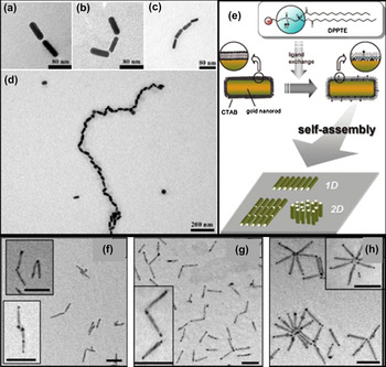
In another study, gold nanorods grown by seed mediation were treated with surfactant (CTAB) and thioctic acid (TA) and allowed to react at room temperature for 4 h and then centrifuged to remove excess CTAB and TA.Reference Chang, Wu, Chen, Ling and Tan54 The carboxyl groups on TA bound to the gold nanorods were activated by adding sulfo-N-hydroxysuccinamide and 1-ethyl-3-(3-dimethylaminopropyl)-carbodiimide in phosphate buffer for 30 min. Antimouse IgG was added to the bifunctional linker-modified gold nanorods for 30 min and centrifuged to remove the supernatant containing unbound antimouse IgG. The derivatized gold nanorods were then incubated with mouse IgG for 3 h. The self-assembled nanorods obtained after incubation were analyzed using UV-Vis spectroscopy and TEM. UV-Vis spectra showed bands at 520 and 720 nm corresponding to transverse plasmon resonance and longitudinal plasmon absorption bands, respectively. The nanorods were 60 nm in length and 20 nm in diameter with CTAB bilayer preferentially bound to the (100) ends, making the (111) ends available for disulfide groups of TA to bind. Highly selective molecular recognition of immune receptor-ligand system (interaction between antimouse IgG and mouse IgG) facilitated the end-to-end assembly between the nanorods resulting in a chain-like formation with distance between individual rods of 10–15 nm arising from the steric effect of immunoglobulin. However, on increasing the concentration to 10 μM of mouse IgG, nonspecific binding was observed and was overcome to obtain the linear assembly by adding the mouse IgG five times in equal quantities with 3-h intervals for incubation. Figures 6(a)–6(d) shows the TEM images of the linked nanorods.
TEM images of (a) di-, (b) tri-, and (c) tetra-gold nanorods assembly. The antimouse IgG provides an anchoring site at the end surface of nanorod and interacts with mouse IgG for assembly. (d) Gold nanorods were assembled into successive chains via stepwise increasing the concentration of mouse IgG. Reprinted with permission from Chang et al.,Reference Chang, Wu, Chen, Ling and Tan54 copyright 2005 Royal Society of Chemistry. (e) Illustration of surface modification of Au NR with DPPTE and 1D and 2D self-assembled configurations of the NR complex on a solid surface. Reprinted with permission from Nakashima et al.,Reference Nakashima, Furukawa, Kashimura and Torimitsu58 copyright 2008 American Chemical Society. TEM images (taken prior to separation) of (f) dimers of NDBs in different conjugation angles, (g) trimers, and (h) flowers (50 nm scale bars). Reprinted with permission from Salant et al.,Reference Salant, Amitay-Sadovsky and Banin57 copyright 2006 American Chemical Society.

Oligonucleotide hybridization was also performed on nanorods obtained with seed-mediated growth.Reference Pan, Ao, Gao, Tian, He and Cui56 After treatment with surfactant CTAB, mercaptoalkyloligonucleotide in PBS buffer was added to the nanorods and allowed to react for 40 h at 25 °C. Target oligonucleotides were added to the mercaptoalkyl probe-modified nanorods and incubated for 48 h at room temperature. TEM analysis showed unexpectedly high proportion of end-to-end linked nanorods separated by 10- to 15-nm spacings, which matched the target oligonucleotide size. The end-to-end linking of the nanorods was due to the preferential binding of the SH group to the (111) ends of the nanorods, whereas a natural preference of the nanorod assembly is side-to-side. Even a mixture of nanosphere and nanorod showed linear assembly on addition of the target oligonucleotide. UV-Vis spectroscopy exhibited a red shift in the absorption bands at 530 and 680 nm on addition of the target as a consequence of the rod-rod and rod-sphere assemblies, which further hybridized to yield larger assemblies. The half width of the absorbance bands was found to increase with increasing incubation time; also the varying size of the rod-rod and rod-sphere assemblies led to different spectral maxima and hence broader bands. The nanoparticle solutions exhibited different colors–nanospheres presented a pink solution, nanorods a jade green solution and rod-sphere mixture exhibited a deep red solution. On addition of the various components, different colors were obtained depending on target concentrations and the system was capable of detecting up to 1 pM of nucleotide.
Salant et al. treated semiconductor nanorods with Au to grow nanorods with gold tips on either ends, referred to as nanorod dumbells (NDBs) owing to their unique shape.Reference Salant, Amitay-Sadovsky and Banin57 The self-assembly of these NDBs was attempted with avidin-biotin-specific interaction. The NDBs were treated with EZ-link biotin HPDP and mixed overnight. Addition of biotin in two different molar ratios (1:1 and 1:2) yielded type 1 and type 2 NDBs, respectively. On addition of avidin to type 1 NDBs in a 1:2 ratio dimers, head to tail binding through the Au tips were obtained. However, this was not observed in unconjugated NDBs, nonbiotinylated NDBs with avidin and CdSe rods without gold tips. Trimers were formed on changing the concentration ratio to 1:2 excess avidin. The avidin-terminated type 1 NDBs were mixed with type 2 NDBs in a ratio 2:1, which resulted in dimers trimers and individual NDBs. The results confirm assembly due to selective conjugation. The differences in the methods of self-assembly were elucidated by separating the dimer and trimer conjugates in solution. The dimer experiment revealed a significant population of monomers and NDBs assembled in head-to-tail manner from nonselective binding before separation. Addition of avidin resulted in an increase in dimer-conjugated NDBs. After separation, the upper fraction was high in monomer content and the lower fraction contained 55% dimers. The trimer experiment revealed 20% trimers before avidin addition and 35% after addition. After separation, the top fraction was found to have 20% trimers and the bottom fraction contained 30% trimers. This experiment elucidates the broad distribution range in combined conjugates and the limitation of avidin-biotin to control NDB chain lengths. In addition to dimers, flower-like assembly of NDBs were observed and suggested to be formed due to several reasons, including tetramer-binding sites in avidin, consequence of more than one biotin-disulfide molecule binding to the NDB Au tips and small diameter of the gold tips and rods. Although present, these structures were relatively less in quantity as compared with the dimers indicating that their formation was not favorable in the presence of dimers. The formation of dimers, trimers and flowerlike structures is shown in Figs. 6(f)–6(h).
In another attempt toward biomolecule-driven self-assembly of nanorods, gold nanorods prepared by seed-mediated growth and treated with CTAB were exposed to polystyrene sulfonate (PSS) solution and centrifuged to remove the excess PSS and redispersed in phosphate buffer saline.Reference Wang, Chen, Wang, Ma and Su55 The modified gold nanorods were then mixed with goat anti-h-IgG solution. After removing the excess unbound antibodies, bovine albumin serum was added to the nanoparticles to block the unbound sites and then mixed with the h-IgG. The UV-Vis spectra showed that the gold nanorods produced a peak at 711 nm, and on addition of PSS there was a red shift to 722 nm due to change in the dielectric function. On addition of the goat anti-h-IgG, further red shift of the longitudinal band to 737 nm was observed owing to the high refractive index of the protein in comparison to PSS. The transverse band of the spectra did not show any wave length shift indicating that the longitudinal band responded more to target biomolecule binding. After addition of h-IgG, the peak intensity of the longitudinal band reduced considerably and red-shifted to 765 nm. This was attributed to the side-to-side assembly driven by antibody-antigen binding. The result also explained the influence of GNR aggregation and refractive index variation on the position, intensity and shape of the LSPR band. The protein detection limit in this method was 60 ng m/L.
As yet another approach to nanorod assembly, gold nanorods were arranged by incorporating surface anchored lipids.Reference Nakashima, Furukawa, Kashimura and Torimitsu58 The nanorods were prepared by chemical reduction of HAuCl4 followed by photoirradiation. 1,2-dipalmitoyl-sn-glycero-3-phosphothioethanol (DPPTE) was dispersed in water with sonication and added to the gold nanorods and the products were centrifuged to remove excess CTAB and DPPTE. A chloroform solution of DPPTE was then added to the nanorod solution and stirred vigorously for 1 h. The color of the nanorods was transferred to the organic phase from the formation of nanorod-DTTPE complex, which was extracted and redispersed in chloroform. 1D assembly of the nanorod-DTTPE complex was carried out by spin coating on to a silicon substrate and drying. 2D assembly was achieved by placing the nanorod-DTTPE complex on to a silicon substrate and evaporating the solvent followed by simultaneous addition of chloroform to the sample and annealing at 60 °C, and repeating the drying process three times. Schematic representations of the 1D and 2D assembly are shown in Fig. 6(e). The initial zeta potential value of the nanorod-lipid complex in water was measured to be 24.0 mV and it decreased with addition of DPPTE, suggesting effective replacement of CTAB by DTTPE. The UV spectra indicated longitudinal and transverse bands at 702 and 521 nm of which the former underwent a red shift to 726 nm as a result of nanorod aggregate formation induced by intermolecular interactions of the lipids attached to the surface. Increase in the DPPTE concentration led to a blue shift in the longitudinal band indicating increased side-to-side interaction of the nanorod complex or due to lipid bilayer formation. SEM images of the ID self-assembly confirmed side-to-side linking of the gold nanorods. The interfacial forces on evaporation result in the side-to-side ordering due to translational entropy, and spin coating was reasoned to reduce the collective density of the nanorods giving rise to the 1D configuration. SEM images of the 2D self-assembly showed that the assembly was dependent on the hydrophilic or hydrophobic nature of the silicon substrate with a uniform interparticle distance of 5.0 nm. Recently, a simple approach for end-to-end assembly of gold nanorods was reported based on the ability of thiamine-rich DNA to specifically and strongly bind to mercury ions (Hg2+) leading to the configuration T-Hg2+-T.Reference Wang, Li, Wang, Sanga and Huang59 The longitudinal band of the absorption spectrum at 748 nm was shifted 759 nm on the addition of Hg2+ ion, indicating the formation of T-Hg2+-T complex. The addition of Cu2+, Ni2+, Zn2+, and Cd2+ ions did not affect the longitudinal band position, confirming the specific binding of Hg2+ ions with the thiamine base pairs. SEM images also clearly indicated end-to-end linking of the nanorods in the presence of mercury ions, which was not evident in the DNA-functionalized gold nanorods.
Reversible end-to-end gold nanorod assembly was also achieved using disulfide polypeptide biomolecules.Reference Walker and Gupta60 Gold nanorods prepared by solution-based approaches were treated with poly vinyl pyrrolidone (PVP) for 24 h with gentle vortex mixing and were then centrifuged to remove excess unbound molecules. Disulfide-modified γ-benzyl-L-glutamate was converted to disulfide-modified poly(L-glutamic acid) (SSPLGA) by debenzylation. The PVP-modified gold nanorods were diluted in ethanol and placed in a scintillation vial and under vortex mixing added with the SSPLGA and allowed to react undisturbed for 24 h for the self-assembly to occur. Irreversible linking was achieved by adding 1,6-hexanedithiol to the PVP-stabilized nanorods in ethanol. UV-spectra of the PVP-modified nanorods did not show any wave length shift of the longitudinal bands. However, the gold nanorods treated with SSPLGA and 1,6-hexanedithiol showed similar wave length shifts due to aggregation of nanorods in the end-to-end fashion in both cases. The difference between the two methods was attributed to the tendency of the SSPLGA to undergo conformational changes with pH variation. At high pH, the polypeptide was deprotonated and hence was in a random coil form, while at low pH it formed a helical structure possessing dipole-dipole and hydrophobic interactions and hence reversible self-assembly was facilitated. Very recently, high aspect ratio gold nanorods were prepared by seed-mediated growth and used to achieve end-to-end assembly by two methods—surfactant control and oligopeptide support.Reference Jain, Roodbeen, Reeler, Vosch, Jensen, Bjørnholm and Nørgaard61 The study demonstrated that cysteine-terminated oligopeptides of different lengths and additional functional groups allowed to react with surfactant-modified gold nanorods enable the establishment of linear end-to-end assembly.
I. Nanorods assembly by solvent evaporation
Solvent evaporation and drying can also strongly contribute to the assembly of nanoparticles. Nikoobakht et al.Reference Nikoobakht, Wang and El-Sayed62 prepared gold nanorods by electrochemical method and treated them with cationic surfactant hexadecyltrimethylammonium bromide (HTAB) and CTAB as cosurfactant and subjected to electrolysis. After extensive dilation and centrifugation, the samples were free from unbound surfactants and diluted with NaCl in a 1:1 ratio and deposited on TEM grids using two methods. In the first method, the nanorod solution was placed on the grid and allowed to dry (1 h), and in the second method the TEM grid was immersed halfway in the nanorod solution and then allowed to dry. The latter method of deposition was favorable in terms of self-assembly and reproducibility. TEM images showed that a 0.001 M concentration solution produced the most ordered structures. Various regions could be differentiated, such as monolayers with random orientations, 2D long nanorod chains, and assemblies with varying thicknesses. The orientations of the nanorod assemblies were shown to be along the (100) and (110) planes. The self-assembly could be attributed to lateral immersion or capillary forces which in turn are related to the interparticle distance, particle radius, surface tension and meniscus deformation of the particles. The study clarified the factors that influence the assembly process, such as the rate of water evaporation, the ionic strength, the surfactant concentration, and the particle size and shape distributions.
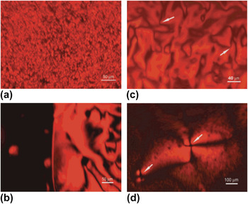
In another study, CdSe nanorods were prepared by pyrolysis of organometallic precursors of Cd and Se in hot surfactant mixture.Reference Li, Walda, Manna and Alivisatos63 Varying surfactant concentration led to nanorods with aspect ratios ranging from 1–15, with widths of 3–7 nm and lengths of 3–70 nm. The CdSe nanorods were modified with amphiphilic molecules with the polar group bound to the nanocrystal surface and protrusion of the alkyl chain, which enhanced the solubility in organic solvents. The liquid crystalline phases were analyzed using birefringence studies. At low nanorod concentrations, complete extinction was observed and the isotropic solution revealed no pattern. As the concentration increased, birefringent red droplets were observed due to the band gap of CdSe nanocrystals and the intensity of the transmitted light indicated orientation of the nanorods. Analysis of the liquid crystalline and isotropic phases, and studying droplets of various sizes, showed how the anisotropy and other optical properties of the liquid crystalline phase of CdSe make them attractive for electrooptical devices. The liquid crystalline phases of CdSe studied with optical microscopy are illustrated in Fig. 7.
Images of liquid crystalline phase in concentrated solution of CdSe nanocrystals under a polarizing optical microscope. Isotropic phase appears dark in these images. All the bright areas in the image correspond to the liquid crystalline phase, with dark lines corresponding to disclinations or the area where the orientation of the nanorods are parallel to either of the polarizers. (a) Droplets of liquid crystalline phase mixed with isotropic phase. (b) Interface between a big liquid crystalline droplet (right) and isotropic phase (left). (c and d) Disclinations of strength of 1/2 (c) and 1 (d), respectively. The arrows point to the disclinations. Reprinted with permission from Li et al.,Reference Li, Walda, Manna and Alivisatos63 copyright 2002 American Chemical Society.

In another study, CdSe nanorod assembly was obtained by slow solvent evaporation method wherein a mica sheet coated with carbon thin film was immersed vertically into CdSe nanorod solution in cyclohexane, allowing the nanorods to deposit as the solvent evaporated.Reference Li and Alivisatos64 TEM images showed structures with complete orientation along with some random multidomain structures, with the CdSe nanorods connected in a head-to-tail manner as linear arrays. Additionally, they contained vortex structures, which were proposed to arise from nematic phases, and also monolayer structures with periodicity in both directions originating from the smectic-A phase. The ability to control the liquid crystalline phase of the nanorods enabled control over their orientation. In another experiment, ZnS nanorod solution was dispersed in a 1:1 mixture of toluene and ethanol and deposited on a carbon-coated copper grid for solvent evaporation. HRTEM analysis showed 1D arrangement of the ZnS nanorods.Reference Li, Li, Yang and Li65 The solvent evaporation process reduced the free volume between the rods and causes them to align side-by-side. The alignment was favored by capillary forces and van der Waals attraction. Three factors primarily affected the aggregation–concentration of nanorods, nature of capping molecules and evaporation rate of the solvent. The 1D assembly was converted into a 2D array by dewetting in toluene-ethanol mixture.
Talapin et al.Reference Talapin, Shevchenko, Murray, Kornowski, Förster and Weller66 reported preparing CdSe and CdS nanorods with narrow size distribution and aspect ratio by use of n-octylphosphonic acid. Copper grids pretreated with carbon were placed inside vials with the colloidal solution. Solvent evaporation was carried out under ambient and reduced pressure conditions. Addition of hexadecylamine resulted in an increased quantity of the nanorods aligned perpendicular to the TEM grid. The CdSe nanorods were observed to be assembled as tracks and superstructures with smectic-A ordering. Orientation of the CdSe nanorods for favorable antiparallel pairing of the dipole moments of individual rods was suggested. High-resolution SEM analysis showed CdSe nanorods assembled as flat sheets with evidence of internal packing inside the sheets. The self-assembled rods also showed specific birefringence, wherein spherical assemblies with maltese crosses were observed confirming the high order of nanorod assembly. Ghezelbash et al.Reference Ghezelbash, Koo and Korgel67 prepared CdSe nanorods by high temperature “multiple injection” arrested precipitation in the presence of tetradecylphosphonic acid and trioctylphosphine and its oxide. When the nanorods were subjected to drop casting from anhydrous chloroform with comparatively low surface coverage, network of “stripes” were observed with parallel alignment to the lattice direction. Increase in the concentration of nanorod solution led to changes in the orientation and alignment, similar to a smectic phase. The ability of the nanorods at high concentrations to form phase boundary regions of high and low nanocrysal concentration leading to phase separation during drying was a factor affecting the observed assembly pattern, along with dipole-dipole interactions and van der Waals forces. In another experiment, CdS nanorods synthesized by pyrolysis route and redispersed in toluene were trapped between a block of highly ordered pyrolytic graphite and gold-coated silicon wafer, protected from contamination and allowed to dry for several hours.Reference Ahmed and Ryan68 SEM analysis showed perpendicular orientation of nanorods as monolayers that were essentially defect-free. However, cracks from supercrystal formation during toluene evaporation were observed at the outer edge of the crystal that showed vertical nanorod alignment. This correlated with the tendency of anisotropic assemblies in the absence of electric field to show parallel to perpendicular orientation from the edges to the center, and hence the mechanism of assembly was driven by supercrystal growth.
Wang et al.Reference Wang, Khoo, Lee and Ma69 synthesized WO3 nanorods by a hydrothermal process and dispersed them in deionized water. The suspension was placed on conductive ITO-coated glass and allowed to dry at room temperature for 48 h. TEM images showed uniform transparent nanorods of ∼100 nm diameter and 2 µm length. HRTEM images of single nanorod showed clear lattice fringes confirming their crystalline nature. High magnification FESEM images also showed compact assembly with a high degree of orientation. The aggregation-deposition mechanism observed for the WO3 nanorods assembly at room temperature is governed by the capillary forces and van der Waals attraction along with large surface tension of water. In a related experiment, n-octadecyltrimethoxysilane patterned self-assembled monolayers on silicon substrates were immersed in hydrophobic gold nanorods, synthesized and suspended in chloroform, and allowed to dry at room temperature.Reference Zhang and Imae70 SEM images at low nanorod concentrations indicated spontaneous formation of perpendicular nanorod assembly in a superlattice packing with hexagonal geometry. The vertical orientation of the nanorods was lost and parallel aggregation in side-by-side arrangement on the substrate was observed with decreasing concentration. At high concentrations, parallel stripes were observed in SEM images with the distance between the stripes ranging between 100–200 μm. Pietrobon et al.Reference Pietrobon, McEachran and Kitaev71 demonstrated self-assembly of silver nanorods, synthesized from thermal regrowth of decaheadral silver nanoparticles with citrate reducing agent, into 3D arrays in smectic phases. This self-assembly has been observed by the notable optical properties with sharp variable plasmon resonances.
Self-assembly of CdSe nanorods into laterally oriented arrays has also been reported.Reference Nobile, Carbonel, Fiore, Cingolani, Manna and Krahne72 Various concentrations of CdSe nanorods in toluene were deposited onto SiO2 substrates and left for solvent evaporation at ambient temperature. Pinning and depinning were observed with the retraction of contact line as solvent evaporation occurred. After complete solvent evaporation, fringes consisting of dense nanorods were observed from SEM analysis. Ribbon-like assembly with side-by-side arrangement was seen. Using a higher concentration of nanorods (1 μM) for deposition yielded self-assembly with very high order. Nanorods aligned in smectic structures with the long axis parallel to circular fringes were displayed in high magnification images along with parallel stripes on the fringes of varying density.
Semiconductor nanorods have also been directed to assemble as 1D and 2D supercrystals by modifying the surface charge. Singh et al.Reference Singh, Gunning, Sanyala and Ryan73 prepared CdSe nanorods capped with phosphonate/TOPO to produce hexagonal perpendicular 2D array assemblies with 7 × 10⁻⁷ mol/L concentration. Elongation of the nanorods was observed with increasing pyridine concentration, and when completely exchanged by pyridine resulted in ring formation as observed through SEM analysis. This was attributed to pinning resulting from the receding solvent causing deposition of material. The occurrence of discrete 1D and 2D array assemblies based on single surfactant or two surfactant systems due to exchange between the pyridine and phosphonate/TOPO. The reduction in center-to-center distance in 1D arrays as compared with 2D arrays was suggested to be a consequence of reduction in the ligand spacing between the rods due to replacement of phosphonate/TOPO with shorter length pyridine.
Yi et al.Reference Yi, Tang, Niu, Han, Houb and Gao74 reported preparing Cu1.94S-ZnS nanorod heterostructures by multistep injection of Zn(acac)2-dodecanethiol into a hot solution of Cu(acac)2-dodecanethiol, generating Cu1.94S nanoparticles. The morphology of the final Cu1.94S-ZnS was tuned by changing molar ratios of Zn(acac)2:Cu(acac)2 and injection times of Zn(acac)2- dodecanethiol and reaction times. Zipper-like self-assembled structures were obtained by dripping tiny drops of the nanorods in toluene on to a carbon-coated copper grid and allowing the solvent to evaporate. TEM images showed zipper-like Cu1.94S-ZnS nanorods self-assembled to form arrays, attributed to electric dipole-dipole interactions.
Binary self-assembly of gold nanorods was achieved by gold nanowire-driven oriented assembly.Reference Sánchez-Iglesias, Grzelczak, Pérez-Juste and Liz-Marzán75 The Au nanorods were transferred to organic phase by treating with cationic surfactant and DTT and dissolved in THF was then mixed with a hexane solution of gold nanowires and deposited onto a substrate and dried to remove the solvent. TEM images showed binary assembly of nanorods between nanowires at various concentrations showcasing the ability of nanowires treated with OA to direct assembly of nanorods in arrays and tune their optical response by altering the distance between nanoparticles. Symmetric circular and semicircular superlattice assembly of multilayer of vertical gold nanorods has also been reported.Reference Xie, Guo, Ji, Guo, Liu, Chen, Wu and Liu76 Gold nanorods prepared by seed-mediated method and treated with CTAB were modified to hydroxyl-terminated hexa(ethylene glycol) undecanethiol (EG6OH) gold nanorods and dropped onto a silicon wafer and dried at 25 °C. SEM images showed ordered circular and semicircular superlattices driven by the altering repulsive interaction among the gold nanorods. This was achieved by replacement of surfactant CTAB with (EG6OH). Solvent-induced nanorod assembly was achieved by treating the nanorods with chloroform and adding isopropanol and allowed to react in nitrogen atmosphere. The nanorod assembly was analyzed with TEM and STEM, which indicated that the semiconductor nanorods were assembled into flat sheets of the order of a few micrometer through solvophobic effect.Reference Hung, Konopliv and Cha77
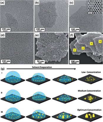
Ng et al.Reference Ng, Udagedara, Rukhlenko, Chen, Tang, Premaratne and Cheng78 reported treating CTAB-modified gold nanorods with a polystyrene-thiol-THF solution with vigorous agitation to obtain PS-modified gold nanorods and then resuspended in chloroform. Horizontal sheets were fabricated by adding the chloroform solution of gold nanorods into a beaker containing water and covered to prevent rapid evaporation. The solidified monolayers present in the water subphase were transferred to TEM lacey grids by a horizontal liftoff technique. Vertical sheets were achieved by a similar procedure with annealing at 60 °C in a water bath. The method resulted in H-sheets and V-sheets of plasmonic ordered superlattices. In more recent studies, semiconductor nanorods such as CdSe were assembled as ordered 3D structures, 2D closed packing perpendicular arrays, or 1D rail tracks.Reference Singh, Gunning, Ahmed, Barrett, English, Garate and Ryan79 The ordering was dependent on a number of factors, such as net charge, dipole moments, nanorod concentration, surface charge, and nature of the solvent. The study revealed that optimization of these factors facilitated assembly with high reproducibility. Figures 8(a)–8(f) show TEM and SEM images of CdSe nanorod assembly at various concentrations. Figure 8(g) shows schematic representations of the drying-mediated nanorod assembly at different concentration. CdSe nanorods with smectic superstructures over millimeter scale were produced by use of capillary flow in drying the droplet of solution. TEM analysis showed the semiconductor nanorods assembled with lamellar-like superstructures and nanorods were aligned side-to-side forming numerous μm-long tracks giving rise to the smectic superstructure.Reference Querner, Fischbein, Heiney and Drndic80 Zanella et al.Reference Zanella, Gomes, Povia, Giannini, Zhang, Riskin, Bael, Hens and Manna81 achieved CdSe nanorod self-assembly over device-scale areas as multilayer vertical nanorod arrangements. CdSe nanorods were precipitated in methanol and redispersed in xylene or orthodichlorobenzene. The substrates were placed on a glass slide inside a petri dish with xylene or orthodichlorobenzene and the sample solution was placed as a drop on the substrate and allowed to dry. SEM images showed multilayer close packing of the nanorods and TEM images confirmed the increasing density of multilayer assembly with increase in concentration.
TEM images of CdS nanorods with concentration (a) 8 × 10⁻⁷ mol/L, (b) 1.4 × 10⁻⁶ mol/L, (c) 2.1 × 10⁻⁶ mol/L, and (d) 3.2 × 10⁻⁶ mol/L. The optimum concentration was found to be 2.1 × 10⁻⁶ mol/L where closed-packed 2D superlattice of CdS nanorods with inset HRTEM image in (c) shows the monodispersity of nanorods. (e–f) HRSEM images showing monolayer and multilayer, vertically oriented CdS nanrods. (g) Schematic representation shows the progression of the nanorod assembly in different stages of droplet drying for different concentrations, the nucleation of the assemblies in the droplet for optimum concentration and further growth process of nanorod assembly is shown in gIII. Reprinted with permission from Singh et al.,Reference Singh, Gunning, Ahmed, Barrett, English, Garate and Ryan79 copyright 2012 Royal Society of Chemistry.

III. CONCLUSIONS
In this review we have highlighted various nanorods assembly methods that have been demonstrated in recent years by manipulating the nanorods surface chemistry, nanorod-ligand interaction, chemical and biochemical strategies and physical forces. While the field is still in its infancy, significant progress has been made in the assembly of nanorods–particularly gold nanorods–and understanding some of the self-directing assembly mechanisms. The work thus far has resulted in the development of a number of well-established protocols with the intent of creating novel architectures utilizing nanorods as primary building blocks. Nevertheless, there are only limited reports on the large-scale assembly of nanorods, which is essential for practical applications. For expanding the development of assembly over large areas, the intrinsic interaction properties of nanorods need to be investigated and exploited. Further in-depth studies of the available surface chemistry are necessary to explore routes for more efficient self-assembly. Ligands with specific binding sites have to be developed that can selectively link nanorods in a controlled manner. Better understanding of the drying kinetics and solvent evaporation phenomena can also open up new avenues for the development of more controlled assembly. Although the assembly of nanorods has seen remarkable progress, there is still much to be discovered and developed, and with many challenges to overcome.
ACKNOWLEDGMENT
This work was supported by the National Science Foundation under Grant No. CHE-1012850.