




No CrossRef data available.
Article contents
A 78 kDa glucose-regulated protein gene of Spirometra erinacei plerocercoid induced by chemical and physiological stresses
Published online by Cambridge University Press: 18 November 2004
Abstract
To adapt to different environmental conditions between poikilothermic and homeothermic hosts, the plerocercoid of Spirometra erinacei (sparganum) might express a variety of biologically active molecules. We have identified a 78 kDa glucose-regulated protein of the sparganum (SpGrp78) by differential display of mRNA, employing RNAs each from sparganum adjusted at 9 °C and 37 °C. A full-length cDNA of 2148 bp encodes for a protein of 651 amino acids with a predicted molecular mass of 71 610 Da and shares molecular characteristics with heat-shock protein 70, including a putative ATP binding site, signal peptide cleavage site and endoplasmic reticulum retention signal. Phylogenetic analysis revealed that SpGrp78 was mostly related to those of Echinococcus multilocularis and E. granulosus. Expression of SpGrp78 mRNA increased approximately 7-fold by inhibition of glycosylation by tunicamycin, 2-fold by temperature-shift from 9 °C to 37 °C and slightly by pH-shift to 4·0 or 5·5. These results suggested that induction of SpGrp78 mRNA is related to the functional role of SpGrp78 as a molecular chaperone when the parasite adapts to a new host environment.
Keywords
Information
- Type
- Research Article
- Information
- Copyright
- © 2004 Cambridge University Press
INTRODUCTION
The sparganum is a plerocercoid of pseudophyllidean tapeworm of the Spirometra species. The type species Spirometra erinaceieuropaei (S. erinacei) is widely distributed throughout Asia and Europe. In clinical conditions, subcutaneous infections are detected sporadically world-wide (Cho, 2003). However, invasion of the central nervous system elicits neurological manifestations such as seizure, headache, hemiplegia, aphasia and progressive deterioration of the mental function (Chang et al. 1987; Kim et al. 1996). Attention should thus be paid to this human-infecting parasite. Biologically, the sparganum has a unique property of low host specificity. It infects a wide variety of intermediate hosts including amphibia, reptiles and mammals. The sparganum might suffer environmental stress especially when it transfers between poikilothermic and homeothermic hosts.
We have hypothesized that the parasite expresses several kinds of biologically active molecules, such as heat-shock proteins (Hsps), anti-oxidant enzymes and proteases, to adapt and to cope with the changing environment of different hosts. One of the plausible candidates is the Hsp70 family, which might play a crucial role in the first line of defence to protect the parasite itself. The Hsp70 families, found in the cytoplasm (Hsp70), nucleus (Hsc70), mitochondrion (Mtp70) and endoplasmic reticulum (Grp78/BiP), are essential for the protection of the cells from various chemical/physiological stresses and are critically involved in the control of cellular differentiation and proliferation and the correct assembly of proteins (Buchner, 1996; Hartl, 1996). The Hsp70 family is divided into 2 major groups, based on their induction patterns: the Hsps and the glucose-regulated proteins (Grps; also known as an immunoglobulin heavy chain binding protein, BiP). Hsp70 has been shown to be a molecule important for survival and adaptation of parasites in the mammalian host environment. Schistosoma mansoni regulates the expression of the Hsp70 gene by a developmentally controlled programme (Neumann et al. 1993; Levy-Holtzman & Schechter, 1996). Differentiation of Trypanosoma cruzi is associated with induction of Hsp70 mRNA (Weiss et al. 1998). BAG1, a small Hsp of Toxoplasma gondii, has also been shown to be specifically expressed in the cyst-forming bradyzoite stage (Bohne, Wirsing & Gross, 1997; Cleary et al. 2002). In cestodes, 2 Grps from Echinococcus granulosus and E. multilocularis (EG-Grp78 and EM-Grp78), which share 99·1% amino acid (aa) sequence identity, have been sequenced (Muhlschlegel et al. 1995); their functional roles, however, remain unexplained.
In the present study, we have characterized a 78 kDa glucose-regulated protein gene of the sparganum (SpGrp78), employing differential display of mRNA. The differential expression of SpGrp78 mRNA in various toxic and stressing conditions have been elucidated. Our results have demonstrated the physiological significance of the SpGrp78 as a defensive mechanism that might protect the parasite from different environmental stresses of the new hosts.
MATERIALS AND METHODS
Collection of Spirometra erinacei plerocercoids (spargana)
Spargana were harvested from the subcutaneous tissues of naturally infected terrestrial snakes, Elaphe rufodorsata and also from BALB/c mice, which had been infected with 5 scoleces, 3 months previously. The worms were washed 5 times with physiological saline at 4 °C. Spargana were stabilized in serum-free minimum essential medium (pH 7·6, MEM; Gibco-BRL, Grand Island, NY, USA) at 9 °C for 18 h prior to use in the experiments. Part of the parasites was stored in liquid nitrogen to be used for protein, total RNA and genomic DNA preparation.
Extraction of RNA
Total RNA was extracted from the sparganum adjusted at 9 °C and 37 °C, as described previously, using guanidium thiocyanate buffer (Chomczynski & Sacchi, 1987). The RNA pellet was washed, air dried and dissolved in distilled water followed by treatment with DNase I (Promega, Madison, WI, USA) for 1 h at 37 °C. The amount and purity of the RNA was determined spectrophotometrically at 260 nm and 280 nm.
Differential display reverse-transcription and polymerase chain reaction (DDRT-PCR)
Differential display was done with the RNAimage kit (GenHunter, Nashville, TN, USA). The first-strand cDNA synthesis was accomplished by RT reactions using 4 μl of 5× reverse transcriptase buffer (125 mM Tris–HCl, 188 mM KCl, 7·5 mM MgCl2, 25 mM dithiothreitol, pH 8·3), 1 μl of Moloney murine leukemia virus (M-MuLV) reverse transcriptase (100 U), 1·6 μl of 250 μM dNTP, 2 μl of oligo-dT primer (T12MG, T12MA, T12MT or T12MC), total RNAs (1 μg) each from the sparganum adjusted at 9 °C and 37 °C and 9·4 μl of D.W. Primers were annealed to the RNA template by incubating the reaction mixture at 65 °C for 5 min and at 37 °C for 60 min in a thermal cycler (Perkin Elmer 9600, Foster City, CA, USA). The reaction was terminated by heating the reactant at 95 °C for 5 min. It was stored at 4 °C until used in PCR.
PCR was performed using combinations of the following anchored primers: 5′-AAGCT11A-3′ (HT11A), 5′-AAGCT11C-3′ (HT11C) and 5′-AAGCT11G-3′ (HT11G) with HAP1 (5′-AAGCTTCGTTGCC-3′), HAP2 (5′-AAGCTTCGACTGT-3′) or randomly designed primers RDP1 (5′-TTGTCGATTGGGAC-3′) and RDP2 (5′-TGTGTCACAG-3′) as upstream primers. Each PCR mixture contained 2 μl of the RT-PCR products, 2 μl of 10× PCR buffer, 2 μl of upstream primer (2 μM), 2 μl of T12MN primer (2 μM), 1·6 μl of dNTP (25 μM), 0·2 μl (1 unit) of AmpliTaq DNA polymerase (Perkin Elmer), 1·0 μl of [α-35S]dATP (1200 Ci/mmol) (Amersham Biosciences, Uppsala, Sweden) and 10 μl of D.W. Reaction mixtures were subjected to 40 cycles of PCR; denaturing at 94 °C for 30 sec, annealing at 40 °C for 2 min, extension at 72 °C for 30 sec and final incubation at 72 °C for 5 min. Resulting products were stored at 4 °C.
[α-35S] dATP-labelled products were separated on a 6% denaturing polyacrylamide gel. Gels were then exposed to Kodak X-AR film overnight at −80 °C and autoradiographed. Differentially expressed bands were gel purified, re-amplified by using the appropriate forward primer and oligo(dT) reverse primer, and cloned into the vector using pCR TRAP Cloning System (GenHunter).The plasmids were analysed by restriction mapping and DNA sequencing using T7 and M-13 reverse primers and an ABI 377 automated DNA sequencer (Perkin Elmer) as described later.
cDNA cloning and terminal deoxynucleotidyl transferase (TdT) reaction
The sparganum cDNA library, which was constructed in λZAPII vector (Stratagene, La Jolla, CA, USA), was screened by the standard method (Sambrook, Russell & Sambrook, 2001) using the nucleotide probe obtained by DDRT-PCR. Approximately 5×10⁵ independent clones were screened by the plaque hybridization method. Pre-hybridization, hybridization and washing conditions were the same as previously described (Sambrook et al. 2001). Single plaques were isolated after consecutive third-round screening. The cDNA inserts were analysed by restriction mapping and DNA sequencing. The nucleotide sequence was determined by the automatic DNA sequencer (Applied Biosystem Model 377, PerkinElmer) using a BigDye Terminator Cycle Sequencing ready reaction kit.
To determine the translation initiation signal of the cloned cDNA, the TdT reaction was performed using 2 specific anti-sense primers, HR1 from 221 nucleotide (5′-GCCGCAGAGAAGGCAAC-3′) and HR2 from 122 nucleotide (5′-CAGGAGTATGTTGTTCCCAG-3′). To add the G-tails at the 5′-end, a total of 1/2 volume of the resulting cDNA was subjected to the TdT reaction for 2 h at 37 °C in 25 μl reaction mixture containing 5× TdT buffer with CoCl2, 500 mM dGTP and 24 U TdT. First round PCR was done in a 25 μl reaction mixture containing 2·5 μl 10× PCR buffer, 5 mM dNTPs, 5 pmole of oligo dC18 primer, 5 pmole of HR1 primer and 2·5 U Taq polymerase. Amplification was carried out in a programmable thermal cycler (PE 9700, Perkin Elmer) for 30 cycles with pre-heating at 95 °C for 2 min, denaturation at 94 °C for 1 min, primer annealing at 55 °C for 1·5 min and extension at 72·5 °C for 2 min with a final incubation for 10 min at 72·5 °C. The amplicon was cleaned with the use of QIAEXII gel extraction kit (Qiagen, Valencia, CA, USA). Second round-PCR was done as previously described, except for primer changing (HR1 to HR2) and cycle shifting 30 to 35 cycles. Resulting products were subcloned into pGEM-T easy vector (Promega).
Sequence alignment and phylogenetic analysis
The full-length cDNA insert was screened against the GenBank database by BLAST N program at the National Center for Biotechnology Information (http://www.ncbi.nlm.nih.gov/BLAST/). A total of 26 sequences with tertiary structures corresponding to the whole sequence were identified and aligned by CLUSTAL W of the BCM search Launcher program (http://dot.imgen.bcm.tmc.edu:9331/multi-align/multi-align.html) (Thompson, Higgins & Gibson, 1994). After optimization by manually trimming and divergence calculation, a phylogenetic analysis was done using PROTDIST and NEIGHBOR of PHYLIP (Felsenstein, 1997) and the phylogram was constructed with TreeView (Page, 1996). Bootstrapping analysis with 1000 replicates was done to evaluate the statistical significance of each node. The PSORT II server program (http://psort.nibb.ac.jp:8800/cpi-bin/runsport.pl) was also used to search the motifs.
Southern blot analysis
High molecular genomic DNAs (3 μg each), isolated from the sparganum following a standard method (Sambrook et al. 2001) were digested with restriction enzymes such as EcoR I, Xho I and Pst I and separated in 0·8% agarose gel and then transferred to Hybond N membrane (Amersham Biosciences). The Southern blot analysis using a 32P-labelled full-length cDNA was carried out following the standard procedure (Sambrook et al. 2001).
Northern blot analysis
The mRNA expression levels of the SpGrp78 were observed under several stressed conditions. After stabilizing the spargana in MEM serum-free medium (pH 7·6) for 18 h at 25 °C (the average temperature of the snake), each of 5 spargana were incubated under different stressed conditions. For temperature-shift experiments, steady-state worms were incubated in pre-warmed or pre-chilled medium at 9 °C, 37 °C and 42 °C, respectively. In a chemical challenging experiment, worms in steady-state were incubated in pre-warmed medium containing tunicamycin (2 mg/ml, Sigma, St Louis, MO, USA) and sodium nitroprusside (10 mg/ml, Sigma). Each solution was diluted with medium to the required concentrations just prior to use. Immediately following the reactions, total RNAs were isolated from individual worms. To observe the SpGrp78 mRNA expression levels, 15 μg total RNA from each of the respective stress conditions was separated on 1·2% formaldehyde denaturing gel and transferred onto Hybond N membrane (Amersham Biosciences). The cDNA was radio-isotope labelled with α-32P dCTP using Rediprime DNA Labelling kit (Amersham Biosciences) and used to probe the membrane in Ultrahyb solution (Ambion, Austin, TX, USA). After hybridization for 18 h at 42 °C, the membrane was thoroughly washed. The membrane was exposed to Kodak X-ray film overnight at −80 °C with intensifying screens. Signal intensities were normalized to the signal from 18S RNA that was derived from ethidium bromide staining. The hybridization signals were quantified using the Kodak Digital Science-1D Image Analysis Software version 2.0.3 (Eastman Kodak, New York, NY, USA).
Western blot analysis
Proteins extracted from each stressed condition were resolved by 6% SDS-PAGE and transferred to nitrocellulose membrane (Schleicher & Schuell, Germany). The membrane was incubated for 1 h with a 1[ratio ]50 dilution of polyclonal mono-specific antibody against Grp78 of human origin, which has an epitope corresponding to aa 24–43 of the amino terminus (Santa Cruz, San Diego, CA, USA). The membrane was subsequently incubated with peroxidase-conjugated anti-goat IgG (Cappel, West Chester, PA, USA) for 1 h. The membrane was washed and developed with ECL Western blotting detection reagents (Amersham Biosciences).
RESULTS
Profile of up-regulated genes of the sparganum under homeothermic conditions
Denaturing polyacrylamide gel electrophoresis of the products amplified by DDRT-PCR identified more than 180 bands that were differentially expressed (data not shown). Nucleic acids contained within these bands were gel-isolated, re-amplified and subjected to dot blotting against total RNAs obtained from each conditioned sparganum. As shown in Table 1, a total of 12 clones, which showed an up-regulatory pattern in the sparganum adjusted at 37 °C, were identified by reverse Northern blot. They included Hsp70s, Grp78s and hypothetical proteins of various organisms, most of which were found to be related to Hsps and Grps. The clones #56-4, 57-6 and 59-3 were found to have the same sequence in which high sequence similarity with Hsps of S. japonicum and E. multilocularis were observed. These clones were confirmed to be up-regulated in the sparganum adjusted at 37 °C by Northern blot analysis (data not shown). We used clone #59-3 (303 bp in size) to isolate a full length cDNA encoding Hsp of the sparganum, because Hsp might play crucial roles in physiological stress, for example, environmental changes of the host from poikilothermic to homeothermic hosts.

Molecular properties of the SpGrp78
A total of 21 clones were obtained after the third round of consecutive screening of a sparganum cDNA library, all of which were found to possess the same sequence in the overlapping regions; however, a translational initiation signal was not identified. To extend the 5′ upstream region, a TdT reaction was performed with the use of cDNA from RT-PCR and gene-specific primers HR1 and HR2 and 6 clones were isolated by colony PCR and sequencing (data not shown). A total of 27 nucleotides present in the clone could be identified, from which we obtained a full-length cDNA sized 2148 bp. A start codon at position 16 of the nucleotide sequence was followed by a single long open reading frame (ORF) of 1953 bp. A stop codon (TAA) was located at position 1969 with a poly (A)+ stretch of 45 nucleotides. The ORF encoded a protein of 651 aa with a predicted molecular mass was 71610 Da (Fig. 1).

Fig. 1. Multiple sequence alignment of deduced amino acid sequence of SpGrp78 with those from other parasites, mouse and human. Percentage similarity is shown at the end of each polypeptide. Black or grey boxes indicate positions of identical amino acids or amino acids that bear biochemical similarity. The arrow indicates the predicted signal peptide cleavage site. Three conserved motifs of all Hsp70 families are underlined. The putative ATP binding site is double underlined. Carboxyl terminal ER retention signal is shown by closed circles ([bull ]). The sequences used for comparison are (1) SpGrp78 in this study; (2) E. multilocularis (GeneBank Accession no. M63604); (3) E. granulosus (M63605); (4) Caenorhabditis elegans (AAB52671); (5) S. japonicum (AF044412); (6) T. gondii (AAC15519); (7) T. cruzi (AAA30201); (8) mouse (AAH50927); and (9) human (AAF13605).
Analysis of the aa sequence revealed typical characteristics of Hsps 70 such as 3 motifs (at aa positions 34–41, 224–231 and 324–330), an ATP-binding site (at aa 157–164), a signal peptide cleavage site (between 20 and 21), coiled-coil structures (between 271–303 and 533–587) and ER retention signal (AKEE, in the C-terminal). Comparison of the predicted aa sequence revealed a significant similarity with other 78 kDa Grps of E. multilocularis, E. granulosus, Caenorhabditis elegans, S. japonicum, T. gondii, T. cruzi, mouse and human by 61·6–78·8% (Fig. 1). We designated this protein as sparganum glucose-regulated protein 78 kDa (SpGrp78).
The genomic distribution of the gene was examined by Southern blot analysis. As shown in Fig. 2A, the 2 or 3 fragments recognized in the genomic DNA digested with restriction enzymes suggested that the gene might be present as a single copy in the genome. In both cDNA and genomic PCR analyses, the products migrated to 2·1 kb without size polymorphism. This result suggested that the gene did not contain an intron sequence in the flanking region (Fig. 2B). Nucleotide sequence data have been submitted to the GenBank data base with the Accession number AY322534.

Fig. 2. Analysis of Southern blot (A) and PCR (B) of genomic DNA from the sparganum. (A) Three μg of genomic DNA were digested with EcoR I, Sal I and Pst I. (B) Genomic PCR was carried out using 50 ng/μl of the sparganum genomic DNA (lane gDNA) and cDNA (lane cDNA) as a template with gene-specific primers.
Molecular phylogenetic analysis of SpGrp78
Molecular phylogenetic analysis was carried out by neighbor-joining algorithm to establish the evolutional and functional relationships of SpGrp78 with other related members. The aa sequence of the SpGrp78 shared a higher degree of similarity with Grp78/BiPs than Mtp70s or Hsp70s (data not shown). A detailed analysis was carried out with the parasite Grp78/BiP to define more precisely its evolutionary relationships. As shown in Fig. 3, the SpGrp78 formed a strong clade with those from other cestode E. multilocularis and E. granulosus with 100% bootstrap value. It could be classified as a sister group of S. japonicum Grp78, which is one of the ancestral forms of Grp78 series of the parasites.

Fig. 3. Phylogenetic analysis of the SpGrp78. The tree was constructed by the neighbor-joining method and rooted with the sequence of E. coli. The sequences used in the construction of the tree included E. coli (GenBank Accession no. K01298), T. brucei (AAC37174), T. cruzi (AAA30201), P. falciparum (Q05866), Eimeria tenella (CAA91253), T. gondii (AAC15519), Zea mays (AAC49899), Nicotina tabacum (Q03684), Arabidopsis thaliana (NP-851119), S. japonicum (AAC00519), E. granulosus (M63605), E. multilocularis (M63604), Suberites domuncula (CAA70695), Xenopus laevis (AAH41200), G. gallus (AAA48785), human (AAF13605), mouse (AAH50927), D. melanogaster (AAN09301), B. mori (BAA32395), Anopheles gambiae (EAA08691), Heterodera glycines (AAM93256), C. elegans (AAB52671), Panagrellus redivivus (AAN15207), Cronartium ribicola (AAK28629), Neurospora crassa (EAA27331) and Pneumocystis carinii (AAB58248). The percentage bootstrap values based on 1000 bootstrap replicates are indicated on each node.
Expression of SpGrp78 by chemical and physiological stresses
The differential expression of SpGrp78 mRNA in various stressing conditions was observed by Northern hybridization. In the temperature up-shift experiments, SpGrp78 mRNA expression was 3-fold higher than steady-state after incubation for 1 and 3 h at 37 °C and for 3 h at 42 °C (lower panel of Fig. 4A). In contrast, expression of SpGrp78 mRNA was not altered by incubating the sparganum at 9 °C for 1, 2 or 3 h. The effect of pH on expression of the SpGrp78 mRNA was observed, in which expression of SpGrp78 mRNA tended to be enhanced at pH 4·0 or 5·5, although the levels were not remarkable (Fig. 4B). An effect of inhibition of protein glycosylation was detected because induction of Grp78 mRNA by glucose deprivation is one of the typical features of Grp78s (Tibbetts et al. 1994). Treatment with tunicamycin markedly induced the expression of SpGrp78 mRNA. An increased expression level of SpGrp78 mRNA was detected after incubation for 1 h, which increased to approximately 7-fold after incubation for 3 h. Sodium nitroprusside, a nitric oxide donor did not influence induction of SpGrp78 mRNA (Fig. 4C). The anterior end of the scolex has been shown to be essential to maintain the viability of the worm (Sohn et al. 1993). To test whether SpGrp78 mRNA expression levels were higher in the scolex than in the tail, total RNAs separately prepared from the sparganum soaked at 37 °C for 3 h were examined. The signal was detected to the same degree which strongly suggested that SpGrp78 was expressed evenly throughout the parasite (Fig. 4D).

Fig. 4. Expression pattern of SpGrp78 induced by various chemical and physiological stresses. (A) Changes in expression pattern of SpGrp78 mRNA induced by temperature shift. After stabilization at 25 °C for 18 h (control), spargana were incubated at 37 °C for 1, or 3 h, or 42 °C for 3 h, respectively. (B) Changes in SpGrp78 mRNA expression at 25 °C induced by pH shift. The parasites were incubated in pH 4·0 or 5·5 medium for 1 or 3 h, respectively. (C) Induction of SpGrp78 mRNA by treatment with tunicamycin and sodium nitroprusside (SNP). (D) Expression of SpGrp78 mRNA according to body compartments. The parasites were incubated for 3 h at 37 °C and then divided into the scolex and tail parts. The upper panel shows ethidium bromide-stained 18S bands and the lower panel exhibits the amount of SpGrp78 mRNA detected by Northern blot analysis.
Expression levels of SpGrp78 protein were analysed after temperature shift and tunicamycin treatment. As shown in Fig. 5, however, expression of SpGrp78 protein was not affected by the temperature shift and tunicamycin treatment as determined by Western blot analysis.

Fig. 5. Western blot analysis. After stabilization at 25 °C, the parasites were incubated for 3 h at 4, 9 and 37 °C. Crude extracts obtained from each shocked sparganum, were resolved by 6% SDS-PAGE and further processed with Western blot, using Grp78 antibody.
DISCUSSION
In the present study, we have screened the differential expression patterns of mRNAs of the sparganum adjusted at different temperatures. Several up-regulated genes have been identified, of which the sparganum glucose-regulated 78 kDa (SpGrp78) has been characterized. The SpGrp78 conserved several motifs of Hsps 70, ATP-binding sites (Hartl, 1996; Vayssier et al. 1999) and signal peptide cleavage site (von Heijine, 1986). ER retention signal (AKEE) that constitutes a series of hydrophobic aa and facilitates the translocation of Grp78 into the ER as well as maintains the protein in the ER lumen, was conserved in the carboxyl terminus (Muhlschlegel et al. 1995). Coiled-coil structures, one of the characteristics of structural proteins, such as myosins and DNA-binding proteins including leucine-zipper, were found at two regions. Two alpha-helices in these regions might bind each other to form a coil in this structure (Lupas, Van & Stock, 1991). Since Hsps might play important roles in the first line of defence, they are stipulated to contribute to the adaptation of the parasites to different conditions of the host environment (Moseley, 1997).
During metamorphogenesis and stage transition of the sparganum, adaptation to the given host environment seems to be one of the most crucial events to overcome. Previous investigations have shown that the parasites undergo marked changes in mitochondrial morphology and components during the environmental adaptation. The parasite might modulate the response of their membrane-bound molecules by altering the membrane composition in response to temperature changes (Kita, Nihei & Tomitsuka, 2003). It was also shown that yeast Grp78 is heat inducible and a heat-shock element is contained in the yeast gene encoding Grp78, KAR2 (Mori et al. 1998). In the case of SpGrp78, it was inducible by heat shock at 37 °C or 42 °C which suggests that SpGrp78 is likely to be more related to yeast Grp78 than mammalian Grp78 in the upstream regulatory region of SpGrp78.
When the steady state is disturbed by changes in the environment, organisms might trigger regulatory mechanisms in order to restore the steady state. In all restoration processes related with physiological condition, ATP plays a central role in cellular activities. Grp78/BiP might act the same way; i.e. in an ATP-dependent manner. This ATP-binding site is also highly conserved in the SpGrp78. Hsps hydrolyse ATP to provide the energy for its role in their conformational change for protein folding, assembly and disassembly and endocytic action (Bukau & Horwich, 1998). A chaperone protein, for example, has been shown to act in vitro as an uncoating ATPase, which used ATP hydrolysis energy to remove the coat from clathrin-coated vesicles. ATP was required to drive the formation of the coatomer complex which did not self-assemble (Hannan, Newmyer & Schmid, 1998). A recent study has also demonstrated that over-expression of arginine kinase of Trypanosoma cruzi can possibly be associated with the adaptation of the parasite to environmental changes by regulating ATP buffering capacity (Pereira et al. 2003).
Grp78/BiP might be induced by glucose deprivation and inhibition of glycosylation, which is an important feature that distinguishes Grp78 from other Hsp70 families. In this study, tunicamycin, which has a structure similar to UDP-N-acetyl glucosamine and inhibits the first stage of the all N-linked oligosaccharide synthesis, was used to observe its effect on the expression of SpGrp78 mRNA. While the levels of Grp78 mRNA increased significantly after tunicamycin treatment, SpGrp78 protein levels remained at a steady state. The apparent incongruity of the SpGrp78 mRNA and protein levels might be a result of a secondary effect of the tunicamycin treatment, since tunicamycin not only inhibits protein glycosylation but also inhibits protein synthesis (Duksin & Mahoney, 1982). However, involvement of an unknown mechanism in this post-transcriptional gene silencing, such as endogenous RNA interference cannot be ruled out. This phenomenon awaits further studies.
Hsp70/Grp78 has been reported to be a reliable diagnostic marker in some parasitic infections, including leishmaniasis, trypanosomiasis, babesiasis, schistosomiasis and filariasis (Skeiky et al. 1995; Krautz et al. 1998; Grijalva, Goodrum & Rowland, 1999) or a parameter useful in prediction of the progress of the disease (Garred et al. 2003). In this study, a recombinant SpGrp78 was expressed in E. coli cells and was used to assess its antigenicity against sera from patients infected with several helminth parasites. Recombinant SpGrp78 did not invoke specific IgE or IgG antibody reactions although it cross-reacted not only with sparganosis sera but also with other parasitic infection sera (data not shown). This result suggested that the epitopes of SpGrp78 recognized by the infected sera are located in the variable carboxyl terminus, like Hsp70 proteins, because the N-terminal peptide sequence of the SpGrp78 is highly conserved with those of other Grp78/BiP (Amorim et al. 1996) but highly variable in the carboxyl terminus. In contrast to Grp78s of other parasites, SpGrp78 could not be used as a diagnostic antigen for human sparganosis.
In conclusion, a glucose-regulated protein gene of the sparganum (SpGrp78) identified in this study encodes for a protein of 651 aa with a predicted molecular mass of 71610 Da and shares molecular characteristics with heat-shock protein 70, including a putative ATP-binding site, a single peptide cleavage site and an endoplasmic reticulum retention signal. The phylogenetic analysis revealed that SpGrp78 was most closely related to those found in other cestodes. SpGrp78 was specifically and significantly induced by the high temperature shift and inhibition of glycosylation; however, an increase of the protein synthesis could not be observed. These results suggest that induction of SpGrp78 mRNA is not directly related with protein synthesis but is related with the functional role of SpGrp78 as a molecular chaperone. It will be interesting to determine which proteins are regulated by SpGrp78 and how SpGrp78 interacts with other proteins as a cellular chaperone.
This work was supported by a KOSEF (KOSEF 96-04-03-05-01-3). The authors are grateful to Dr June Traicoff for editing the manuscript.
References
REFERENCES
AMORIM, A. G.,
CARRINGTON, M.,
MILES, M. A.,
BARKER, D. C. &
DE ALMEIDA, M. L. (
1996).
Identification of the C-terminal region of 70 kDa heat shock protein from Leishmania (Viannia) braziliensis as a target for the humoral immune response.
Cell Stress Chaperones
1,
177–187.2.3.CO;2>CrossRefGoogle Scholar
BOHNE, W.,
WIRSING, A. &
GROSS, U. (
1997).
Bradyzoite-specific gene expression in Toxoplasma gondii requires minimal genomic elements.
Molecular and Biochemical Parasitology
85,
89–98.CrossRefGoogle Scholar
BUCHNER, J. (
1996).
Supervising the fold: functional principles of molecular chaperones.
FASEB Journal
10,
10–19.CrossRefGoogle Scholar
BUKAU, B. &
HORWICH, A. L. (
1998).
The Hsp70 and Hsp60 chaperone machines.
Cell
92,
351–366.CrossRefGoogle Scholar
CHANG, K. H.,
CHO, S. Y.,
CHI, J. G.,
KIM, W. S.,
HAN, M. C.,
KIM, C. W.,
MYUNG, H. &
CHOI, K. S. (
1987).
Cerebral sparganosis: CT characteristics.
Radiology
165,
505–510.CrossRefGoogle Scholar
CHO, S. Y. (
2003).
Pseudophyllidean tapeworms: diphyllobothriasis and sparganosis. In
Oxford Textbook of Medicine, 4th Edn (
ed. Warrell, D. A., Cox, T. M. & Firth, J. D.), pp.
828–829.
Oxford University Press,
Oxford.
CHOMCZYNSKI, P. &
SACCHI, N. (
1987).
Single-step method of RNA isolation by acid guanidium thiocyanate-phenol-chloroform extraction.
Analytical Biochemistry
162,
156–159.CrossRefGoogle Scholar
CLEARY, M. D.,
SINGH, U.,
BLADER, I. J.,
BREWER, J. L. &
BOOTHROYD, J. C. (
2002).
Toxoplasma gondii asexual development: identification of developmentally regulated genes and distinct patterns of gene expression.
Eukaryotic Cell
1,
329–340.CrossRefGoogle Scholar
DUKSIN, D. &
MAHONEY, W. C. (
1982).
Relationship of the structure and biological activity of the natural homologues of tunicamycin.
Journal of Biological Chemistry
257,
3105–3109.Google Scholar
FELSENSTEIN, J. (
1997).
PHYLIP version 3.5c. University of Washington,
Seattle, WA, USA.
GARRED, P.,
NIELSEN, M. A.,
KURTZHALS, J. A.,
MALHOTRA, R.,
MADSEN, H. O.,
GOKA, B. Q.,
AKANMORI, B. D.,
SIM, R. B. &
HVIID, L. (
2003).
Mannose-binding lectin is a disease modifier in clinical malaria and may function as opsonin for Plasmodium falciparum-infected erythrocytes.
Infection and Immunity
71,
5245–5253.CrossRefGoogle Scholar
GRIJALVA, M. J.,
GOODRUM, K. J. &
ROWLAND, E. C. (
1999).
Immunological characterization of antigens released by Trypanosoma cruzi-infected cells.
Journal of Parasitology
85,
663–671.CrossRefGoogle Scholar
HANNAN, L. A.,
NEWMYER, S. L. &
SCHMID, S. L. (
1998).
ATP- and cytosol-dependent release of adaptor proteins from clathrin-coated vesicles: a dual role for Hsc70.
Molecular Biology of the Cell
9,
2217–2229.CrossRefGoogle Scholar
HARTL, F. U. (
1996).
Molecular chaperones in cellular protein folding.
Nature, London
381,
571–579.CrossRefGoogle Scholar
KIM, D. G.,
PAEK, S. H.,
CHANG, K. H.,
WANG, K. C.,
JUNG, H. W.,
KIM, H. J.,
CHI, J. G. &
HAN, D. H. (
1996).
Cerebral sparganosis: clinical manifestations, treatment, and outcome.
Journal of Neurosurgery
85,
1066–1071.CrossRefGoogle Scholar
KITA, K.,
NIHEI, C. &
TOMMITSUKA, E. (
2003).
Parasite mitochondria as drug target: diversity and dynamic changes during the life cycle.
Current Medical Chemistry
10,
2535–2548.CrossRefGoogle Scholar
KRAUTZ, G. M.,
PETERSON, J. D.,
GODSEL, L. M.,
KRETTLI, A. U. &
ENGMAN, D. M. (
1998).
Human antibody responses to Trypanosoma cruzi 70-kD heat-shock proteins.
American Journal of Tropical Medicine and Hygiene
58,
137–143.CrossRefGoogle Scholar
LEVY-HOLTZMAN, R. &
SCHECHTER, I. (
1996).
Expression of different forms of the heat-shock factor during the life cycle of the parasitic helminth Schistosoma mansoni.
Biochimica et Biophysica Acta
1317,
1–4.CrossRefGoogle Scholar
LUPAS, A.,
VAN DYKE, M. &
STOCK, J. (
1991).
Predicting coiled coils from protein sequences.
Science
252,
1162–1164.CrossRefGoogle Scholar
MORI, K.,
OGAWA, N.,
KAWAHARA, T.,
YANAGI, H. &
YURA, Y. (
1998).
Palindrome with spacer of one nucleotide is characteristic of the cis-acting unfolded protein response element in Saccharomyces cerevisiae.
Journal of Biological Chemistry
273,
9912–9920.CrossRefGoogle Scholar
MOSELEY, P. L. (
1997).
Heat shock proteins and heat adaptation of the whole organism.
Journal of Applied Physiology
83,
1413–1417.CrossRefGoogle Scholar
MUHLSCHLEGEL, F.,
FROSCH, P.,
CASTRO, A.,
APFEL, H.,
MULLER, A. &
FROSCH, M. (
1995).
Molecular cloning and characterization of an Echinococcus multilocularis and Echinococcus granulosus stress protein homologous to the mammalian 78 kDa glucose regulated protein.
Molecular and Biochemical Parasitology
74,
245–250.CrossRefGoogle Scholar
NEUMANN, S.,
ZIV, E.,
LANTNER, F. &
SCHECHTER, I. (
1993).
Regulation of HSP70 gene expression during the life cycle of the parasitic helminth Schistosoma mansoni.
European Journal of Biochemistry
212,
589–596.CrossRefGoogle Scholar
PAGE, R. D. (
1996).
Tree view: an application to display phylogenetic trees on personal computers.
Computerized Applied Biosciences
12,
357–358.CrossRefGoogle Scholar
PEREIRA, C. A.,
ALONSO, G. D.,
IVALDI, S.,
SILBER, A. M.,
ALVES, M. J.,
TORRES, H. N. &
FLAWIA, M. M. (
2003).
Arginine kinase overexpression improves Trypanosoma cruzi survival capability.
FEBS Letter
554,
201–205.CrossRefGoogle Scholar
SAMBROOK, J.,
RUSSELL, D. W. &
SAMBROOK, J. (
2001).
Molecular Cloning: A Laboratory Manual, 3rd Edn.
Cold Spring Harbor Laboratory Press,
New York, USA.
SKEIKY, Y. A.,
BENSON, D. R.,
GUDERIAN, J. A.,
WHITTLE, J. A.,
BACELAR, O.,
CARVALHO, E. M. &
REED, S. G. (
1995).
Immune responses of leishmaniasis patients to heat shock proteins of Leishmania species and humans.
Infection and Immunity
63,
4105–4114.Google Scholar
SOHN, W. M.,
HONG, S. T.,
CHAI, J. Y. &
LEE, S. H. (
1993).
Infectivity of the sparganum treated by praziquantel, gamma-irradiation and mechanical cutting.
Korean Journal of Parasitology
31,
135–139.CrossRefGoogle Scholar
THOMPSON, J. D.,
HIGGINS, D. G. &
GIBSON, T. J. (
1994).
CLUSTAL W: improving the sensitivity of progressive multiple sequence alignment through sequence weighting, positions-specific gap penalties and weight matrix choice.
Nucleic Acids Research
22,
4673–4680.CrossRefGoogle Scholar
TIBBETTS, R. S.,
KIM, I. Y.,
OLSON, C. L.,
BARTHEL, L. M.,
SULLIVAN, M. A.,
WINQUIST, A. G.,
MILLER, S. D. &
ENGMAN, D. M. (
1994).
Molecular cloning and characterization of the 78-kilodalton glucose-regulated protein of Trypanosoma cruzi.
Infection and Immunity
62,
2499–2507.Google Scholar
VAYSSIER, M.,
LE GUERHIER, F.,
FABIEN, J. F.,
PHILIPPE, H.,
VALLET, C.,
ORTEGA-PIERRES, G.,
SOULE, C.,
PERRET, C.,
LIU, M.,
VEGA-LOPEZ, M. &
BOIREAU, P. (
1999).
Cloning and analysis of a Trichinella britovi gene encoding a cytoplasmic heat shock protein of 72 kDa.
Parasitology
119,
81–93.CrossRefGoogle Scholar
VON HEIJNE, G. (
1986).
A new method for predicting signal sequence cleavage sites.
Nucleic Acids Research
14,
4683–4690.CrossRefGoogle Scholar
WEISS, L. M.,
MA, Y. F.,
TAKVORIAN, P. M.,
TANOWITZ, H. B. &
WITTNER, M. (
1998).
Bradyzoite development in Toxoplasma gondii and the hsp70 stress response.
Infection and Immunity
66,
3295–3302.Google Scholar

Table 1. BLAST search of up-regulated genes by DDRT-PCR and reverse Northern blot from the sparganum adjusted at 37 °C

Fig. 1. Multiple sequence alignment of deduced amino acid sequence of SpGrp78 with those from other parasites, mouse and human. Percentage similarity is shown at the end of each polypeptide. Black or grey boxes indicate positions of identical amino acids or amino acids that bear biochemical similarity. The arrow indicates the predicted signal peptide cleavage site. Three conserved motifs of all Hsp70 families are underlined. The putative ATP binding site is double underlined. Carboxyl terminal ER retention signal is shown by closed circles ([bull ]). The sequences used for comparison are (1) SpGrp78 in this study; (2) E. multilocularis (GeneBank Accession no. M63604); (3) E. granulosus (M63605); (4) Caenorhabditis elegans (AAB52671); (5) S. japonicum (AF044412); (6) T. gondii (AAC15519); (7) T. cruzi (AAA30201); (8) mouse (AAH50927); and (9) human (AAF13605).
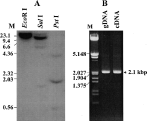
Fig. 2. Analysis of Southern blot (A) and PCR (B) of genomic DNA from the sparganum. (A) Three μg of genomic DNA were digested with EcoR I, Sal I and Pst I. (B) Genomic PCR was carried out using 50 ng/μl of the sparganum genomic DNA (lane gDNA) and cDNA (lane cDNA) as a template with gene-specific primers.
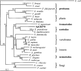
Fig. 3. Phylogenetic analysis of the SpGrp78. The tree was constructed by the neighbor-joining method and rooted with the sequence of E. coli. The sequences used in the construction of the tree included E. coli (GenBank Accession no. K01298), T. brucei (AAC37174), T. cruzi (AAA30201), P. falciparum (Q05866), Eimeria tenella (CAA91253), T. gondii (AAC15519), Zea mays (AAC49899), Nicotina tabacum (Q03684), Arabidopsis thaliana (NP-851119), S. japonicum (AAC00519), E. granulosus (M63605), E. multilocularis (M63604), Suberites domuncula (CAA70695), Xenopus laevis (AAH41200), G. gallus (AAA48785), human (AAF13605), mouse (AAH50927), D. melanogaster (AAN09301), B. mori (BAA32395), Anopheles gambiae (EAA08691), Heterodera glycines (AAM93256), C. elegans (AAB52671), Panagrellus redivivus (AAN15207), Cronartium ribicola (AAK28629), Neurospora crassa (EAA27331) and Pneumocystis carinii (AAB58248). The percentage bootstrap values based on 1000 bootstrap replicates are indicated on each node.

Fig. 4. Expression pattern of SpGrp78 induced by various chemical and physiological stresses. (A) Changes in expression pattern of SpGrp78 mRNA induced by temperature shift. After stabilization at 25 °C for 18 h (control), spargana were incubated at 37 °C for 1, or 3 h, or 42 °C for 3 h, respectively. (B) Changes in SpGrp78 mRNA expression at 25 °C induced by pH shift. The parasites were incubated in pH 4·0 or 5·5 medium for 1 or 3 h, respectively. (C) Induction of SpGrp78 mRNA by treatment with tunicamycin and sodium nitroprusside (SNP). (D) Expression of SpGrp78 mRNA according to body compartments. The parasites were incubated for 3 h at 37 °C and then divided into the scolex and tail parts. The upper panel shows ethidium bromide-stained 18S bands and the lower panel exhibits the amount of SpGrp78 mRNA detected by Northern blot analysis.
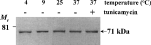
Fig. 5. Western blot analysis. After stabilization at 25 °C, the parasites were incubated for 3 h at 4, 9 and 37 °C. Crude extracts obtained from each shocked sparganum, were resolved by 6% SDS-PAGE and further processed with Western blot, using Grp78 antibody.
 You have
Access
- 3
- Cited by