Introduction
Independent mobility in society requires not only sufficient motor function, but also the ability to navigate in both familiar and unfamiliar surroundings. Spatial navigation involves both route-planning and way-finding, and can be defined as the ability to determine and maintain a route from one place to another (Gallistel, Reference Gallistel1990). Failure in spatial navigation may therefore lead to topographical disorientation and getting lost. Navigational skills decline with age (Cushman et al., Reference Cushman, Stein and Duffy2008), and this decline is even more pronounced in individuals with AD, where spatial disorientation is considered one of the earliest signs of the disease (Pai and Jacobs, Reference Pai and Jacobs2004). Studies have found that approximately 50% of community-dwelling people with AD experience navigational impairment (Pai and Jacobs, Reference Pai and Jacobs2004; deIpolyi et al., Reference deIpolyi, Rankin, Mucke, Miller and Gorno-Tempini2007). This is likely because spatial navigation impairment is related to atrophy of the hippocampus and parahippocampal gyrus (O’Keefe and Nadel, Reference O’Keefe and Nadel1978; deIpolyi et al., Reference deIpolyi, Rankin, Mucke, Miller and Gorno-Tempini2007), where the first neural losses are observed in patients with AD (Braak and Braak, Reference Braak and Braak1991). Loss of confidence in navigational skills is likely to hamper activities of daily living (ADL). This was shown in a study where older drivers with self-perceived navigational impairments reported that they avoided unfamiliar routes and places and drove less than those who experienced no navigational problems (Burns, Reference Burns1999). Thus, navigational ability is therefore an important issue to address with regard to impact on daily activities, as well as in the light of early identification of AD.
Descriptions of a continuum between healthy aging and AD involve at-risk states referred to as SCI and MCI (Reisberg et al., Reference Reisberg, Shulman, Torossian, Leng and Zhu2010). MCI is characterized by a measurable decline in cognition, but still largely preserved functional abilities (Winblad et al., Reference Winblad2004), although some decrements in complex ADL are reported (Hesseberg et al., Reference Hesseberg, Bentzen, Ranhoff, Engedal and Bergland2013). While MCI has a relatively well-established position in the continuum between healthy aging and AD, SCI is considered a more heterogeneous condition, where there is no objective evidence of cognitive or functional impairments despite personal concern (Jessen et al., Reference Jessen2010). The risk of cognitive decline and progression to MCI and AD is higher for individuals with SCI than for healthy controls (Reisberg et al., Reference Reisberg, Shulman, Torossian, Leng and Zhu2010), but the estimated time for progression may be as long as 15 years (Reisberg et al., Reference Reisberg2008). It is important to acknowledge that a substantial portion of people with SCI, as well as about half of them with MCI, remain cognitively stable and even cease to worry about their cognitive function. However, studies suggest that changes in spatial abilities occur before patients fill the criteria for AD. In a longitudinal study, visuospatial abilities began to decline three years prior to a clinical diagnosis of AD (Johnson et al., Reference Johnson, Storandt, Morris and Galvin2009). In cross-sectional studies navigational impairments have also been reported in patients with MCI (deIpolyi et al., Reference deIpolyi, Rankin, Mucke, Miller and Gorno-Tempini2007; Hort et al., Reference Hort, Laczo, Vyhnalek, Bojar, Bures and Vlcek2007; Benke et al., Reference Benke, Karner, Petermichl, Prantner and Kemmler2013), while patients with SCI have shown no impairment of their navigational skills relative to healthy controls (Kalova et al., Reference Kalova, Vlcek, Jarolimova and Bures2005; Hort et al., Reference Hort, Laczo, Vyhnalek, Bojar, Bures and Vlcek2007). However, literature on spatial navigational abilities in the continuum between healthy and AD is still scarce, and a reason might be that research is hampered by the lack of established consensus on how to conduct assessments of spatial navigation with real-life applicability.
Real-life spatial navigation is continuously and dynamically conducted in complex surroundings, and assessments that are ecologically valid are therefore difficult to perform in both clinical and research settings. Studies aimed to reflect real-life complexity of spatial navigation have assessed the ability to navigate in hospital settings (deIpolyi et al., Reference deIpolyi, Rankin, Mucke, Miller and Gorno-Tempini2007; Benke et al., Reference Benke, Karner, Petermichl, Prantner and Kemmler2013) or in advanced virtual reality (VR) settings (Cushman et al., Reference Cushman, Stein and Duffy2008). In clinical practice however, it is more common to use pencil-and-paper tests, probably because tests using real-life environments consume time and space, and VR equipment is seldom available. The pencil-and-paper tests typically target visuoconstruction and figure copying skills, which do represent one aspect of spatial abilities; however, the relationship between these tests and real-life navigation is not clear (Moffat, Reference Moffat2009). Both the traditional VR tests and the pencil-and-paper tests lack the multisensory processes related to actual real-life navigation, where we hear, see, and move our bodies in relation to our surroundings. In one study of walking on a treadmill with and without support, the increased postural demands of walking without support was found to influence the performance on a VR-based spatial navigation task in cognitively healthy persons (Lovden et al., Reference Lovden, Schellenbach, Grossman-Hutter, Kruger and Lindenberger2005). This study by Lovden et al. indicates a dual task effect on the way-finding ability during motor activity in healthy people. We suggest that this approach may be useful to detect spatial navigation impairments also in patients with cognitive impairment and dementia.
The FMT (Sanders et al., Reference Sanders, Holtzer, Lipton, Hall and Verghese2008), utilizes this approach by combining a two-dimensional maze task with walking and was presented as a clinical test of spatial navigation with real-life applicability. The combined walking and navigation in the FMT may provide an opportunity to identify navigational impairments at a very early stage of cognitive impairment. It should be kept in mind that spatial navigation is one of several interrelated cognitive domains, and successful navigation requires contributions from other cognitive processes such as visual perception, learning, memory, and executive functions (Moffat, Reference Moffat2009). In a sample of cognitively healthy elderly subjects, the FMT was associated primarily with executive function, as well as memory function (Sanders et al., Reference Sanders, Holtzer, Lipton, Hall and Verghese2008). Findings from previous studies, using other measures of spatial abilities, have not been consistent regarding the contribution from standard cognitive test batteries, or demographical factors, on performance of navigation tasks (deIpolyi et al., Reference deIpolyi, Rankin, Mucke, Miller and Gorno-Tempini2007; Benke et al., Reference Benke, Karner, Petermichl, Prantner and Kemmler2013), which leaves a concern that navigational impairments may go undetected.
The FMT is a relatively new test, so we believe it is important to develop an understanding of which other cognitive domains contribute to the performance on the FMT. The aims of this study were to explore (1) whether there are any differences with regard to performance on the FMT between patients with SCI and MCI and between patients with MCI and mild AD; and (2) which sociodemographic factors and tests in the routine cognitive test battery used in memory clinics are related to the performance on the FMT.
Methods
Participants
This cross-sectional study recruited patients from a larger study focused on gait and balance in patients with cognitive impairment (Tangen et al., Reference Tangen, Engedal, Bergland, Moger and Mengshoel2014). For that study, the following inclusion criteria were used: (1) ability to walk without a walking device; (2) be home-dwelling; (3) be able to follow instructions in Norwegian; and (4) have a tentative diagnosis of SCI, MCI, or mild to moderate AD. The exclusion criteria were moderate to severe pain when walking, other dementia disorders, other severe neurological disease, or severe hearing and vision impairment. In this study, moderate AD was an additional exclusion criterion. The patients’ eligibility was determined based on clinical judgment of information from the patients, carers, staff at the memory clinic and medical records. Patients from the Memory Clinic at Oslo University Hospital were consecutively enrolled from January 2011 to August 2012. Furthermore, we included seven patients with SCI during November 2013. All patients provided their informed written consent, and the study was approved by the Regional Committee for Medical and Health Research Ethics in the Southeast of Norway.
Demographic and clinical information
All participants were included in the Norwegian Dementia Registry (NDR), and data from this registry and the patients’ medical records were used to obtain the demographic characteristics, medical information, and cognitive assessments. A very experienced geriatric psychiatrist (KE) blinded to the FMT results reviewed all of the tentative diagnoses after the patients’ first visit to the memory clinic. Patients with subjective memory complaints that did not meet the criteria for MCI were classified as having SCI. For the MCI diagnosis, we applied the Winblad criteria: self and/or informant-reported cognitive impairment and one or more results more than1.5 SD below the normative means in the cognitive test battery, but still having no or minimal functional impairment (Winblad et al., Reference Winblad2004). We used the International Classification of Diseases-ten diagnostic criteria for research to diagnose AD and to determine if the AD status was mild or moderate (patients with the latter were excluded) (World Health Organization, 1993). The assessment of gait speed (10-m test) was conducted in a quiet corridor at the memory clinic, with the patients instructed to walk at their usual pace. Timing began when the patient began to walk and ended when they crossed the 10-m line.
Cognitive assessments
The MMSE was used to assess global cognition (Folstein et al., Reference Folstein, Folstein and McHugh1975), which can be scored from 0–30, with higher scores indicating better performance. The TMT-A was used to evaluate attention and processing speed, and the TMT-B was used to examine executive function and set-shifting ability (switching between multiple tasks) (Reitan, Reference Reitan1955). Attempts on the TMTs were interrupted after 5 min, although patients were allowed to continue if they insisted (no performances exceeding 6 min are reported). The Clock Drawing test (CDT) was utilized to evaluate visuoconstructive abilities, applying a dichotomized score of correct versus incorrect based on the Shulman scoring system (5 vs. ≤4) (Shulman, Reference Shulman2000). The learning aspect of memory was evaluated with the Word List Memory test from the Consortium to Establish a Registry for Alzheimer's disease (scored 0–30, where 30 is best) (Fillenbaum et al., Reference Fillenbaum2008).
Floor Maze Test procedure
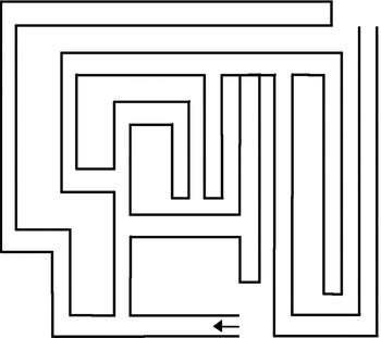
The FMT was created based on the illustration in the original paper (Sanders et al., Reference Sanders, Holtzer, Lipton, Hall and Verghese2008); we used a 7 × 10 foot solid dark blue wax cloth with white tape indicating the lines of the maze (Figure 1). The same physical therapist conducted all of the FMT assessments. The patients were positioned at the entry of the maze and then given instructions. Three components of the FMT were timed: (1) planning time (PT), the time from finishing the instructions until the patient started to walk; (2) immediate maze time (IMT), the time spent walking through the maze from entry to exit; and (3) delayed maze time (DMT), the time spent walking through the maze a second time 10 min after the initial performance, with other walking tests conducted between the two maze walks. Corrections during the walk were counted, and if a patient asked for advice during the walk, the initial instructions were repeated. Two different outcomes were utilized on the FMT: timed performances for the PT, IMT, and DMT and a dichotomous score of error-free performance versus performance with errors or discontinuation of the IMT. Psychometric properties are yet not established for the FMT.
The Floor Maze Test. The start is indicated by the arrow.

Statistical analysis
Data were analyzed using the IBM SPSS Statistics version 20 (IBM Corp., Armonk, NY, USA) with a 5% level of significance. Group differences on the demographic variables with a normal distribution were analyzed by a one-way analysis of variance, Kruskal–Wallis tests were used for variables with a skewed distribution, and χ 2 statistics were used for categorical variables. Comparisons between the patients that were included and omitted from the regression analyses were conducted by t-tests (age, education, and gait speed), by Mann–Whitney U-test (MMSE), and χ 2 tests (sex and comorbidities). Data are presented as the mean and SD, the median and first and third quartiles (Q1 and Q3), or as a frequency.
To analyze group differences on the FMT, we applied χ 2 statistics for the dichotomous variable of error-free versus with error performances on the IMT and DMT, and we applied Mann–Whitney U-tests to analyze the differences in the timed FMT components between the SCI and MCI groups and between the MCI and AD groups.
The relationship between the FMT and the independent variables were analyzed using multiple regression models. Because the timed performances for the PT, IMT, and DMT had skewed distributions, these variables were log transformed prior to the regression analysis. Correlation analyses were performed to check for collinearity among the independent variables. A multiple linear regression analysis was performed by entering all of the independent variables into the model and performing a manual stepwise backward regression analysis, removing the least significant variables until only the significant variables were left. The regression coefficients and confidence intervals were back transformed by the formula (exp (estimate) – 1) × 100%, and are reported as percentage (%).To detect a large effect size (> 0.35) with 80% power and a significance level of 0.05 for nine independent variables in the regression analyzes, we estimated that 54 patients would be required, thus we had the necessary sample for these analysis. As this was an exploratory study, we did not perform power analyzes for the group comparisons.
Results
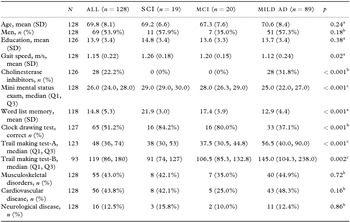
As presented in Table 1, 128 patients (69 men; 53.9%) were included: 19 with SCI, 20 with MCI, and 89 with mild AD. The mean (SD) age was 69.8 (8.1) years, and the median (Q1, Q3) MMSE was 26.0 (24, 28). There were no significant differences between the groups with regard to age, sex, or education (p > 0.18). Sixteen patients (15 with AD and 1 with SCI, mean (SD) age 71.3 (10.7) and MMSE 23.3 (4.1)) gave up to complete the IMT, and had thus no valid results for the IMT and the DMT, and we also excluded them from the analyses of the PT component. Five patients did not complete the DMT due to time constraints.
Demographic characteristics of the patients (n = 128)

aOne-way analysis of variance; bχ 2 test; c Kruskal–Wallis test.
SD, standard deviation; (Q1, Q3), 1st and 3rd quartiles; SCI, subjective cognitive impairment; MCI, mild cognitive impairment; AD, Alzheimer's disease. p = level of significance.
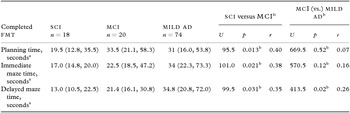
The Fisher's exact test we ran to evaluate the difference in the percentage of error-free performance on the IMT between the SCI group and the MCI group (84.2% vs. 75% error-free performance) indicated no significant differences (p = 0.70). The percentage of error-free performance did significantly differ (x 2(1, N = 109) = 7.3, p = 0.007) between the groups of MCI and mild AD (75% vs. 41.6% error-free performance).
The SCI group was significantly faster than the MCI group on all three FMT components (Table 2): PT (p = 0.013), IMT (p = 0.021), and DMT (p = 0.031), and the effect sizes ranged from 0.35 (DMT) to 0.40 (PT). The MCI group was significantly faster than the AD group on the DMT (p = 0.02), but not on the PT (p = 0.57) or IMT (p = 0.12). There were no significant differences regarding time used as a whole between the PT and IMT (p = 0.46) or between the IMT and DMT (p = 0.39).
In the regression analysis (Table 3), we had one or more missing pieces of data in 30 patients, primarily related to the TMT-B. We did not impute missing data, as these were not missing at random. Thus, the regression analysis consisted of 82 (64%) of the 128 patients enrolled in the study: 15 with SCI, 19 with MCI, and 48 with mild AD. The sample in the regression analysis had significantly higher educational levels (p = 0.030), better MMSE scores (p < 0.001), and faster walking rates (p < 0.001) than those patients who were omitted. There were no significant differences in their age, sex, or medical conditions (p > 0.05).
Group comparisons of the Floor Maze Test performances

amedian (1st quartile, 3rd quartile); bMann–Whitney U-test.
SCI, subjective cognitive impairment; MCI, mild cognitive impairment; AD, Alzheimer's disease; IMT, immediate maze time; FMT, Floor Maze Test. p = level of significance, r = effect size.
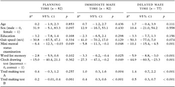
Simple linear regression analysis between the three Floor Maze Test components (log transformed) and the sociodemographic factors, gait speed, and cognitive assessments

aUnstandardized coefficients are back transformed by the formula (exp (estimate) − 1) × 100%, and reported as percentage (%).
CI, confidence interval. p = level of significance
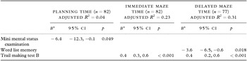
The PT was moderately correlated with the DMT (r = 0.30, p = 0.002), but not with the IMT (r = 0.15, p = 0.11), while the IMT and DMT were highly correlated (r = 0.65, p < 0.001). All independent variables had correlations below r = 0.7; therefore, none were omitted from the analyses. In the multiple linear regression analysis, the MMSE was the only variable that was significantly associated with the PT, adjusted R 2 = 0.04; F1,80 = 4.0, p = 0.049) (Table 4). TMT-B was the only variable significantly associated with the IMT (adjusted R 2 = 0.23; F1,80 = 25.3, p <0.001), and it also contributed to the final DMT (adjusted R 2 = 0.31; F2,74 = 18.3, p <0.001) model together with the Word List Memory test. The B-coefficient of 0.4% indicates that if the performance on the TMT-B increases with 10 sec, the corresponding change on IMT is 4%. The sociodemographic factors were not significantly associated with any of the three FMT components. The estimated effect size (f 2 = R 2/(1−R 2)) of the multiple regression model was small for the PT (f 2 = 0.12), while the IMT (f 2 = 0.41) and DMT (f 2 = 0.56) effect sizes were large (Cohen, Reference Cohen1992).
Final multiple regression models of the associations between the three Floor Maze Test components (log transformed) and independent variables: cognitive tests, sociodemographic factors, and gait speed

aUnstandardized coefficients are back transformed by the formula (exp(estimate) − 1) × 100%, and reported as percentage (%).
CI, confidence interval. Variables age, sex, education, gait speed, clock drawing test, and trail making test-A did not contribute to any of the final models. p = level of significance.
Discussion
In this study, patients with MCI were slower on all the three components of the FMT than the patients with SCI, while the patients with mild AD completed the IMT with errors or gave up more often than the patients with MCI. Executive function, as measured by the TMT-B, was significantly associated with both the IMT and DMT. None of the sociodemographic variables were significantly associated with the FMT components.
We found few indications of spatial navigation impairments in the SCI group; since all but one of our SCI patients were able to complete the IMT without errors, and the SCI group was also faster than the MCI group on the FMT components. These results are in concordance with previous studies that have reported no deficits in spatial navigation in patients with SCI (Kalova et al., Reference Kalova, Vlcek, Jarolimova and Bures2005; Hort et al., Reference Hort, Laczo, Vyhnalek, Bojar, Bures and Vlcek2007). We found more deficits of spatial navigation in the patients with mild AD than in those with MCI. This is in line with findings from previous studies (deIpolyi et al., Reference deIpolyi, Rankin, Mucke, Miller and Gorno-Tempini2007; Cushman et al., Reference Cushman, Stein and Duffy2008; Benke et al., Reference Benke, Karner, Petermichl, Prantner and Kemmler2013), although studies focused on the subgroups of MCI have found no differences between amnestic MCI (primarily memory impairments) and AD patients regarding spatial navigation (Hort et al., Reference Hort, Laczo, Vyhnalek, Bojar, Bures and Vlcek2007; Laczo et al., Reference Laczo2012). However, the patients in the MCI group were not faster than the patients in the AD group on the PT or IMT. So, the patients in the MCI group were generally able to solve the navigational task, but their performances were slow, which may reflect increased effort to carry out the tasks. This is in line with the idea that the continuum from healthy persons to MCI, and then to AD also involves functional limitations, including spatial skills (Hort et al., Reference Hort, Laczo, Vyhnalek, Bojar, Bures and Vlcek2007; Johnson et al., Reference Johnson, Storandt, Morris and Galvin2009; Benke et al., Reference Benke, Karner, Petermichl, Prantner and Kemmler2013). Our results corroborate findings from previous studies that indicate navigational impairments occur before patients fulfill the diagnostic criteria for AD, but they may not be a central feature of SCI.
In this study, we found a significant association between the FMT and the tests in a standard cognitive test battery. Executive function, measured by the TMT-B, was significantly associated with both the IMT and DMT. Memory, measured by the Word List Memory test, was associated with the DMT, and global cognition (assessed by MMSE) was associated with PT. These findings largely agree with the original study (Sanders et al., Reference Sanders, Holtzer, Lipton, Hall and Verghese2008), where the executive function and attention factor were related to all three components of the FMT. Our findings also agree with a study where both executive function and memory were predictors of poor performance on a route learning test in patients with MCI or AD (Benke et al., Reference Benke, Karner, Petermichl, Prantner and Kemmler2013). Other studies have identified executive function as important for “getting lost behavior” (assessed by questionnaire) in home-dwelling patients with AD (Chiu et al., Reference Chiu, Algase, Liang, Liu and Lin2005). Problem solving and maintaining attention are related to way-finding and spatial abilities, but they are also central features of the executive function (Passini et al., Reference Passini, Rainville, Marchand and Joanette1995).
Memory was significantly associated with the DMT, but not the IMT, which contrasts with Sanders’ study that found the opposite (Sanders et al., Reference Sanders, Holtzer, Lipton, Hall and Verghese2008). However, it is reasonable for memory impairments to be more related to the DMT than the IMT in our sample, which is characterized by impaired memory function. It is also interesting to note that our patients devoted the same amount of time to all of the components of the FMT. This stands in contrast to Sanders’ cognitively healthy sample, which spent less time on the IMT component than on PT, and less time on the DMT component than on the IMT, indicating a learning process occurred throughout the test.
The PT and IMT were not correlated, and there was a clear difference in the explained variance between the PT (4%) and the IMT/DMT (23/31%) in our study. This point to two different factors. First, the PT and IMT/DMT measure two different aspects of spatial navigation. There is likely a substantial difference between planning a walk standing at a fixed point and actually executing the planned walk while rotating the map as turns in the maze are made. Second, although we found significant associations between the cognitive tests and FMT, we should be careful not to overestimate their importance. The explained variance is relatively low, indicating our standard cognitive test battery does not sufficiently capture impairments in spatial navigation.
None of the demographic factors were independently associated with any of the FMT components. Several studies have found an age-effect on navigation, where younger people perform better than those that are older. However, these studies typically compared individuals in their twenties to people 60–80 years old (Cushman et al., Reference Cushman, Stein and Duffy2008; Taillade et al., Reference Taillade2013). Our patients were all between 51 and 83 years old, which may explain why no age-effect was seen in our study. Our results also agree with the results from Benke's study, where none of the demographic factors (age, sex, and education) were predictors for route learning performance in patients referred to a memory clinic (Benke et al., Reference Benke, Karner, Petermichl, Prantner and Kemmler2013).
A shortcoming of this study is that the cross-sectional design prevents us from drawing conclusion related to decline of navigational abilities. Further, all patients were recruited from a memory clinic, and this limits the generalizability of our findings to the populations normally seen by specialist outpatient clinics. In addition, the SCI and the MCI groups were small, and the heterogeneity in these two conditions should lead to a cautious interpretation of the findings regarding group differences. However, recruiting patients from a memory clinic is also a strength of this study, because we have a sample consistently examined with the same diagnostic protocol. We also believe patients with SCI recruited from memory clinics may have a higher risk for future cognitive decline than individuals with SCI in population-based studies.
We acknowledge that even though the FMT involves walking, it is obviously still far from a test of real-life spatial navigation, given the lack of use of landmarks, a limited-sized surface, and only being two-dimensional. However, the FMT consumes little time and is an inexpensive test that does not require advanced equipment, and our study has shown that it is feasible for patients with a mild degree of cognitive impairment. Nevertheless, future studies are needed to validate the FMT against real-life navigational tasks and to provide normative values to increase the interpretability of results.
Conclusion
The performance on the FMT worsened with the increasing severity of cognitive impairment. The FMT was associated primarily with executive function and memory; however, the explained variance was relatively low, suggesting the standard cognitive test battery does not capture impaired spatial navigation. Sustained participation in both social and physical activities is important for people with dementia, and therefore we believe it is important to identify those individuals who experience impaired spatial navigation. However, our findings need to be confirmed in future larger cohort studies.
Conflict of interest
None.
Description of authors’ roles
G. G. Tangen, K. Engedal, A. Bergland, O. Hansson and A. M. Mengshoel contributed to the conception and design of the study. G. G. Tangen and T. A. Moger undertook the analyses. G. G. Tangen made the first draft of the manuscript, and, K. Engedal, A. Bergland, T. A. Moger, O. Hansson and A. M. Mengshoel contributed to revising the manuscript critically for important intellectual content. All authors contributed and approved the final manuscript before submission.
Acknowledgment
The authors thank the participants for their cooperation in the study. The research was conducted at the Memory Clinic at Oslo University Hospital, Oslo, Norway.