INTRODUCTION
Plasmodium knowlesi is a simian malaria parasite of the long tail (Macaca fascicularis) and pig tail (Macaca nemestrina) macaques in South-east Asia. It is unique amongst the genus, exhibiting a 24-h asexual erythrocytic cycle, but it has the morphological appearance of Plasmodium malariae (Coatney et al. Reference Coatney, Collins, Warren and Contacos1971). It was utilized in malariotherapy for neurosyphilis from its discovery in the 1930s until the mid-1950s (Knowles and Gupta, Reference Knowles and Gupta1932; Ciuca, Reference Ciuca, Chelarescu, Sofletes, Constantinescu, Teritaenu, Cortez, Balanovschi and Ilies1955). Two cases of naturally-acquired human infections, from Western Malaysia, were reported in the literature (Chin et al. Reference Chin, Contacos, Coatney and Kimball1965; Fong et al. Reference Fong, Cadigan and Coatney1971), but screening in the same area failed to find further human cases (Warren et al. Reference Warren, Cheong, Fredericks and Coatney1970).
Although interest in naturally occurring human infections then waned, the ability of P. knowlesi to induce fatal infections in Rhesus macaques stimulated further research in non-human primate models examining immunological and pathophysiological aspects of malaria [see recent review (Butcher and Mitchell, Reference Butcher and Mitchell2016)] and for pharmacological studies including testing of novel agents [see for examples (Singh et al. Reference Singh, Misra, Ray, Basu and Bami1951a , Reference Singh, Ray, Basu and Nair b ; Singh and Dutta, Reference Singh and Dutta1981; Ding et al. Reference Ding, Li, Shi and Ning1991)].
In 2000, Singh et al. began investigating the unusual clinical manifestations of what was presumed to be P. malariae infections in the Malaysian state of Sarawak. The group's findings led to a profound shift in our understanding of the epidemiology and clinical course of knowlesi malaria. Plasmodium knowlesi proved to be a common naturally occurring infection in humans in Sarawak (Singh et al. Reference Singh, Kim Sung, Matusop, Radhakrishnan, Shamsul, Cox-Singh, Thomas and Conway2004). Further studies have led to the recognition that it is a potentially fatal emerging infectious disease that occurs throughout South-east Asia (Cox-Singh et al. Reference Cox-Singh, Davis, Lee, Shamsul, Matusop, Ratnam, Rahman, Conway and Singh2008).
In this review, we summarize the key human studies of knowlesi malaria with particular reference to clinical features and management. Unless stated, the disease described relates to infections in adults.
HISTORICAL BACKGROUND
Before P. knowlesi was recognized as a naturally-occurring human pathogen, infections with P. malariae were commonly reported in Sarawak. Some of the affected patients were acutely unwell and had high parasitaemias, raising the possibility of a virulent P. malariae strain (Singh et al. Reference Singh, Kim Sung, Matusop, Radhakrishnan, Shamsul, Cox-Singh, Thomas and Conway2004). To investigate this further, polymerase chain reaction (PCR) studies conducted by Singh et al. found species-specific primers for P. malariae and the other human malaria species were all negative. When species primers were expanded to include local primate malaria species (P. knowlesi, Plasmodium cynomolgi, Plasmodium inui and Plasmodium fieldi), the causative organism was confirmed as P. knowlesi.
Further investigations in the Kapit region of Sarawak revealed that knowlesi malaria was common in adults (Singh et al. Reference Singh, Kim Sung, Matusop, Radhakrishnan, Shamsul, Cox-Singh, Thomas and Conway2004). In 141 samples, diagnosed by microscopy as P. malariae, none were confirmed by PCR, while 106 were found to be P. knowlesi mono-infections. All 106 patients were symptomatic, with an acute febrile illness of median duration of 4 days and a wide range of parasitaemias (geometric mean 2641 parasites µL−1 [range 80–117 600]). Infections were successfully treated with conventional courses of chloroquine or quinine.
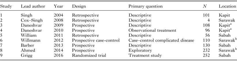
In 2004, there was a single-case report of a Thai male with knowlesi malaria who had spent time in the forested region of Prachuap Khiri Khan in southern Thailand (Jongwutiwes et al. Reference Jongwutiwes, Putaporntip, Iwasaki, Sata and Kanbara2004). These early reports indicated the need for further research and, over the past 10 years, a number of case reports, retrospective case series and prospective clinical studies have informed our understanding of the clinical disease caused by P. knowlesi. A summary of key clinical studies is shown in Table 1.
Summary of key clinical studies in knowlesi malaria

a,bOverlapping cohorts.
EPIDEMIOLOGY
Most cases of knowlesi malaria occur in the states of Sabah and Sarawak in Malaysian Borneo, but the infection has been found in other parts of Malaysia as well as in Singapore, Thailand, Indonesia, Cambodia, Myanmar, Philippines, Vietnam and the Andaman and Nicobar Islands. There is, therefore, a large reservoir of knowlesi malaria in South-east Asia, but the risk of infection varies according to the geographical area (Moyes et al. Reference Moyes, Henry, Golding, Huang, Singh, Baird, Newton, Huffman, Duda, Drakeley, Elyazar, Anstey, Chen, Zommers, Bhatt, Gething and Hay2014; Sunish et al. Reference Sunish, Shriram, De and Vijayachari2015; Shearer et al. Reference Shearer, Huang, Weiss, Wiebe, Gibson, Battle, Pigott, Brady, Putaporntip, Jongwutiwes, Lau, Manske, Amato, Elyazar, Vythilingam, Bhatt, Gething, Singh, Golding, Hay and Moyes2016).
Genetic evidence suggests that knowlesi malaria is a zoonosis (Lee et al. Reference Lee, Divis, Zakaria, Matusop, Julin, Conway, Cox-Singh and Singh2011), with few reported clusters of human cases and no definitive evidence of human-to-human transmission (Lee et al. Reference Lee, Divis, Zakaria, Matusop, Julin, Conway, Cox-Singh and Singh2011; Barber et al. Reference Barber, William, Grigg, Menon, Auburn, Marfurt, Anstey and Yeo2013a ; Grigg et al. Reference Grigg, Barber, Marfurt, Imwong, William, Bird, Piera, Aziz, Boonyuen, Drakeley, Cox, White, Cheng, Yeo, Auburn and Anstey2016a ). Recent modelling studies estimate the risk of human-to-human transmission to be very low (Imai et al. Reference Imai, White, Ghani and Drakeley2014; Brock et al. Reference Brock, Fornace, Parmiter, Cox, Drakeley, Ferguson and Kao2016).
To date, no study has comprehensively investigated risk factors for P. knowlesi infection, although a case control study is currently underway in Sabah (Grigg et al. Reference Grigg, William, Drakeley, Jelip, von Seidlein, Barber, Fornace, Anstey, Yeo and Cox2014b ). However, interactions between the rural population, vectors and natural hosts are common in these areas along with otherwise successful human malaria control programmes. Visitors to these regions are also at risk if engaging in activities that take them into the forest or forest fringe (Cramer, Reference Cramer2015).
Age
Knowlesi malaria is predominantly a disease of adults with children accounting for only 6–14% of cases. This reflects both low endemicity and age-specific differences in forest exposure (Daneshvar et al. Reference Daneshvar, Davis, Cox-Singh, Rafa'ee, Zakaria, Divis and Singh2009; Barber et al. Reference Barber, William, Jikal, Jilip, Dhararaj, Menon, Yeo and Anstey2011; Rajahram et al. Reference Rajahram, Barber, William, Grigg, Menon, Yeo and Anstey2016). In prospective studies of adults, the mean age of patients was 45 years (Daneshvar et al. Reference Daneshvar, Davis, Cox-Singh, Rafa'ee, Zakaria, Divis and Singh2009; Barber et al. Reference Barber, William, Grigg, Menon, Auburn, Marfurt, Anstey and Yeo2013a ). In southern Vietnam, where knowlesi malaria is less prevalent and typically occurs as a co-infection with other Plasmodium species, patients tend to be younger (mean age 15 years) (Marchand et al. Reference Marchand, Culleton, Maeno, Quang and Nakazawa2011). In Sabah, malaria notification data showed that patients with knowlesi malaria were older than those with falciparum or vivax malaria (William et al. Reference William, Jelip, Menon, Anderios, Mohammad, Awang Mohammad, Grigg, Yeo, Anstey and Barber2014).
Gender
In adult disease, there is evidence of regional variation in gender distribution but with a male predominance. In Sarawak, men accounted for 62% of 213 acute hospital admissions with knowlesi malaria (Singh et al. Reference Singh, Kim Sung, Matusop, Radhakrishnan, Shamsul, Cox-Singh, Thomas and Conway2004; Daneshvar et al. Reference Daneshvar, Davis, Cox-Singh, Rafa'ee, Zakaria, Divis and Singh2009), while, in Sabah, men accounted for 74% of acute admissions and 78% of uncomplicated infections (Barber et al. Reference Barber, William, Grigg, Menon, Auburn, Marfurt, Anstey and Yeo2013a ; Grigg et al. Reference Grigg, William, Menon, Dhanaraj, Barber, Wilkes, von Seidlein, Rajahram, Pasay, McCarthy, Price, Anstey and Yeo2016b ). The marked male preponderance in Sabah is supported by Malaysian Department of Health data from Sabah showing that infections in women comprised only 19% of the 4217 knowlesi infections recorded between 2010 and 2014 (Rajahram et al. Reference Rajahram, Barber, William, Grigg, Menon, Yeo and Anstey2016). Such gender differences most likely reflect exposure-related activities. The limited data from children show an even gender distribution (Barber et al. Reference Barber, William, Jikal, Jilip, Dhararaj, Menon, Yeo and Anstey2011).
Occupations, dwellings and forest activities
In the Kapit region of Sarawak, most patients with knowlesi malaria are Iban (ethnic minority) farmers or loggers, with 87% of patients reporting recent activities in the jungle or forest fringes (Daneshvar et al. Reference Daneshvar, Davis, Cox-Singh, Rafa'ee, Zakaria, Divis and Singh2009). In Sabah, farming or plantation work accounted for 40% of the occupations of patients with the disease, but 92% had some forest exposure in the preceding 4 weeks (Barber et al. Reference Barber, William, Grigg, Menon, Auburn, Marfurt, Anstey and Yeo2013a ). In Sabah, 50% of cases reported recent proximity to monkeys, significantly more than in non-knowlesi malaria cases (Barber et al. Reference Barber, William, Grigg, Menon, Auburn, Marfurt, Anstey and Yeo2013a ). In Kapit, 72% of patients reported recent sightings of monkeys (47% both long- and pig-tailed macaques, 22% long-tailed macaques alone and 3% pig-tailed macaques only) (Daneshvar et al. Reference Daneshvar, Davis, Cox-Singh, Rafa'ee, Zakaria, Divis and Singh2009). Recent studies have identified distinct genomic clusters of knowlesi malaria, each associated with their natural macaque hosts, although the clinical significance of this in humans is uncertain (Divis et al. Reference Divis, Singh, Anderios, Hisam, Matusop, Kocken, Assefa, Duffy and Conway2015; Pinheiro et al. Reference Pinheiro, Ahmed, Millar, Sanderson, Otto, Lu, Krishna, Rayner and Cox-Singh2015).
In Cambodia, knowlesi malaria was found in two of 1475 patients in a cross-sectional study of patients with febrile illnesses, accounting for 0·3% of the 754 malaria cases (Khim et al. Reference Khim, Siv, Kim, Mueller, Fleischmann, Singh, Divis, Steenkeste, Duval, Bouchier, Duong, Ariey and Menard2011). Both cases were men aged in their forties who reported hunting in the forest. All cases in travellers have involved jungle exposure (Cramer, Reference Cramer2015).
CLINICAL FEATURES
Asymptomatic infection
Knowlesi infections can vary in presentation from asymptomatic parasitaemia to severe and fatal disease. The prevalence of asymptomatic infections in at-risk communities is unknown (Van den Eede et al. Reference Van den Eede, Van, Van Overmeir, Vythilingam, Duc, Hung le, Manh, Anne, D'Alessandro and Erhart2009; Marchand et al. Reference Marchand, Culleton, Maeno, Quang and Nakazawa2011). However, in a case–control study in Sabah, estimated prevalence in the population was 6·9% (95% CI 5·6–8·4), with 112 of the 1147 asymptomatic participants selected from the same village as symptomatic cases having detectable knowlesi DNA in their blood (Fornace et al. Reference Fornace, Nuin, Betson, Grigg, William, Anstey, Yeo, Cox, Ying and Drakeley2016). While the true prevalence cannot be extrapolated, these data suggest that the burden of silent infections is high. Whether these cases become symptomatic, and at risk of complications are currently unknown.
Symptoms
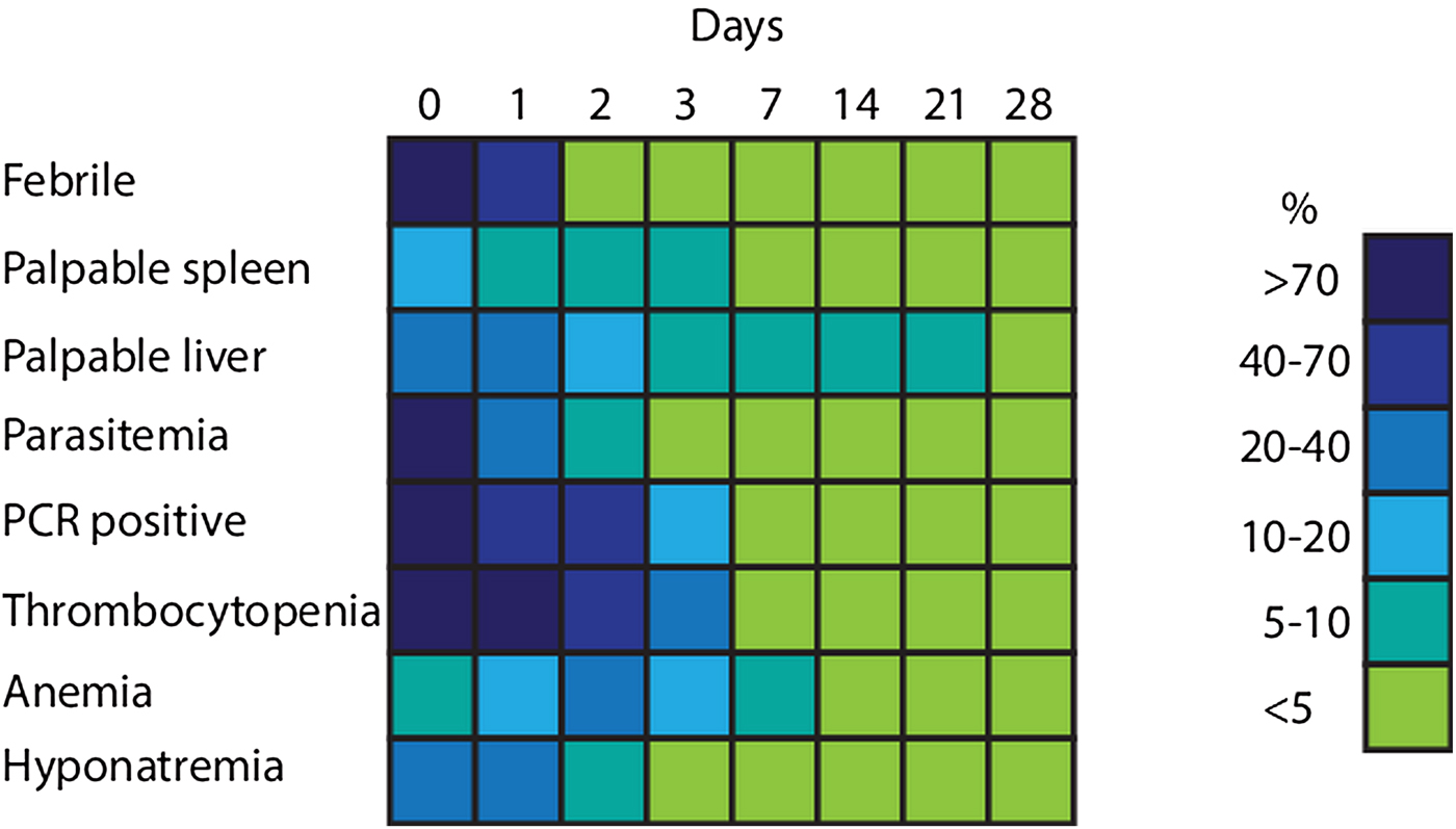
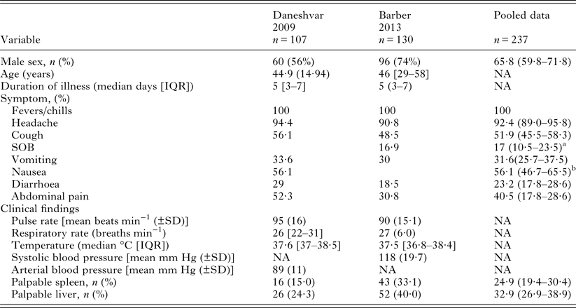
Prospective studies have investigated the symptoms caused by P. knowlesi in adults presenting to hospital with acute infection. A schematic summary of the course of uncomplicated knowlesi malaria is shown in Fig. 1 (Daneshvar, Reference Daneshvar2013a ). Knowlesi malaria is typically characterized by a 5-day history of fever and chills as well as other non-specific symptoms, including headache, myalgia, respiratory and gastrointestinal complaints in over half of patients (see Table 2) (Daneshvar et al. Reference Daneshvar, Davis, Cox-Singh, Rafa'ee, Zakaria, Divis and Singh2009; Barber et al. Reference Barber, William, Grigg, Menon, Auburn, Marfurt, Anstey and Yeo2013a ). Symptoms may pre-date diagnosis by several weeks (Daneshvar et al. Reference Daneshvar, Davis, Cox-Singh, Rafa'ee, Zakaria, Divis and Singh2009; Figtree et al. Reference Figtree, Lee, Bain, Kennedy, Mackertich, Urban, Cheng and Hudson2010).
Schematic representation of the clinical course of uncomplicated Plasmodium knowlesi infections (adapted from Daneshvar, Reference Daneshvar2013b ).

Clinical characteristic of patients from prospective observational studies admitted to hospital with Plasmodium knowlesi infections
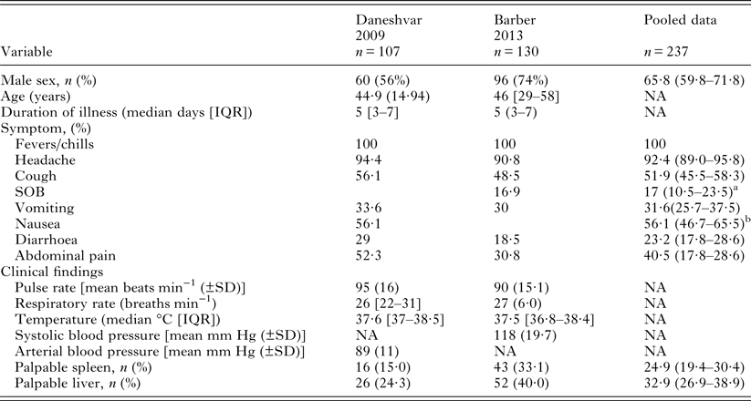
Data are %, % (95% CI), mean (SD) or median [interquartile range].
NA, not available.
a Denominator 130.
b Denominator 107.
Failure to consider malaria as a diagnosis, possibly due to declining malaria incidence in areas where P. knowlesi is transmitted, together with the mistaken belief that parasites on blood films were P. malariae have impeded prompt diagnosis and appropriate treatment (Rajahram et al. Reference Rajahram, Barber, William, Menon, Anstey and Yeo2012). The presence of abdominal pain has led to an erroneous diagnosis of acute gastroenteritis with subsequent treatment delays (Cox-Singh et al. Reference Cox-Singh, Davis, Lee, Shamsul, Matusop, Ratnam, Rahman, Conway and Singh2008; Rajahram et al. Reference Rajahram, Barber, Yeo, Tan and William2013). Prospective studies in Sarawak and Sabah have shown abdominal pain to be present in 31–52% of cases with a greater prevalence in severe cases (90 vs 48%; P < 0·001). This symptom is also positively associated with parasitaemia [odds ratio (OR) 1·25 [log increase] P = 0·026] (Daneshvar et al. Reference Daneshvar, Davis, Cox-Singh, Rafa'ee, Zakaria, Divis and Singh2009; Barber et al. Reference Barber, William, Grigg, Menon, Auburn, Marfurt, Anstey and Yeo2013a ). Other significant symptoms may include breathlessness and vomiting, both being associated with higher parasite counts (Daneshvar et al. Reference Daneshvar, Davis, Cox-Singh, Rafa'ee, Zakaria, Divis and Singh2009).
Clinical signs
As summarized in Table 2, fever, tachycardia and an increased respiratory rate are found commonly while hepatomegaly and splenomegaly can be detected in 24–40 and 15–33% of patients, respectively (Daneshvar et al. Reference Daneshvar, Davis, Cox-Singh, Rafa'ee, Zakaria, Divis and Singh2009; Barber et al. Reference Barber, William, Grigg, Menon, Auburn, Marfurt, Anstey and Yeo2013a ). Clinical signs associated with complicated disease may be present, with up to one in ten patients presenting with hypotension unresponsive to fluid replacement or signs of respiratory compromise (Daneshvar et al. Reference Daneshvar, Davis, Cox-Singh, Rafa'ee, Zakaria, Divis and Singh2009; Barber et al. Reference Barber, William, Grigg, Menon, Auburn, Marfurt, Anstey and Yeo2013a , Reference Barber, William, Grigg, Piera, Yeo and Anstey b ).
Unlike falciparum malaria, abnormal neurological findings are not a prominent feature of knowlesi malaria. In the largest series of well-characterized severe disease, no coma or convulsions were seen (Barber et al. Reference Barber, William, Grigg, Menon, Auburn, Marfurt, Anstey and Yeo2013a ). There have been single case reports of focal neurological signs (Daneshvar et al. Reference Daneshvar, Davis, Cox-Singh, Rafa'ee, Zakaria, Divis and Singh2009) and coma (Glasgow coma scale 6/15 with asymmetric pupils, normal tone, reduced reflexes and down going plantar reflexes) (Rajahram et al. Reference Rajahram, Barber, William, Grigg, Menon, Yeo and Anstey2016) but, in the absence of specialized diagnostic tests such as lumbar puncture and neuroimaging, the underlying aetiology could not be characterized in either case. Ophthalmoscopy has shown retinal haemorrhages in 18% of cases with no difference between severe and non-severe cases, but an association with thrombocytopenia (Govindasamy et al. Reference Govindasamy, Barber, Ghani, William, Grigg, Borooah, Dhillon, Dondorp, Yeo, Anstey and Maude2015).
Laboratory tests
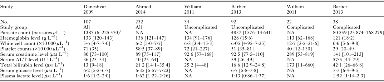
Laboratory tests are frequently abnormal in patients with knowlesi malaria and may range from mild abnormalities to evidence of end organ damage. Laboratory findings are summarized in Table 3.
Summary of admission laboratory findings in prospective studies in adults with Plasmodium knowlesi monoinfections

Data are median [interquartile range], mean (±SD) unless stated.
NA, not available.
a Geometric mean (range).
Platelets
Thrombocytopenia has been observed in nearly all cases of knowlesi malaria infection published in the literature (Singh and Daneshvar, Reference Singh and Daneshvar2013). Platelet counts and their nadir are inversely associated with parasitaemia (Daneshvar et al. Reference Daneshvar, Davis, Cox-Singh, Rafa'ee, Zakaria, Divis and Singh2009; Barber et al. Reference Barber, William, Grigg, Menon, Auburn, Marfurt, Anstey and Yeo2013a ). The platelet count may be a predictor of severity, with ⩽45 000 platelets µL−1 found in 71% of severe cases [OR 5·27; 95% confidence interval (CI) 1·59–17·4] (Willmann et al. Reference Willmann, Ahmed, Siner, Wong, Woon, Singh, Krishna and Cox-Singh2012). Despite thrombocytopenia, bleeding is rarely reported. This contrasts with dengue in which some degree of bleeding associated with thrombocytopenia was observed in 84% of adults with uncomplicated infections (Trung et al. Reference Trung, Thao le, Dung, Ngoc, Hien, Chau, Wolbers, Tam, Farrar, Simmons and Wills2012). Since there was no thrombocytopenia in two of six splenectomized patients with knowlesi malaria, splenic sequestration may be an underlying mechanism (Barber et al. Reference Barber, William, Grigg, Menon, Auburn, Marfurt, Anstey and Yeo2013a , Reference Barber, Grigg, William, Yeo and Anstey2016; Bird et al. Reference Bird, Parameswaran, William, Khoo, Grigg, Aziz, Marfurt, Yeo, Auburn, Anstey and Barber2016; Boo et al. Reference Boo, Lim, Chin, Lim and Hoo2016).
Anaemia
Severe anaemia (haemoglobin < 70 g L−1) is uncommon, occurring in only 5% of cases with severe knowlesi malaria (Willmann et al. Reference Willmann, Ahmed, Siner, Wong, Woon, Singh, Krishna and Cox-Singh2012; Barber et al. Reference Barber, William, Grigg, Menon, Auburn, Marfurt, Anstey and Yeo2013a ). However, moderate anaemia (haemoglobin < 100 g L−1) is seen in 5% of patients on admission, with a mean drop in haemoglobin of 13 g L−1 during hospitalization leading to a third of patients developing moderate anaemia (Daneshvar et al. Reference Daneshvar, Davis, Cox-Singh, Rafa'ee, Zakaria, Divis and Singh2009; Barber et al. Reference Barber, William, Grigg, Menon, Auburn, Marfurt, Anstey and Yeo2013a , Reference Barber, William, Grigg, Piera, Yeo and Anstey b ). Recovery by day 28 is usual. However, based on more recent WHO age-specific criteria for anaemia, between 62 and 75% of patients with uncomplicated knowlesi infection developed anaemia during 28 days of post-treatment follow-up with approximately 30% of patients still being anaemic at day 28 (Grigg et al. Reference Grigg, William, Menon, Dhanaraj, Barber, Wilkes, von Seidlein, Rajahram, Pasay, McCarthy, Price, Anstey and Yeo2016b ).
Data from Sarawak indicated that erythrocytes are normocytic (median mean red cell volume 85 fL), while the change in haemoglobin in the first 24–48 h is correlated with admission parasitaemia (Daneshvar, Reference Daneshvar2013b ). The absolute fall in red blood cell concentration is greater than that which could be could be accounted for by parasite burden alone, probably reflecting reticuloendothelial removal of non-parasitized red blood cells as seen, but not fully understood in falciparum and vivax malaria (Looareesuwan et al. Reference Looareesuwan, Davis, Pukrittayakamee, Supanaranond, Desakorn, Silamut, Krishna, Boonamrung and White1991; Perkins et al. Reference Perkins, Were, Davenport, Kempaiah, Hittner and Ong'echa2011). In a logistic regression model, respiratory distress was inversely associated with the admission haemoglobin concentration (Daneshvar et al. Reference Daneshvar, Davis, Cox-Singh, Rafa'ee, Zakaria, Divis and Singh2009).
White cells
In patients with knowlesi malaria, the median total white cell count is consistently about 6000 µL−1, the neutrophil count 3500 µL−1 and lymphocyte count 1500 µL−1 (Daneshvar et al. Reference Daneshvar, Davis, Cox-Singh, Rafa'ee, Zakaria, Divis and Singh2009; Willmann et al. Reference Willmann, Ahmed, Siner, Wong, Woon, Singh, Krishna and Cox-Singh2012; Barber et al. Reference Barber, William, Grigg, Menon, Auburn, Marfurt, Anstey and Yeo2013a ). The median neutrophil count on admission is greater in severe vs uncomplicated knowlesi malaria and it is also associated with a higher serum creatinine, the development of respiratory distress, and higher interleukin-1ra (IL-1ra) and interleukin-10 (IL-10) concentrations (Cox-Singh et al. Reference Cox-Singh, Singh, Daneshvar, Planche, Parker-Williams and Krishna2011; Willmann et al. Reference Willmann, Ahmed, Siner, Wong, Woon, Singh, Krishna and Cox-Singh2012; Barber et al. Reference Barber, William, Grigg, Menon, Auburn, Marfurt, Anstey and Yeo2013a ). While the total white cell count does not change significantly during treatment, the neutrophil count falls and the lymphocyte count rises (Daneshvar et al. Reference Daneshvar, Davis, Cox-Singh, Rafa'ee, Zakaria, Divis and Singh2009).
Renal function
Impaired renal function has been a consistent feature of severe disease (Cox-Singh et al. Reference Cox-Singh, Davis, Lee, Shamsul, Matusop, Ratnam, Rahman, Conway and Singh2008; Daneshvar et al. Reference Daneshvar, Davis, Cox-Singh, Rafa'ee, Zakaria, Divis and Singh2009; William et al. Reference William, Menon, Rajahram, Chan, Ma, Donaldson, Khoo, Frederick, Jelip, Anstey and Yeo2011; Willmann et al. Reference Willmann, Ahmed, Siner, Wong, Woon, Singh, Krishna and Cox-Singh2012; Barber et al. Reference Barber, William, Grigg, Menon, Auburn, Marfurt, Anstey and Yeo2013a ). Acute kidney injury defining severe malaria (creatinine > 265 µm L−1) occurred in three (2·8%) patients in Kapit and 16/17 (94%) of severe cases in Sarikei (Daneshvar et al. Reference Daneshvar, Davis, Cox-Singh, Rafa'ee, Zakaria, Divis and Singh2009; Willmann et al. Reference Willmann, Ahmed, Siner, Wong, Woon, Singh, Krishna and Cox-Singh2012). In the largest study, which included 38 patients with severe knowlesi malaria, acute kidney injury was present in 9 (24%) (Barber et al. Reference Barber, William, Grigg, Menon, Auburn, Marfurt, Anstey and Yeo2013a ).
Liver function
A mild hepatitis may be observed, with a raised serum alanine aminotransferase (ALT) (47%) and total serum bilirubin (18%) (Daneshvar, Reference Daneshvar2013b ). A serum bilirubin of ⩾43 µ m L−1 was recorded in 3% of patients in Sarawak (Daneshvar et al. Reference Daneshvar, Davis, Cox-Singh, Rafa'ee, Zakaria, Divis and Singh2009), while jaundice was present in 20/38 (53%) of patients with severe knowlesi malaria in Sabah (Barber et al. Reference Barber, William, Grigg, Menon, Auburn, Marfurt, Anstey and Yeo2013a ). The serum bilirubin normalizes within days, suggesting a major contribution from malaria-associated haemolysis, but the increase in serum ALT may persist for weeks (Daneshvar et al. Reference Daneshvar, Davis, Cox-Singh, Rafa'ee, Zakaria, Divis and Singh2009). Serum albumin concentrations <35 g dL−1 were observed in 35/107 (32·7%) patients, and were lower in women (Daneshvar, Reference Daneshvar2013b ). Serum albumin concentrations on admission are lower in severe knowlesi infections than in uncomplicated disease (Barber et al. Reference Barber, William, Grigg, Menon, Auburn, Marfurt, Anstey and Yeo2013a ). Synthetic liver function appears preserved, with few cases of abnormal clotting reported.
Diagnosis
A blood film for malaria parasites is essential in all patients with suspected knowlesi malaria. While there are limitations with the accuracy of species differentiation, alternative accessible rapid diagnostic tests are yet to be developed, while the standard rapid diagnostic tests applied to knowlesi malaria lack sensitivity and specificity (Barber et al. Reference Barber, William, Grigg, Piera, Yeo and Anstey2013b ; Foster et al. Reference Foster, Cox-Singh, Mohamad, Krishna, Chin and Singh2014; Grigg et al. Reference Grigg, William, Barber, Parameswaran, Bird, Piera, Aziz, Dhanaraj, Yeo and Anstey2014a ). Careful blood film examination is required since low parasitaemias are frequent. In the Kapit cohort, for example, 30% of patients had a parasitaemia <500 µL−1 or <0·01% (Daneshvar et al. Reference Daneshvar, Davis, Cox-Singh, Rafa'ee, Zakaria, Divis and Singh2009). Confirmation by PCR is recommended, and while unlikely to influence the acute treatment, may improve the management of Plasmodium vivax, which may be misdiagnosed as P. knowlesi.
Severe disease
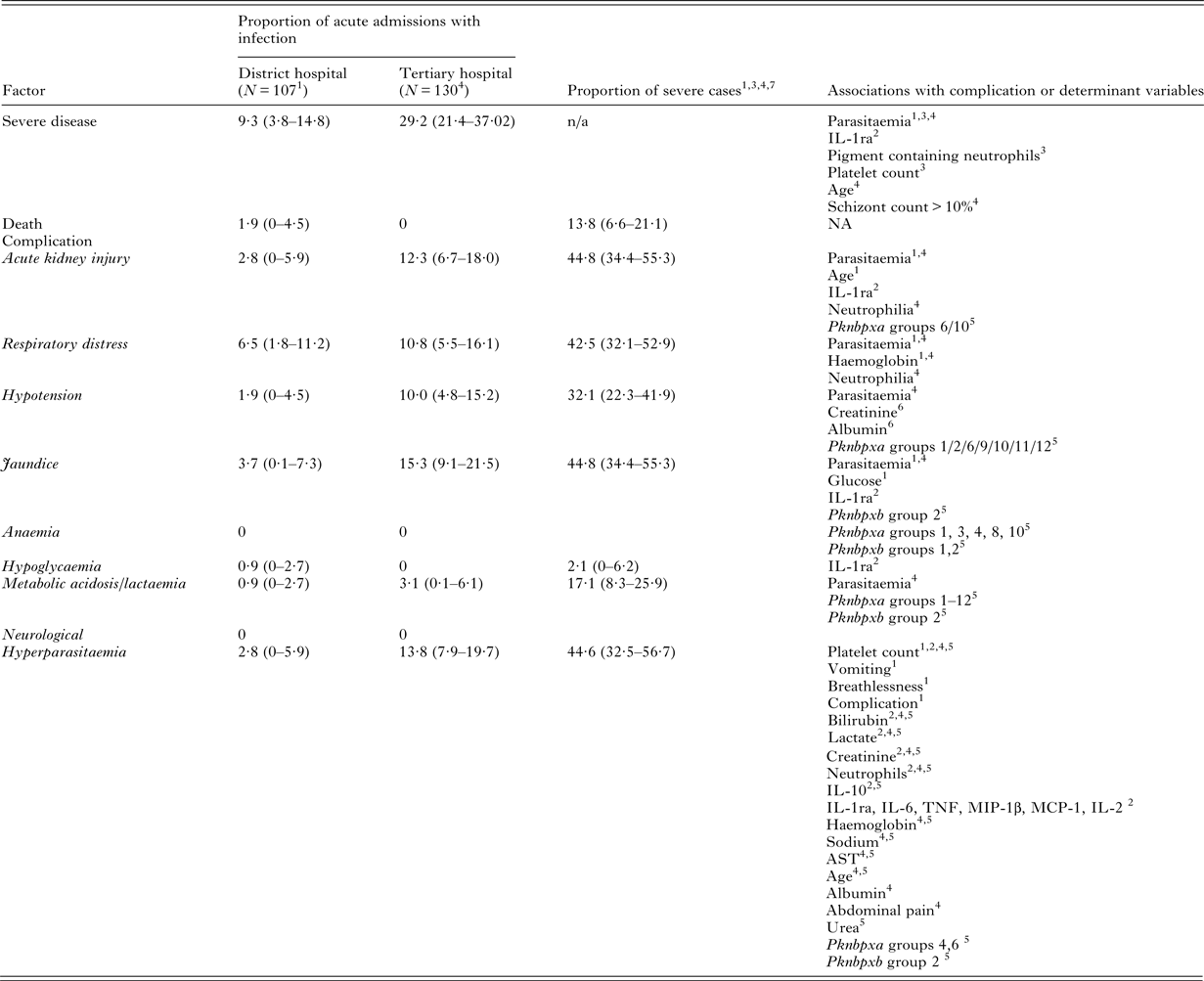
Patients may present with severe disease or develop complications during the treatment. Table 4 and Supplementary Table S1 give an overview of the features of severe disease and the associations identified in the larger retrospective and prospective studies.
Severe disease in adults with Plasmodium knowlesi malaria

Data show % (95% confidence intervals). Severe criteria used were: Hyperparasitaemia was defined as >100 000 parasites µL−1. Hypotension was defined as systolic blood pressure <80 mm Hg. Acute kidney injury was defined as a serum creatinine level >265 µ m L−1 despite rehydration. Jaundice was defined as a serum bilirubin >43 µ m L−1; (Study 4 – bilirubin >43 µ m L−1 plus parasitaemia >20 000 parasites µL−1 and/or creatinine >132 µ m L−1). Hypoglycaemia was defined as a serum glucose <2·2 mm L−1. Lactaemia/acidosis was defined as >5 mm L−1; (Study 4 – bicarbonate <15 mm L−1 or lactate >5 mm L−1). Respiratory distress was defined as a respiratory rate >30 breaths min−1 plus oxygen saturation <94% on room air and/or pulmonary infiltrates visible on a chest radiograph. NA, not available. Breakdown of criteria per study is shown in Supplementary Table S1.
Pknbp, P. knowlesi normocyte binding protein (associations of Pknbp are within group alleles); IL, interleukin; TNF, tumour necrosis factor; IL-1ra, interleukin 1 receptor antagonist; MIP-1, Macrophage Inhibitory Protein-1β; MCP-1, Monocyte Chemoattractant Protein-1.
Studies are 1Daneshvar et al. (Reference Daneshvar, Davis, Cox-Singh, Rafa'ee, Zakaria, Divis and Singh2009), 2Cox-Singh et al. (Reference Cox-Singh, Singh, Daneshvar, Planche, Parker-Williams and Krishna2011), 3Willmann et al. (Reference Willmann, Ahmed, Siner, Wong, Woon, Singh, Krishna and Cox-Singh2012), 4Barber et al. (Reference Barber, William, Grigg, Menon, Auburn, Marfurt, Anstey and Yeo2013a , Reference Barber, William, Grigg, Piera, Yeo and Anstey b ), 5Ahmed et al. (Reference Ahmed, Pinheiro, Divis, Siner, Zainudin, Wong, Lu, Singh-Khaira, Millar, Lynch, Willmann, Singh, Krishna and Cox-Singh2014), 6Daneshvar (Reference Daneshvar2013b ), 7William et al. (Reference William, Menon, Rajahram, Chan, Ma, Donaldson, Khoo, Frederick, Jelip, Anstey and Yeo2011).
Prospective clinical descriptive studies indicate severe knowlesi malaria occurs in between 9 and 29% of acute admissions depending on the referral population, with case fatality rates ranging from 0 to 1·9% (Daneshvar et al. Reference Daneshvar, Davis, Cox-Singh, Rafa'ee, Zakaria, Divis and Singh2009; Barber et al. Reference Barber, William, Grigg, Menon, Auburn, Marfurt, Anstey and Yeo2013a ). These are only estimates, and the true prevalence of severe disease has proved difficult to define. From the ACT-KNOW study, a treatment study in consecutive patients with uncomplicated knowlesi cases based in Sabah, 26 patients were excluded due to severe knowlesi malaria and a further 21 who had an admission parasite count >20 000 µL−1 (Grigg et al. Reference Grigg, William, Menon, Dhanaraj, Barber, Wilkes, von Seidlein, Rajahram, Pasay, McCarthy, Price, Anstey and Yeo2016b ). This suggests an estimated prevalence of 8·6% (95% CI 5·4–11·8; 26/299), consistent with that seen in Kapit (Daneshvar et al. Reference Daneshvar, Davis, Cox-Singh, Rafa'ee, Zakaria, Divis and Singh2009).
The first fatal cases reported were four patients from Sarawak aged 39–69 years (Cox-Singh et al. Reference Cox-Singh, Davis, Lee, Shamsul, Matusop, Ratnam, Rahman, Conway and Singh2008). Since then a further 24 deaths, all adults aged 31–84 years, have been reported in the literature (Daneshvar et al. Reference Daneshvar, Davis, Cox-Singh, Rafa'ee, Zakaria, Divis and Singh2009; Cox-Singh et al. Reference Cox-Singh, Hiu, Lucas, Divis, Zulkarnaen, Chandran, Wong, Adem, Zaki, Singh and Krishna2010; William et al. Reference William, Menon, Rajahram, Chan, Ma, Donaldson, Khoo, Frederick, Jelip, Anstey and Yeo2011; Rajahram et al. Reference Rajahram, Barber, William, Menon, Anstey and Yeo2012, Reference Rajahram, Barber, Yeo, Tan and William2013, Reference Rajahram, Barber, William, Grigg, Menon, Yeo and Anstey2016; Willmann et al. Reference Willmann, Ahmed, Siner, Wong, Woon, Singh, Krishna and Cox-Singh2012). Further, malaria case notification data from Sabah between 2010 and 2014 revealed 13 deaths in 4225 cases or a case fatality rate of 0·3% (95% CI 0·14–0·46) (Rajahram et al. Reference Rajahram, Barber, William, Grigg, Menon, Yeo and Anstey2016).
In the most recent WHO treatment guidelines, special references for knowlesi malaria reflect reported phenotypes in adult disease, along with evidence of end organ damage at relatively low parasitaemias (see Table 4) (WHO treatment guidelines 2015). There are no validation studies of these clinical and laboratory features.
Hyperparasitaemia
In prospective studies, the range of parasitaemia associated with clinical disease ranges from 6 to 584 000 µL−1 (geometric mean 1387 µL−1) (Daneshvar et al. Reference Daneshvar, Davis, Cox-Singh, Rafa'ee, Zakaria, Divis and Singh2009; Barber et al. Reference Barber, William, Grigg, Menon, Auburn, Marfurt, Anstey and Yeo2013a ), but up to 24% parasitaemias have been found in individual cases. In receiver operator characteristic (ROC) analysis, hyperparasitaemia was highly specific (100%) but had poor sensitivity (30%) for severe disease, with patients with low parasitaemias presenting with severe disease (Daneshvar et al. Reference Daneshvar, Davis, Cox-Singh, Rafa'ee, Zakaria, Divis and Singh2009). Despite this, parasitaemia was associated with respiratory distress and serum creatinine concentrations, and inversely with haemoglobin and platelet counts. Willmann et al. found that 8% of patients with hyperparasitaemia did not have other features of severe disease, while 9/17 (52%) patients with severe disease had a parasite count of <100 000 µL−1 (Willmann et al. Reference Willmann, Ahmed, Siner, Wong, Woon, Singh, Krishna and Cox-Singh2012). In ROC analysis of severe disease, the parasite count area under the curve was 0·85 (95% CI 0·75–0·94) with sensitivity, specificity, positive predictive value (PPV) and negative predictive value (NPV) of 71, 80, 28 and 96%, respectively, at a cutoff ⩾20 000 µL−1. At ⩾35 000 µL−1, the sensitivity was 64% and the NPV 95%. Therefore, one in 20 patients with a parasite count <35 000 µL−1 could be misdiagnosed as not having complicated disease based on parasite density alone.
The largest prospective study was conducted in Sabah and included 38 patients with knowlesi malaria whose risk of severe malaria increased by 11-fold with a parasitaemia ⩾20 000 µL−1 with sensitivity, specificity, PPV, NPV of 75, 79, 47 and 97%, respectively (Barber et al. Reference Barber, William, Grigg, Menon, Auburn, Marfurt, Anstey and Yeo2013a ).
The strong association between parasite burden and severe disease in this predominantly non-sequestering species of malaria affecting mainly non-immune individuals is perhaps unsurprising, but an important finding linking parasite burden with pathophysiology. Indeed, evidence of disease has been linked to reticulocyte binding-like protein family – P. knowlesi normocyte-binding proteins (Pknbp)xa and Pknbpxb (Ahmed et al. Reference Ahmed, Pinheiro, Divis, Siner, Zainudin, Wong, Lu, Singh-Khaira, Millar, Lynch, Willmann, Singh, Krishna and Cox-Singh2014). Within haplotype groups, various markers of disease severity were found (see Table 4).
Acute kidney injury
In pooled data from severe knowlesi malaria cases from three prospective studies and a large retrospective study, AKI was a common feature of severe disease, occurring in 44·8% (95% CI 34·4–55·3), (Daneshvar et al. Reference Daneshvar, Davis, Cox-Singh, Rafa'ee, Zakaria, Divis and Singh2009; William et al. Reference William, Menon, Rajahram, Chan, Ma, Donaldson, Khoo, Frederick, Jelip, Anstey and Yeo2011; Willmann et al. Reference Willmann, Ahmed, Siner, Wong, Woon, Singh, Krishna and Cox-Singh2012; Barber et al. Reference Barber, William, Grigg, Menon, Auburn, Marfurt, Anstey and Yeo2013a ). Blackwater fever has been reported in knowlesi malaria (William et al. Reference William, Menon, Rajahram, Chan, Ma, Donaldson, Khoo, Frederick, Jelip, Anstey and Yeo2011; Barber et al. Reference Barber, William, Grigg, Menon, Auburn, Marfurt, Anstey and Yeo2013a ). Dilated congested renal cortices, acute tubular necrosis and a large burden of parasitized red blood cells and pigment deposition in capillaries was observed at post-mortem of another case of knowlesi infection (Cox-Singh et al. Reference Cox-Singh, Hiu, Lucas, Divis, Zulkarnaen, Chandran, Wong, Adem, Zaki, Singh and Krishna2010). Parasitaemia, neutrophilia, high IL-1ra and age have all been associated with increased serum creatinine concentrations (Daneshvar et al. Reference Daneshvar, Davis, Cox-Singh, Rafa'ee, Zakaria, Divis and Singh2009; Cox-Singh et al. Reference Cox-Singh, Singh, Daneshvar, Planche, Parker-Williams and Krishna2011; Barber et al. Reference Barber, William, Grigg, Menon, Auburn, Marfurt, Anstey and Yeo2013a ).
Respiratory distress
Respiratory distress in severe malaria is a broad definition for use in resource-limited settings, and may include pulmonary complications and metabolic acidosis. Pulmonary oedema under current WHO guidelines is defined as that confirmed radiologically or an oxygen saturation <92% on air with a respiratory rate of >30 min−1 with chest in-drawing (children) and crackles on auscultation (WHO, 2015). In a patient series from Sarawak, respiratory complications were defined as a respiratory rate >30 min−1 plus oxygen saturation <94% on air and/or pulmonary infiltrates on chest radiography. Seven (6·5%) patients met these criteria, five on admission and a further two during treatment (Daneshvar et al. Reference Daneshvar, Davis, Cox-Singh, Rafa'ee, Zakaria, Divis and Singh2009). In the prospective study from Sabah, respiratory distress occurred in 14 (11%) patients (Barber et al. Reference Barber, William, Grigg, Menon, Auburn, Marfurt, Anstey and Yeo2013a ). Of these, two patients met the criteria for an acute respiratory distress syndrome (ARDS) defined by partial pressure of arterial oxygen:fraction of inspiratory oxygen <200 mm Hg, but neither required mechanical ventilation.
Parasitaemia [OR 1·65; 95% CI 1·18–2·06) and neutrophilia (controlling for parasitaemia) were both found to be associated with respiratory distress (Barber et al. Reference Barber, William, Grigg, Menon, Auburn, Marfurt, Anstey and Yeo2013a ). While a similar proportion of patients with falciparum had respiratory distress (31%) no association was found between parasitaemia and respiratory distress among these patients. In Sarawak, respiratory distress was associated with parasitaemia and inversely associated with admission haemoglobin concentrations (Daneshvar et al. Reference Daneshvar, Davis, Cox-Singh, Rafa'ee, Zakaria, Divis and Singh2009).
Hypotension
Hypotension (systolic blood pressure ⩽80 mm Hg) is a feature of severe knowlesi malaria, affecting 2–10% of acute knowlesi admissions studied (Daneshvar et al. Reference Daneshvar, Davis, Cox-Singh, Rafa'ee, Zakaria, Divis and Singh2009; Barber et al. Reference Barber, William, Grigg, Menon, Auburn, Marfurt, Anstey and Yeo2013a ). Hypotension was reported in 24% (95% CI 14–34) of prospectively studied severe knowlesi patients (Daneshvar et al. Reference Daneshvar, Davis, Cox-Singh, Rafa'ee, Zakaria, Divis and Singh2009; Fatih et al. Reference Fatih, Siner, Ahmed, Woon, Craig, Singh, Krishna and Cox-Singh2012; Willmann et al. Reference Willmann, Ahmed, Siner, Wong, Woon, Singh, Krishna and Cox-Singh2012; Barber et al. Reference Barber, William, Grigg, Menon, Auburn, Marfurt, Anstey and Yeo2013a ). A lower mean arterial blood pressure has been shown to be associated with lower serum albumin, and raised serum creatinine and parasitaemia (Daneshvar, Reference Daneshvar2013b ).
Jaundice
Jaundice or hyperbilirubinaemia (⩾43 or ⩾51 µ m L−1) as a criterion for severe malaria has been interpreted as having varying importance, with the most recent WHO guidelines linking it to parasitaemia >100 000 µL−1 in falciparum malaria (WHO, 2000, 2010, 2015). Due to concerns over poor sensitivity of parasitaemia at this level in knowlesi malaria, the presence of jaundice with a parasitaemia of ⩾20 000 µL−1 is recommended (WHO, 2015). In the studies of knowlesi malaria, varying criteria have been used. Parasitaemia and bilirubin are highly correlated (Daneshvar et al. Reference Daneshvar, Davis, Cox-Singh, Rafa'ee, Zakaria, Divis and Singh2009; Barber et al. Reference Barber, William, Grigg, Menon, Auburn, Marfurt, Anstey and Yeo2013a ).
Other features
Lactic acidosis has been found in five (10%; 95% CI 1·5–20) severe infections in the prospective studies. The median serum lactate concentrations measured in acute knowlesi infection is 1·5 mm L−1, higher in severe vs uncomplicated cases and positively associated with parasitaemia (Daneshvar et al. Reference Daneshvar, Davis, Cox-Singh, Rafa'ee, Zakaria, Divis and Singh2009; Barber et al. Reference Barber, William, Grigg, Menon, Auburn, Marfurt, Anstey and Yeo2013a ). Hypoglycaemia is rare, occurring in 2% of patients with severe knowlesi malaria and often in the context of multiple organ failure.
Comparisons with falciparum and vivax malaria
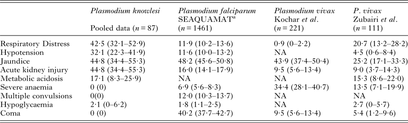
Complicated adult knowlesi infections share similarities with complicated falciparum malaria and the increasingly recognized burden of severe malaria in non-falciparum species especially that caused by P. vivax (Baird, Reference Baird2013). In studies in which knowlesi infections have been observed alongside falciparum and vivax malaria, the numbers of cases are relatively small. Ministry of Health data from Sabah, indicates that the case fatality rate between 2010 and 2014 for knowlesi, falciparum and vivax infection in persons >15 years were 3·37, 4·17 and 1·02 per 1000 notifications, respectively (Rajahram et al. Reference Rajahram, Barber, William, Grigg, Menon, Yeo and Anstey2016). Although confounded by referral bias to the study centre, Barber et al. found knowlesi, falciparum and vivax infections to be complicated in 29, 10·6 and 16·3% of cases, with an increased odds of developing complications in knowlesi when compared with falciparum malaria (OR 2·96; 95% CI 1·19–7·38) (Barber et al. Reference Barber, William, Grigg, Menon, Auburn, Marfurt, Anstey and Yeo2013a ). The distribution of criteria for severity did not significantly differ across Plasmodium species although this may be confounded by the limited numbers in the non-knowlesi groups.
The variation in criteria for severity among adult patients with varying species of malaria based on some of the larger published studies are shown in Table 5 (Dondorp et al. Reference Dondorp, Nosten, Stepniewska, Day and White2005; Zubairi et al. Reference Zubairi, Nizami, Raza, Mehraj, Rasheed, Ghanchi, Khaled and Beg2013; Kochar et al. Reference Kochar, Das, Kochar, Middha, Acharya, Tanwar, Pakalapati, Subudhi, Boopathi, Garg and Kochar2014). Notwithstanding between-study differences, the absence of neurological involvement and the presence of severe anaemia in knowlesi malaria are noteworthy. The frequency of jaundice, metabolic acidosis and hypoglycaemia appear consistent across studies, while acute kidney injury, respiratory distress and hypotension appear greater in knowlesi malaria. Understanding these differences may lead to progression in our knowledge of the species-specific malaria pathophysiology.
Comparison of severe criteria in patients with complicated knowlesi, falciparum and vivax malaria

Data are percentage (95% CI).
NA, not available.
a Dondorp et al. (Reference Dondorp, Nosten, Stepniewska, Day and White2005).
Special circumstances
Children and pregnant women
In Sabah, there were 373/4225 (8·8%) reported cases of knowlesi infection among children between 2010 and 2014 (Rajahram et al. Reference Rajahram, Barber, William, Grigg, Menon, Yeo and Anstey2016). This percentage is similar to that in the Kapit cohort (6·6%), a report by Barber et al. (9–14%) and the 9% of patients aged between 1 and 12 years recruited to the ACT-KNOW study (Barber et al. Reference Barber, William, Jikal, Jilip, Dhararaj, Menon, Yeo and Anstey2011; Singh and Daneshvar, Reference Singh and Daneshvar2013; Grigg et al. Reference Grigg, William, Menon, Dhanaraj, Barber, Wilkes, von Seidlein, Rajahram, Pasay, McCarthy, Price, Anstey and Yeo2016b ). The clinical spectrum of disease among children is difficult to define because it has not been studied prospectively. However, there have been no reported deaths in children with knowlesi malaria in Sabah between 2010 and 2014 (Rajahram et al. Reference Rajahram, Barber, William, Grigg, Menon, Yeo and Anstey2016).
In the most detailed retrospective description of 16 children, the median admission parasitaemia was 2240 parasites µL−1 (Barber et al. Reference Barber, William, Jikal, Jilip, Dhararaj, Menon, Yeo and Anstey2011). Mild anaemia was common (56%). All but one patient had thrombocytopenia. The clinical response to treatment was similar to that described in adults. In Kapit, the median parasitaemia in eight children was 940 (range 240–26 270) µL−1, two were mildly anaemic, two had a mild transaminitis, and one was mildly jaundiced (Singh and Daneshvar, Reference Singh and Daneshvar2013). In the child with a parasite count of 26 270 µL−1, retinal haemorrhages and a petechial rash were observed. More studies are warranted to understand the full spectrum of disease, and in particular the modifiable risk factors for children acquiring this infection. There are few data on knowlesi malaria in pregnancy (Barber et al. Reference Barber, Bird, Wilkes, William, Grigg, Paramaswaran, Menon, Jelip, Yeo and Anstey2015).
Travellers
There is a clear risk of infection to travellers who visit knowlesi-endemic areas (Cramer, Reference Cramer2015). There have been reports of knowlesi malaria in 17 adults who visited South-east Asia (Malaysian Borneo, Philippines, Thailand and/or Indonesia) (Chin et al. Reference Chin, Contacos, Coatney and Kimball1965; Kantele et al. Reference Kantele, Marti, Felger, Muller and Jokiranta2008; Bronner et al. Reference Bronner, Divis, Farnert and Singh2009; Ennis et al. Reference Ennis, Teal, Habura, Madison Antenucci and Keithly2009; Kuo et al. Reference Kuo, Chiang, Chan, Tsai and Ji2009; Figtree and Hudson, Reference Figtree and Hudson2010; Tang et al. Reference Tang, Salas, Ali-Tammam, Del Carmen Martinez, Lanza, Arroyo and Rubio2010; Van Hellemond and Van Genderen, Reference Van Hellemond and Van Genderen2010; Berry et al. Reference Berry, Iriart, Wilhelm, Valentin, Cassaing, Witkowski, Benoit-Vical, Menard, Olagnier, Fillaux, Sire, Le Coustumier and Magnaval2011; Hoosen and Shaw, Reference Hoosen and Shaw2011; Link et al. Reference Link, Bart, Verhaar, van Gool, Pronk and Scharnhorst2012; Ehrhardt et al. Reference Ehrhardt, Trein, Kremsner and Frank2013; Orth et al. Reference Orth, Jensen, Holtfreter, Kocheril, Mallach, MacKenzie, Muller-Stover, Henrich, Imwong, White, Haussinger and Richter2013; Tanizaki et al. Reference Tanizaki, Ujiie, Kato, Iwagami, Hashimoto, Kutsuna, Takeshita, Hayakawa, Kanagawa, Kano and Ohmagari2013; Cordina et al. Reference Cordina, Culleton, Jones, Smith, MacConnachie, Coyne and Alexander2014; Kroidl et al. Reference Kroidl, Seilmaier, Berens-Riha, Bretzel, Wendtner and Loscher2015; Mackroth et al. Reference Mackroth, Tappe, Tannich, Addo and Rothe2016). All presented within 2 weeks of returning home and few took antimalarial chemoprophylaxis. Parasite counts varied from <0·1 to 2·9%, with complications seen in two cases (Van Hellemond and Van Genderen, Reference Van Hellemond and Van Genderen2010; Orth et al. Reference Orth, Jensen, Holtfreter, Kocheril, Mallach, MacKenzie, Muller-Stover, Henrich, Imwong, White, Haussinger and Richter2013). Treatment was with a variety of antimalarial regimens, but no deaths were reported.
An accurate map showing where there is a risk of knowlesi malaria will assist in informing travellers including regions where knowlesi cases are likely but yet to be reported (Shearer et al. Reference Shearer, Huang, Weiss, Wiebe, Gibson, Battle, Pigott, Brady, Putaporntip, Jongwutiwes, Lau, Manske, Amato, Elyazar, Vythilingam, Bhatt, Gething, Singh, Golding, Hay and Moyes2016). In Poland, screening for knowlesi malaria in returned travellers from endemic areas did not yield any positive diagnoses (Biernat et al. Reference Biernat, Lass, Pietkiewicz, Szostakowska, Wroczynska, Kuna and Nahorski2015). Nevertheless, knowlesi malaria should be considered in any patient returning from an endemic area with a febrile illness, even if initial blood films or rapid diagnostic tests for malaria are negative.
TREATMENT
There is a paucity of antimalarial drug treatment studies in human knowlesi malaria. However, patients respond well to a wide range of antimalarial drugs (Singh and Daneshvar, Reference Singh and Daneshvar2013). This is not surprising for zoonotic malaria in which previous drug studies in simian models and during induced infections for neurosyphilis showed a good response to a variety of treatments. Prompt recognition and treatment of severe disease have improved the care of patients in Sabah, where reported mortality from knowlesi malaria has reduced (Barber et al. Reference Barber, William, Grigg, Menon, Auburn, Marfurt, Anstey and Yeo2013a ). It is likely that the availability of the artemisinin derivatives has contributed to this.
Uncomplicated knowlesi malaria
There are two key studies that have provided accurate parasite clearance data, an observational study of chloroquine and primaquine treatment and a randomized controlled trial of artesunate–mefloquine vs chloroquine in uncomplicated knowlesi malaria (ACT-KNOW) (Daneshvar et al. Reference Daneshvar, Davis, Cox-Singh, Rafa'ee, Zakaria, Divis and Singh2010; Grigg et al. Reference Grigg, William, Menon, Dhanaraj, Barber, Wilkes, von Seidlein, Rajahram, Pasay, McCarthy, Price, Anstey and Yeo2016b ). In the first, all patients without severe disease were given 10 mg chloroquine kg−1 followed by three doses of 5 mg kg−1 at 6, 24 and 48 h in addition to 2 days treatment with 15 mg of primaquine daily after 24 h in G6PD-replete patients. Within the limitation of a small sample size, median 50 and 90% parasite clearance times (PCT) were rapid (3·1 and 10·3 h, respectively) and the parasite reduction ratio (PRR) at 24 h was 99·3%. Total parasite clearance at 24 h was observed in 33%. There were no treatment failures within 28 days by microscopy or PCR. Unfortunately, low admission parasite densities and rapid parasite clearance made accurate quantification of parasitaemia difficult in some patients.
Subsequently, a randomized controlled study of combined artesunate–mefloquine vs chloroquine was conducted (Grigg et al. Reference Grigg, William, Menon, Dhanaraj, Barber, Wilkes, von Seidlein, Rajahram, Pasay, McCarthy, Price, Anstey and Yeo2016b ). The primary endpoint of the proportion of patients who had cleared parasites at 24 h was significantly higher in the artesunate–mefloquine group compared with the chloroquine group (84 vs 55%, respectively). However, parasite clearance at 48 h was similar (100% for artesunate–mefloquine vs 98% after chloroquine). There were differences between the PCT between the chloroquine arm in this study and those previously reported, but the use of the more robust and now universally standardized WWARN algorithm may in part account for this (Daneshvar et al. Reference Daneshvar, Davis, Cox-Singh, Rafa'ee, Zakaria, Divis and Singh2010; Flegg et al. Reference Flegg, Guerin, White and Stepniewska2011). One patient in the artesunate–mefloquine arm died on day 24 due to a hospital acquired pneumonia. Side-effect profiles were similar in the two arms, although there was a trend to less gastrointestinal disturbance in the artesunate–mefloquine group. One patient developed an acute psychosis attributed to mefloquine in the artesunate–mefloquine group.
As P. knowlesi has a short (24 h) erythrocytic cycle, the parasitaemia can increase relatively rapidly. It is thus important to institute treatment promptly to reduce parasite burden. Around one quarter of patients experience a transient rise in parasitaemia within the first 6 h when treated with chloroquine and primaquine (Daneshvar et al. Reference Daneshvar, Davis, Cox-Singh, Rafa'ee, Zakaria, Divis and Singh2010). In the ACT-KNOW study 9 and 24% were noted to have a lag-phase for the artesunate–mefloquine and chloroquine groups, respectively (Grigg et al. Reference Grigg, William, Menon, Dhanaraj, Barber, Wilkes, von Seidlein, Rajahram, Pasay, McCarthy, Price, Anstey and Yeo2016b ). The more rapid effect of artesunate–mefloquine may be beneficial, and likely reflects the broader stage specificity of artesunate compared with chloroquine. Indeed, there was faster clearance of ring forms (PCT50 8·6 vs 13·8 h) but equivalent clearance of trophozoites (PCT50 7·6 vs 7·9 h) in the artesunate–mefloquine vs chloroquine groups (Grigg et al. Reference Grigg, William, Menon, Dhanaraj, Barber, Wilkes, von Seidlein, Rajahram, Pasay, McCarthy, Price, Anstey and Yeo2016b ).
In the Kapit study, 5·6% (95% CI 0–32) of patients had gametocytes on admission microscopy, and the estimated slope of the log-transformed parasitaemia-time curve was 0·06 compared with the overall 0·22 for asexual forms (Daneshvar et al. Reference Daneshvar, Davis, Cox-Singh, Rafa'ee, Zakaria, Divis and Singh2010). In the ACT-KNOW study 85 of 100 patients had gametocytes detected by use of RT–PCR pks25 mRNA, while gametocytes were observed in only 16 (19%) of these patients by microscopy (Grigg et al. Reference Grigg, William, Menon, Dhanaraj, Barber, Wilkes, von Seidlein, Rajahram, Pasay, McCarthy, Price, Anstey and Yeo2016b ). Gametocyte persistence to day 7 was found in 6 and 4% of patients in artesunate–mefloquine and chloroquine treatment arms, respectively, with no evidence of treatment inducing gametocyte development.
Complicated knowlesi malaria
In the first of the four fatal knowlesi cases reported, treatment did not commence for 72 h and, when started, oral chloroquine and sulfadoxine–pyrimethamine were given (Cox-Singh et al. Reference Cox-Singh, Davis, Lee, Shamsul, Matusop, Ratnam, Rahman, Conway and Singh2008). The third fatal case was recognized as having malaria, but given chloroquine and sulfadoxine–pyrimethamine. Finally, the fourth patient was initially given the same combination, but intravenous quinine was added soon after admission. Whether earlier treatment would have changed the outcome in all of these cases is unknown. It is, however, likely that a rapid diagnosis of malaria, recognition of severity, initiating appropriate antimalarial treatment, resuscitation, monitoring and anticipation of complications gives the best chance of recovery.
In Sabah, a large number of severe cases with a high proportion of fatalities have been reported (William et al. Reference William, Menon, Rajahram, Chan, Ma, Donaldson, Khoo, Frederick, Jelip, Anstey and Yeo2011). All patients received either intravenous quinine or intravenous artesunate. Of those receiving quinine, 5/16 (31·3%) patients died, while 1/6 (16·7%) receiving artesunate died. While there was no statistically significant difference, this may reflect a lack of power for the comparison and it is still likely that that the preferred treatment for severe knowlesi patients should include a parenteral artemisinin derivative. In an area with falciparum malaria, endemic chloroquine resistance and an inability to reliably and rapidly differentiate knowlesi from falciparum malaria, the treatment of knowlesi malaria is recommended to contain an artemisinin derivative for both uncomplicated and complicated disease (WHO working party). In a prospective study from Sabah of severe knowlesi cases involving this management strategy, no patients died (Barber et al. Reference Barber, William, Grigg, Menon, Auburn, Marfurt, Anstey and Yeo2013a ). Patients included were admitted directly or referred from regional sites, and given oral ACT or IV artesunate at diagnosis, including 36/38 (95%) with severe knowlesi and 39/98 (42%) with uncomplicated disease. In addition to prompt institution of artemisinin-based therapy, increased awareness of the diagnosis and its implications, accurate identification of malaria species, and improved nursing care are likely to have contributed to the favourable outcome, and possibly the noted decline in knowlesi case fatalities in adults observed in Sabah (Rajahram et al. Reference Rajahram, Barber, William, Grigg, Menon, Yeo and Anstey2016).
Drug resistance
There have been no reports of resistance to conventional antimalarial drugs in prospective studies of knowlesi malaria. However, in patients undergoing malariotherapy for syphilis, there was rapid parasite clearance after intramuscular and oral quinine, but quinacrine often failed to control the parasitaemia (van Rooyen and Pile, Reference van Rooyen and Pile1935). Quinacrine is closely related to mefloquine, which was associated with treatment failure in a recent case report, while P. knowlesi is relatively insensitive to mefloquine ex vivo and resistance can be easily induced in animal models (Gutteridge and Trigg, Reference Gutteridge and Trigg1971; Lau et al. Reference Lau, Tan, Chin, Fong, Noraishah and Rohela2011; Fatih et al. Reference Fatih, Staines, Siner, Ahmed, Woon, Pasini, Kocken, Singh, Cox-Singh and Krishna2013). Furthermore, the background innate parasite resistance to mefloquine is high (Singh and Dutta, Reference Singh and Dutta1981). Recently ABC C transporter genes, which include PkMRP1 and PkMDR2 [orthologues of P. falciparum PfMDR2 and PfMRP1 (markers of mefloquine resistance)], show considerable variation being polymorphic and dimorphic implying natural selection pressures at these loci in P. knowlesi (Pinheiro et al. Reference Pinheiro, Ahmed, Millar, Sanderson, Otto, Lu, Krishna, Rayner and Cox-Singh2015).
Concluding remarks
In the past 12 years, knowlesi malaria has become established as a recognized cause of malaria in humans, capable of inducing a wide spectrum of disease severity that includes death. There have been a number of studies that have characterized the features and clinical course of the infection in adults, including comparisons with the features of other more common human plasmodial infections. Nevertheless, many pathophysiological, clinical, epidemiological, and public health questions remain unanswered, especially in children and pregnant women.
There has been a 12 year gap between the identification of knowlesi malaria as an important human pathogen and publication of the first randomized treatment study. Further pharmacological and treatment studies are needed. These studies may employ innovative molecular methods of parasite quantification to better define the response to treatment in an infection characterized by the diagnostic limitations of microscopy, low presenting parasitaemias and rapid clearance times. Such an approach may lead to early identification of the treatment failure that has plagued management of other human malarias. At a more basic level, improvements in local health care will continue to play an essential role in improving clinical outcomes.
SUPPLEMENTARY MATERIAL
The supplementary material for this article can be found at http://doi.org/10.1017/S0031182016002638.
ACKNOWLEDGEMENTS
T.M.E.D. is supported by a National Health and Medical Research Council of Australia Practitioner Fellowship.
FINANCIAL SUPPORT
This work received no specific grant from any funding agency, commercial or not-for-profit sectors.
DECLARATIONS OF INTERESTS
None.