

Introduction
Rudolf Schlechter (Reference Schlechter1915) described Clemensiella with a single validly published species, Clemensiella mariae (Schltr.) Schltr. Clemensiella was considered one of three genera of Apocynaceae – Asclepiadoideae endemic to the Philippines (Madulid, Reference Madulid1991). The other two genera, Dolichostegia Schltr. and Quisumbingia Merr., have since been put under synonymy of the genera Dischidia R.Br. (Livshultz, Reference Livshultz2003) and Sarcolobus R.Br. (Omlor, Reference Omlor1998), respectively. All of them belong to the tribe Marsdenieae (Omlor, Reference Omlor1998). Clemensiella can no longer be considered a Philippine endemic either since specimens unambiguously assignable to this genus have been collected in Sumatra and Borneo. Omlor (Reference Omlor1998) was the first to note that the specimen de Wilde & de Wilde-Duyfjes 14377 (K) from Sumatra represents a species of Clemensiella, although he was uncertain whether it should be considered as conspecific with C. mariae. We have since identified two additional collections of this same species, one from Sumatra, the other from Sarawak, Malaysian Borneo. These three specimens represent a new species closely allied to Clemensiella mariae but clearly distinguishable by characters of the inflorescence and corolla. Recently, we also obtained living material of Clemensiella mariae; the plant flowered in the greenhouse at the Department of Plant Systematics, Bayreuth, thereby offering an opportunity to investigate this interesting and poorly known species in more detail. In the present paper we (i) discuss the taxonomic affinities of Clemensiella, (ii) emend the descriptions of Clemensiella and C. mariae, (iii) lectotypify C. mariae, and (iv) describe a new species, C. omlori.
Taxonomic Relationships of Clemensiella
The placement of Clemensiella within the tribe Marsdenieae of Apocynaceae – Asclepiadoideae was never in doubt – Schlechter (Reference Schlechter1915) originally placed it here, and assumed that Marsdenia R.Br. and especially Telosma Coville were the most closely related genera. Omlor (Reference Omlor1998) in his generic revision of the tribe supposed, in contrast, that Clemensiella is rather isolated within Marsdenieae without any direct connection to any other genus. He nevertheless pointed out that the pollinaria are most similar to those of Marsdenia, whereas the habit is very Hoya-like.
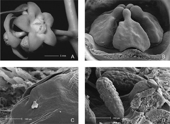
Clemensiella possesses a number of characters that are uncommon among genera of Marsdenieae: epiphytic growth habit (occasionally reported in Clemensiella mariae), adventitious roots (known only in C. mariae), persistent inflorescences, and valvate corolla lobes. The combination of these four characters occurs in only two other genera of the tribe: Hoya R.Br. and Dischidia. These four characters, along with the robust dimensions of the plant, the fleshy, shallow corolla, and the fleshy corona lobes, are the basis for the misinterpretation of Clemensiella mariae as a species of Hoya (Kloppenburg & Siar, Reference Kloppenburg and Siar2006). However, the corona and gynostegium of the two genera are quite different. The corona lobes of Clemensiella do not have the Hoya-characteristic fold on the underside of the corona lobes (recently interpreted as a fusion of a basal process of the staminal corona lobe and an ‘anther skirt’; Kunze & Wanntorp, Reference Kunze and Wanntorp2008). The pollinia are clavate and not elongatedly ellipsoid as is usual in Hoya, and they do not possess any sign of a sterile pellucid germination crest typical for most species of Hoya (Figs 3, 4). In addition, the corpuscles are small and narrowly oblong in Clemensiella, but massive and rhomboid to broadly oblong in Hoya (Wanntorp, Reference Wanntorp2007). Caudicles are extremely long, ribbon-shaped and geniculate in Clemensiella (Figs 3, 4), and not short and broad as in Hoya.
Several characters of Clemensiella also link it to other genera of Marsdenieae. The persistent inflorescences, present in Clemensiella, Hoya and Dischidia, are also shared with several species of Marsdenia, including the type species, Marsdenia tinctoria R.Br. The shape of the corona lobes of Clemensiella is very reminiscent of several Marsdenia species, for example Marsdenia gonoloboides Schltr. (illustrated in Forster, Reference Forster1995), but their massive size relative to the anthers (Fig. 4) is unknown among species of Marsdenia. The pollinarium structure and the depressed, umbonate style-head of Clemensiella, along with the very short guide-rails, oriented at 45°, of Clemensiella mariae, are reminiscent of the flowers of Sarcolobus, another genus of Southeast Asian-Australasian distribution (see Forster, Reference Forster1991). The surface of the style-head umbo differs between the two genera: it is just a little uneven in Clemensiella and not papillate as in Sarcolobus.
The seeds of Clemensiella, obovate with a small wing, and a long, apically attached coma, are typical of most Marsdenieae except Dischidia and Hoya which have more cylindrical seeds with wings highly reduced or absent (Omlor, Reference Omlor1998). A ciliate seed margin, limited to rather scattered hairs in Clemensiella, is very unusual, otherwise known only in a few species of Dischidia such as D. acutifolia Hook.f. (Livshultz, pers. obs.). A ciliate seed margin has also been reported from Finlaysonia Wall. (Forster, Reference Forster1989) and Raphionacme namibiana Venter & Verh. of Apocynaceae – Periplocoideae (Venter & Verhoeven, Reference Venter and Verhoeven1986). In the latter two taxa, however, there is no apically attached, parachute-like coma, whereas in Clemensiella and Dischidia acutifolia the marginal hairs co-occur with an apical coma.
Generic delimitation in Marsdenieae, particularly in drawing the boundaries of Marsdenia, remains problematic (Forster, Reference Forster1995; Omlor, Reference Omlor1998). However, Clemensiella possesses a unique combination of characters that places it outside the range of morphological variation in Marsdenia. The presence of valvate corolla lobes is a potential synapomorphy for Clemensiella, Dischidia and Hoya. A preliminary phylogeny of chloroplast markers supports Clemensiella as sister to a Hoya–Dischidia clade (Liede-Schumann & Meve, unpubl.). This indicates that the ecology, physiology and anatomy of the occasionally epiphytic Clemensiella may hold important clues as to how the epiphytic growth habit evolved in the common ancestor of Dischidia and Hoya.
Taxonomy
Clemensiella Schltr., Repert. Spec. Nov. Regni Veg. 13: 566 (Reference Schlechter1915), substitute name for Clemensia Schltr., Repert. Spec. Nov. Regni Veg. 13: 542 (Reference Schlechter1915), nom. illeg. (non Clemensia Merr. 1908, Meliaceae). – Type: Clemensiella mariae (Schltr.) Schltr. (Clemensia mariae Schltr.).
Large, herbaceous to slightly woody twiners with white latex. Leaves opposite, blades thinly coriaceous to subsucculent, elliptic, basally rounded and slightly peltate, apically acuminate, margins slightly revolute, with 1(–4) colleters adaxially on the base of the midvein, discolorous, venation pinnate with 5–8 pairs of secondaries diverging at 60–70° to the midvein, brochidodromous. Inflorescences extra-axillary, solitary, much shorter than adjacent leaves, few-flowered, umbelliform, development bostrychoid; peduncles succulent, persistent. Flowers nectariferous; calyx with minute, axillary colleters; corolla rotate to campanulate, fused for c.1/2–2/3 total length, succulent, adaxially papillose, glabrous; corolla lobes valvate in bud, triangular; corolline corona absent; gynostegial corona of free staminal lobes attached to the back of the anthers, longer than the gynostegium and connivent over it, lobes succulent, laterally broadened and thickened, triangular to ovate, acuminate; pollinia acropetal to corpusculum in situ, clavate, without sterile pellucid regions, basally attached to the caudicles; caudicles geniculate, flattened, broadened at the insertion of the corpusculum; corpusculum narrowly oblong, much smaller than pollinia; style-head 5-angled, depressed, umbonate. Mericarps single, fusiform, 2-ribbed, light brown, smooth, glabrous. Seeds obovate, light brown, marginally winged, sparsely ciliate; micropylar coma well-developed, white.
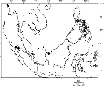
Distribution.Indonesia, Malaysia, Philippines (Fig. 1).
Distribution of Clemensiella: C. mariae (▲); C. omlori (•). Presence of Clemensiella mariae in Leyte and Mindanao is based on specimens cited by Schlechter (Reference Schlechter1915) that are no longer extant; occurrence on Bohol is based on a specimen cited by Omlor (Reference Omlor1998), Ramos 43106 (BM n.v.). Map constructed using Online Map Creation (www.aquarius.geomar.de).

Key to the species
1a. Corolla rotate, gynostegium exserted for 1/3–1/2 of its length, bud apices flattened-obtuse, peduncles 1–4 cm long 1. C. mariae
1b. Corolla campanulate, gynostegium apex barely exserted from the corolla throat, bud apices subobtuse to acute, peduncles 0.2–1 cm long 2. C. omlori
1. Clemensiella mariae (Schltr.) Schltr., Repert. Spec. Nov. Regni Veg. 13: 566 (Reference Schlechter1915), substitute name for Clemensia mariae Schltr., Repert. Spec. Nov. Regni Veg. 13: 543 (Reference Schlechter1915). – Type: Philippines, [Lanao del Sur]: Mindanao, Lake Lanao, Camp Keithley, iv 1906, Mary S. Clemens 512 (holo B, destroyed). Philippines, Leyte: Dagami, viii 1912, Ramos 15307 (paratype, not traced). Philippines, Laguna: Luzon, San Antonio, ix–x 1912, Ramos 15962 (lecto P!, designated here; isolecto BM n.v., K n.v.). Figs 2,3.
Clemensiella dischidioides Elmer in Merrill, Enum. Philipp. Fl. Pl. 3: 356 (Reference Merrill1923); Elmer, Leafl. Philipp. Bot. 10: 3549 (Reference Elmer1938), nom. nud.
Hoya viracensis Kloppenb. & Siar, Fraterna 19(4): 5 (Reference Kloppenburg and Siar2006), syn. nov. – Type: Philippines, Catanduanes: Virac, Brgy. Kalatagan, secondary forest, soil clay loam, 2 xii 1991, Barbon, Garcia & Alvarez PPI 5658 (holo PNH n.v.; iso BISH n.v., BRIT!, CAHUP n.v.).
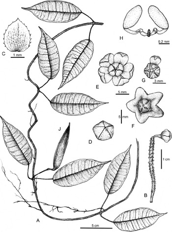
Clemensiella mariae Schltr. A, habit of plant, with adventitious roots; B, pedunculate inflorescence; C, sepal; D, flower bud (scale as for E and F); E, top view of opening flower; F, top view of open flower; G, top view of gynostegium with corona removed (including one recurved staminal lobe); H, pollinarium; J, ripe follicle. (Drawn by B. Diego from Laurente 001, 002, 003 and 004 (University of Santo Tomas Herbarium, Manila, Philippines).)

Clemensiella mariae Schltr. A, nectariferous flowers; B, corona in lateral view (SEM); C, guide-rail in lateral view, corona lobes removed (SEM); D, pollinarium, attached to the style-head (SEM). (All from ex hort. P. Gozon (UBT).)

Twiners up to 6 m tall, glabrous or nearly so. Taproots slender, occasionally with unpaired adventitious roots on internodes when in contact with substrate. Stems 3–5[–10 basally] mm diameter, hollow, green, shiny. Leaf blades thinly coriaceous, elliptic, basally rounded and slightly peltate, apically acuminate, 6–11 × 3–6 cm, adaxially shiny, minutely ciliate, petioles 1–2 cm long, shiny. Inflorescences 3–7-flowered, peduncles held perpendicular to the stems, 10–40 × c.2 mm diameter, slightly conical, rachis unbranched, up to 3 cm long. Flowers scentless; pedicels c.5 mm long, conical, sepals broadly ovate, c.2 mm long, appressed to the corolla tube; bud shape thickly lenticular with flattened apex; corolla rotate, 1.5–2 cm diameter, sympetalous for c.1/2 of length, adaxially bright yellow to orange-brown, occasionally spotted purplish, abaxially light green, occasionally spotted or tinged purplish; corolla lobes ovoid-triangular, c.6 × 4 mm at base, with median depression, recurved, margins revolute; gynostegial corona with discoid outline, concealed in corolla tube for 1/3–1/2 of its length, c.5 mm diameter, white, lobes c.3 × 2 mm at base; anthers triangular, c.1 × 1.2 mm, guide-rails c.0.25 mm long, spreading at an angle of 45°; anther connective appendages triangular-lanceolate, hyaline, c.0.3 × 0.3 mm, covering style-head completely; pollinia positioned horizontally on style-head in situ, c.0.6 × 0.25 mm, ovate in cross-section; caudicles convexly recurved, c.0.5 mm long; corpusculum 0.15 × 0.05 mm, brown; style-head c.2 mm diameter. Mericarps ± pendent, c.5–8 × 0.5–0.7 cm, with small ridges along dorsal and ventral sutures, broadening towards tip; seeds c.6.5 × 3 mm, wing up to 0.5 mm broad, with scattered white cilia, coma c.3 cm long, white.
Distribution. Philippines: Luzon (Laguna, Sorsogon), Catanduanes, Leyte, Mindanao (Lanao del Sur), Bohol (Fig. 1).
Ecology. Vigorous climber in forests (including secondary forests) and forest edges at low to medium altitudes (c.0–800 m). Sometimes epiphytic on shrubs (Shorea sp.) and small trees, or growing on rocks with little soil.
Chromosomes. Root tips were pretreated in 0.002 m 8-hydroxychinoline solution (Tjio & Levan, Reference Tjio and Levan1950), stained with carmine (Snow, Reference Snow1963), and squashed. We counted 2n = 22 chromosomes at the somatic metaphases (voucher: ex hort. P. Gozon (UBT)). On average the chromosomes are 1.27 μm long (measurement based on the analysis of four complete metaphase plates).
This count represents the first for Clemensiella. Because 94% of the investigated taxa of Asclepiadoideae, and 100% of Marsdenieae, have been found to have 2n = 22 chromosomes this result was expected. The average chromosome length of 1.27 μm is also within the range of what has been reported for Marsdenieae (min. 0.91 – max. 1.44 μm), measured from 21 species in eight genera. However, it is above the range of 0.91–1.06 μm documented for seven species of Hoya (Albers & Meve, Reference Albers, Meve, Endress and Stevens2001; Meve, unpubl. data). As pointed out by Albers & Meve (Reference Albers, Meve, Endress and Stevens2001), larger chromosomes are ancestral in Asclepiadoideae.
Specimens examined. Philippines. Sorsogon: Luzon, Irosin, Mt. Balusan, vi 1916, Elmer 16374 (BISH, L, P, US) and vii 1916, Elmer 16801 (BISH, C, L, P, US); Luzon, Irosin, road going to Lake Balusan, 9 ix 2005, Laurente 001 and 002, and 31 xii 2005, Laurente 003, and 30 iii 2006, Laurente 004 (CAHUP, LBC, University of Santo Tomas Herbarium, Manila); Luzon, Irosin, SW of Balusan National Park, 11 vii 1996, Garcia & Fernando 25051 (PNH); s.c. and s.n. (ex hort. P. Gozon) (UBT).
The holotype, which originated in Mindanao, was lost in the Berlin herbarium (B) during World War II. Of the two paratypes from Leyte and Luzon (Ramos 15307 and 16592, respectively), we found a sheet of Ramos 16592 at P, here selected as the lectotype. Omlor (Reference Omlor1998) cited sheets of Ramos 16592 from BM and K, here designated as isolectotypes. A second name under Clemensiella, C. dischidioides (see a specimen under http://sciweb.nybg.org/science2/hcol/vasc/index.asp), circulated via herbarium specimen exchange by Elmer, has never been validly published, because both Merrill (Reference Merrill1923) and Elmer (Reference Elmer1938) realised early enough that it is a specimen of C. mariae.
Recently, Kloppenburg & Siar (Reference Kloppenburg and Siar2006) described a collection of Clemensiella mariae from the island of Catanduanes as a new species of Hoya, H. viracensis Kloppenb. & Siar. In citing the type specimen we have transcribed the collection number, locality, and habitat data from the (newly spotted) isotype at BRIT; these differ somewhat from the citation in the protologue. The date on the specimen is written as ‘02/12/91’ which is interpreted as 2 December 1991. In his report on Philippine Hoya, Gozon (Reference Gozon2006: 18, fig. 14) included a photograph of Clemensiella mariae of unknown provenance labelled as ‘Dregea (?)’. The most recent collections were made by the second author in Sorsogon Prov., Irosin, Balusan Lake for her MSc thesis on Philippine Marsdenieae (Laurente, Reference Laurente2008). Altogether, Clemensiella mariae seems to be rarely collected and documented, but it is not a narrow endemic, occurring throughout the Philippine archipelago (Fig. 1).
2. Clemensiella omlori Livsh. & Meve, sp. nov.
Clemensiella mariae similaribus sed pedunculis brevioribus, corollis campanulatis, alabastris cum apicem subobtusis ad acutis. – Type: Indonesia, Aceh: Gunung Leuser Reserve, Camp Simpang and vicinity, 3–5 km upstream Lau [stream] Ketambe, c.35 km NW of Kutatjane, 400–600 m, 19 viii 1972, de Wilde & de Wilde-Duyfjes 14377 (holo L!; iso K, scan!). Fig. 4.
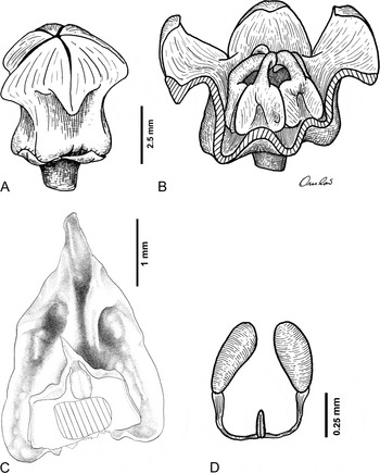
Clemensiella omlori Livsh. & Meve. A, bud; B, flower in lateral view, parts of the corolla removed; C, anther with corona lobe, adaxial view; region of retinacle depicted with diagonal cross-hatching; D, pollinarium. (A, B and D drawn from de Wilde & de Wilde-Duyfjes 14377 (K). C drawn from de Wilde & de Wilde-Duyfjes 14377 (L). A, B and D drawn by R. Omlor; C drawn by Jennifer Prevatt; B and D from Omlor (Reference Omlor1998), with permission.)

Twiners to 10 m tall (fide de Wilde & de Wilde-Duyfjes 14377). Roots unknown, no evidence of adventitious roots. Stems 2–3.5 mm diameter, internodes 10–18 cm long, pith solid. Leaf blades elliptic, basally rounded and slightly peltate, 8.1–11.1 × 3.5–6 cm, acumen 0.7–1.2 cm long, margins ciliate, petioles 2.2–5.9 cm long × 1–1.6 mm diameter, with line of short adpressed hairs on adaxial side. Inflorescences up to 12-flowered, peduncles 2–10 × 1.2–1.5 mm diameter, rachis unbranched. Flowers with pedicels 3.3–3.8 × 1.3 mm diameter, sparsely puberulent, sepals ovate, obtuse, c.1.5 × 1 mm, sparsely puberulent or ciliate; bud shape cylindrical with (sub)obtuse to acute apex, with 5 sharp downward-pointed projections at bases of corolla lobes; corolla campanulate, pale yellow to yellow-green (fide de Wilde & de Wilde-Duyfjes 14377, Anderson & Bisset S20257), c.9–12 mm diameter, throat c.3.5 mm diameter, tube with 5 rounded ribs opposite the lobes at base, tapering upwards, 3–5 × 4.5–5.5 mm diameter at base; corolla lobes 3.2–3.4 × 3.3–3.9 mm. Gynostegial corona barely exserted from corolla throat, c.2 × as tall as gynostegium, 2.9–4 × 2–2.8 mm; anthers 1.4–1.9 × 1–1.9 mm, guide-rails 0.4–0.7 mm long, ± parallel to the vertical axis of the gynostegium; anther connective appendages triangular, hyaline, c.1/3–1/2 total anther length, 0.6–0.8 × 0.3–0.6 mm, adpressed to and covering the style-head apex; pollinia c.0.4 × 0.15 mm; caudicles ± equal to pollinia in length, pale yellow; corpusculum c.0.1–0.15 × 0.05 mm, brown; style-head c.0.8 × 1.6 mm diameter. Fruit unknown.
Distribution. Known from two disjunct populations in North Sumatra and Aceh, Indonesia, and Sarawak, Malaysian Borneo (Fig. 1).
Ecology. A climber on trees in riparian rain forest (de Wilde & de Wilde-Duyfjes 14377) and a pendulous vine growing epilithically on damp limestone cliffs (Anderson & Bisset S20257), at elevations from c.60–600 m.
Chromosomes. Unknown.
Paratypes. Indonesia. North Sumatra: Sibaulangit [Sibolangit], Betimaesdal, 18 iv 1918, Lörzing 5648 (L!). Malaysia. Sarawak: Seburan, Bau, 15 vii 1964, Anderson & Bisset S20257 (K, scan!, L!).
This species is very similar to the Philippine endemic Clemensiella mariae. The most obvious distinguishing characters are to be found in the corolla, both in bud and at anthesis. Clemensiella mariae has thickly lenticular (to slightly obovoid) buds with flattened to obtuse apices. In Clemensiella omlori the bases of the corolla lobes form sharp downward-pointed projections in bud that mark an abrupt transition zone between the basally inflated, cylindrical tube and the lobes which form a subobtuse to acute apex (Fig. 4). At anthesis, the corolla of Clemensiella mariae is rotate with the gynostegium exserted from the corolla throat for 1/3–1/2 of its length while that of C. omlori is campanulate with the tips of the corona lobes barely exserted from the throat (compare Figs 2 and 4). The orientation of the corolla lobes is depicted as somewhat erect in Fig. 4, but many flowers on herbarium specimens have rotate or reflexed corolla lobes. The guide-rails of Clemensiella omlori are approximately vertical (parallel to the long axis of the gynostegium) while those of C. mariae spread at an angle of 45° (Fig. 3C). Clemensiella omlori also has shorter peduncles, typically less than 1 cm long, while those of C. mariae are 1–4 cm long.
The cited specimens were first identified as ‘Hoya sp.’, ‘Secamone sp.’, and ‘Asclepiadaceae gen. et sp. indet.’. There are likely other collections of this species in the world's herbaria.
This species is named in honour of Dr Ralf Omlor (Mainz, Germany) who first recognised it and discussed some of the differences between it and Clemensiella mariae in his generic revision of Marsdenieae (Omlor, Reference Omlor1998).
Acknowledgements
We thank David Goyder for locating and imaging specimens of Clemensiella omlori at K, the curators of BISH, BRIT, C, K, L, P, PNH and US for loan of specimens to and/or hosting visits by the fourth author, T. Nyhuus for sending us living material of Clemensiella mariae, Boy Diego for drawing C. mariae, and Ralf Omlor and Jennifer Prevatt for drawing the figures of C. omlori.