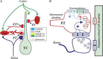
The thalamus serves as the obligatory pathway through which sensory information must pass en route to the neocortex. It is well accepted that the thalamus is not a passive relay, but based on synaptic connectivity, intrinsic properties of neurons, and multiple intrathalamic connectivity, the transfer of information through the thalamus is a dynamic process. Sensory information enters the thalamus via sensory afferents that innervate two subtypes of neurons: thalamocortical neurons that send efferents to primary sensory neocortex and intrinsic inhibitory interneurons with dendrites and axons that remain in the thalamus (Fig. 1). In addition to the sensory inputs, the thalamus receives extensive innervation from corticothalamic neurons in layer VI of the neocortex. These corticothalamic efferents innervate thalamocortical neurons, intrinsic interneurons, and the thalamic reticular nucleus (TRN) neurons.
(A) Schematic diagram of visual thalamic circuitry showing multiple inhibitory inputs. Axonal outputs (F1 terminals) arise from local inhibitory interneurons (Int) and TRN neurons. Presynaptic dendrites (F2 terminals) of interneurons are innervated by retinogeniculate axons that also innervate the dendrite of thalamocortical neuron (TC). This is a simplified schematic in which other thalamic inputs to this circuit have been omitted for clarity. Adapted from Govindaiah and Cox (Reference Govindaiah and Cox2006). (B) Schematic of triad. Retinogeniculate terminals form monosynaptic excitatory synapse on thalamocortical cell dendrite and disynaptic inhibitory input via the F2 terminal. Diagram illustrates the multiple transmitter systems that have been shown to alter the output of putative F2 terminals.

Inhibition within the primary visual thalamus, the dorsal lateral geniculate nucleus (dLGN), primarily arises from intrinsic interneurons and TRN neurons with a sparser input from the pretectum. In this review, we will focus on the intrinsic interneurons. These interneurons are somewhat unique because these cells provide inhibitory output to thalamocortical neurons via dendrodendritic outputs (Guillery, Reference Guillery1969; Ralston, Reference Ralston1971; Famiglietti & Peters, Reference Famiglietti and Peters1972; Lieberman, Reference Lieberman1973; Hamos et al., Reference Hamos, Van Horn, Raczkowski, Uhlrich and Sherman1985; Montero, Reference Montero1986; Ohara & Lieberman, Reference Ohara and Lieberman1993). These presynaptic dendrites are referred to as F2 terminals from ultrastructural analyses and have been identified in different thalamic nuclei across multiple species including cat ventrobasal (Ralston & Herman, Reference Ralston and Herman1969), cat dLGN (Guillery, Reference Guillery1969; Famiglietti, Reference Famiglietti1970), rat dLGN (Grossman et al., Reference Grossman, Lieberman and Webster1973; Lieberman, Reference Lieberman1973; Parnavelas et al., Reference Parnavelas, Mounty, Bradford and Lieberman1977), mouse dLGN (Rafols & Valverde, Reference Rafols and Valverde1973), monkey dLGN, and cat medial geniculate (Jones & Rockel, Reference Jones and Rockel1971; Morest, Reference Morest1971). In addition, these interneurons may also give rise to classical axonal outputs (F1 terminals); however, it is important to note that multiple light-level anatomical studies have indicated the presence of axon-like structures, subsequent ultrastructural confirmation is lacking (Morest, Reference Morest1971; Grossman et al., Reference Grossman, Lieberman and Webster1973; Lieberman, Reference Lieberman1973; Parnavelas et al., Reference Parnavelas, Mounty, Bradford and Lieberman1977; Perreault et al., Reference Perreault, Qin, Heggelund and Zhu2003).
Synaptic transmission typically occurs by suprathreshold excitation near the soma that in turn generates an action potential, which can propagate into the axonal arbor producing neurotransmitter release from axonal terminals. The dendrites of a neuron are typically responsible for the integration of afferent synaptic input properties, and this process can be quite complex given that a variety of voltage-dependent channels within the dendritic arbor can shape the integrative process (Johnston & Narayanan, Reference Johnston and Narayanan2008). The intrinsic interneurons in the thalamus have dendrites that may also contain synaptic vesicles, and therefore, can provide dendrodendritic synaptic output. These dendrodendritic outputs (F2 terminals) are part of a triadic arrangement that includes three components: retinogeniculate axon terminal, distal presynaptic dendrite of the interneuron, and the proximal postsynaptic dendrite of the thalamocortical neuron. The retinogeniculate axon innervates the proximal dendrite of the thalamocortical neuron forming a monosynaptic excitatory synapse and also innervates the distal dendrite of the interneuron. This innervation of the interneuron is presynaptic to the thalamocortical neuron and thus gives rise to disynaptic inhibition of that local region of the thalamocortical neuron dendrite (Fig. 1). Considering the close proximity of the monosynaptic and disynaptic innervation of the thalamocortical neurons, the interneuron is situated to provide robust regulation of retinogeniculate excitation of thalamocortical neurons.
These intrinsic interneurons are thought to primarily provide feedforward inhibition in the retinogeniculate pathway. These cells are primarily excited by sensory afferents and have been shown to shape receptive fields of thalamocortical neurons, augment stimulus selectivity, increase sensory coding, and refine temporal precision of action potential discharge (Sillito & Kemp, Reference Sillito and Kemp1983; Holdefer et al., Reference Holdefer, Norton and Godwin1989; Wang et al., Reference Wang, Wei, Vaingankar, Wang, Koepsell, Sommer and Hirsch2007, Reference Wang, Vaingankar, Sanchez, Sommer and Hirsch2011). Within the thalamocortical circuit, this linear role of the interneuron is complicated in that these cells also receive innervation from corticothalamic neurons (Dubin & Cleland, Reference Dubin and Cleland1977; Li et al., Reference Li, Wang and Bickford2003; Jurgens et al., Reference Jurgens, Bell, McQuiston and Guido2012), inhibitory GABAergic inputs (Pape & McCormick, Reference Pape and McCormick1995; Williams et al., Reference Williams, Turner, Anderson and Crunelli1996; Van Horn et al., Reference Van Horn, Erisir and Sherman2000), as well as multiple neuromodulator inputs (see Neuromodulator regulation: Altering the state of output). The functional significance of these different inputs onto the interneurons is unclear at this time, but will likely impact how retinogeniculate information is processed through this circuit and thereby influence visual processing.
F2 terminals give rise to transient and persistent inhibition
Although these F2 terminals were first identified nearly 50 years ago, the function of F2 terminal-mediated inhibition has remained speculative for many years (Guillery, Reference Guillery1969; Ralston, Reference Ralston1971; Hamos et al., Reference Hamos, Van Horn, Raczkowski, Uhlrich and Sherman1985; Montero, Reference Montero1986). Numerous studies have shed light on the role of inhibition in sensory processing; however, the functional contribution of F2 terminals to sensory processing has remained unclear considering that pharmacological manipulation could not distinguish between axonal output and dendritic output of intrinsic interneurons. Most studies involving local interneurons typically have focused on postsynaptic alterations in excitability (e.g., alteration in membrane potential, action potential discharge rates) produced by different neurotransmitters and neuromodulators. More recently, by way of creative pharmacological manipulations and/or modern technological approaches, multiple studies have shed light on the regulation of F2 terminal activity. These studies have revealed that output of these dendrodendritic synapses is quite complicated and are regulated by a variety of neurotransmitter/modulators, including glutamate, acetylcholine, serotonin, and dopamine (Cox & Sherman, Reference Cox and Sherman2000; Munsch et al., Reference Munsch, Freichel, Flockerzi and Pape2003, Reference Munsch, Yanagawa, Obata and Pape2005; Govindaiah & Cox, Reference Govindaiah and Cox2009; Antal et al., Reference Antal, Acuna-Goycolea, Pressler, Blitz and Regehr2010) (Fig. 1). Currently, the functional role of F2 terminal-mediated inhibition in sensory processing remains speculative. We suspect that with increased knowledge of how F2 terminal output is regulated, the function of the triad and role of F2 terminal-mediated inhibition is within sight.
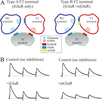
The primary input to the triad is the glutamate-containing retinogeniculate axon terminal. While initial studies isolating the output of F2 terminals focused on the role of mGluRs, there is growing evidence that F2 terminals are regulated by the activation of ionotropic glutamate receptors (iGluRs) and that this mechanism may actually be more common than the metabotropic glutamate receptor (mGluR)-dependent regulation of F2 terminals (Cox & Sherman, Reference Cox and Sherman2000; Blitz & Regehr, Reference Blitz and Regehr2005; Acuna-Goycolea et al., Reference Acuna-Goycolea, Brenowitz and Regehr2008; Crandall & Cox, Reference Crandall and Cox2012, Reference Crandall and Cox2013). A variety of approaches including stimulation of synaptic afferents, focal application of glutamate agonists, and photo-uncaging of caged glutamate have demonstrated that activation of iGluRs produce a short-lasting, robust increase in putative F2 terminal output (Crandall & Cox, Reference Crandall and Cox2012, Reference Crandall and Cox2013). As predicted, the iGluR-dependent inhibitory response has a short latency and relatively short duration (<100 ms) (Acuna-Goycolea et al., Reference Acuna-Goycolea, Brenowitz and Regehr2008). The iGluR-dependent response is also evoked by single stimuli, which may shed light on the function of this mechanism considering that activation of the mGluR-dependent response requires tetanic activity of the retinogeniculate pathway. By use of focal glutamate uncaging, there appears to be two distinct types of F2 terminals: (1) the predominant F2 terminal is stimulated only by iGluR activation and (2) F2 terminals that are activated by iGluRs and mGluRs in an activity dependent manner; at low stimulation frequencies, only iGluRs are engaged producing a short-lasting response, and at higher frequencies, mGluRs are engaged producing a longer lasting response (Crandall & Cox, Reference Crandall and Cox2013).
By taking advantage of this distinct distribution of mGluRs in the cat dLGN (Godwin et al., Reference Godwin, Van Horn, Erisir, Sesma, Romano and Sherman1996), pharmacological activation of type 5 mGluRs (mGluR5) produced a robust, long-lasting increase in inhibitory activity within cat thalamocortical neurons (Cox et al., Reference Cox, Zhou and Sherman1998; Cox & Sherman, Reference Cox and Sherman2000). Subsequent studies indicated similar actions of mGluR activation in rat thalamocortical neurons as well (Govindaiah & Cox, Reference Govindaiah and Cox2006). An initial interpretation was that mGluR activation of F2 terminals could provide local inhibitory function related to the triads, and this distal dendritic activity of interneurons could occur independent of somatic activity of the interneuron. The general mGluR agonist, ACPD, which produced a robust increase in F2 terminal output, produced minimal alteration in membrane potential from somatic recordings from local interneurons further supporting the hypothesis of independence of distal dendritic actions from the somatic activity (Pape & McCormick, Reference Pape and McCormick1995; Cox et al., Reference Cox, Zhou and Sherman1998). Subsequent studies found that activation of group I mGluRs (mGluR1, mGluR5) depolarized interneurons, and activation of group II mGluRs (mGluR2, mGluR3) hyperpolarizes the interneurons (Govindaiah & Cox, Reference Govindaiah and Cox2006; Pressler & Regehr, Reference Pressler and Regehr2013). It is unclear if subsets of afferent inputs can selectively activate group I versus group II mGluRs. If so, this could have important implications on how we interpret the local vs. global influence of interneurons on thalamic process.
Engaging mGluRs following synaptic activation of focal glutamate produces a distinctly different type of inhibition compared to that mediated by iGluR activation. Synaptic activation of mGluRs requires tetanic stimulation of the optic tract that leads to a frequency-dependent increase in mGluR5-dependent activation of F2 terminals, resulting in a long-lasting increase in inhibition in the postsynaptic thalamocortical neuron from rats (Govindaiah & Cox, Reference Govindaiah and Cox2004). However, when recording from these interneurons, this same tetanic stimulation paradigm only produced a short-lasting depolarization in the somatic recordings that was completely blocked by iGluR antagonists, supporting the notion that dendritic events can occur independent of somatic actions in the interneurons (Govindaiah & Cox, Reference Govindaiah and Cox2004, Reference Govindaiah and Cox2006). However, it is important to note that other studies have shown that optic tract stimulation can evoke an excitatory postsynaptic potential (EPSP) with a lasting calcium-dependent depolarizing plateau (∼100 ms duration) and at higher intensity a much longer lasting plateau response (>10 s duration) in mouse dLGN interneurons (Acuna-Goycolea et al., Reference Acuna-Goycolea, Brenowitz and Regehr2008; Pressler & Regehr, Reference Pressler and Regehr2013). These authors hypothesized that the prolonged increase in inhibitory output from such stimulation resulted from the backpropagation of a calcium-dependent current into the dendritic arbor that in turn could stimulate gamma-aminobutyric acid (GABA) release from F2 terminals. Currently, it is unclear if this lasting potential is due to different subtypes of dLGN interneurons or if this could be due to different species: mouse versus rat/cat. Thus, this requires additional studies considering that these two different mechanisms could have very distinct downstream impact on thalamocortical processing (see Multiprocessing versus unitary processing characteristics of F2 terminal outputs).
Neuromodulator regulation: Altering the state of output
In addition to the activation of F2 terminals by mGluR activation, a number of other neuromodulators have been shown to alter this inhibitory output. Multiple monoamines have been found to increase the output of F2 terminals in the dLGN. Serotonin, via 5HT2 receptors, increases F2 terminal output via a calcium-dependent manner similar to that produced by mGluR5 activation (Munsch et al., Reference Munsch, Freichel, Flockerzi and Pape2003). Activation of D2 dopamine receptors also increases the inhibition arising from F2 terminals in thalamocortical neurons (Munsch et al., Reference Munsch, Yanagawa, Obata and Pape2005). By contrast, acetylcholine, via activation of M2 receptors, has been shown to significantly decrease the inhibitory output of intrinsic interneurons presumably through decreased output of F2 terminals (Cox & Sherman, Reference Cox and Sherman2000; Munsch et al., Reference Munsch, Freichel, Flockerzi and Pape2003). With respect to the discussion above, the actions of these neuromodulators would result in a slow, lasting alteration in F2 terminal activity. Thus, the overall action would be quite similar to the time course we observe with mGluR activation.
Clearly, the excitation of F2 terminals is an activity-dependent process in which the iGluR-dependent inhibition can occur at low frequency activity of retinogeniculate afferents, and with higher frequency activity, mGluR-dependent activation of F2 terminals is engaged. While the impact of these specific forms of inhibition is unclear, we speculate that these would provide distinct forms of inhibition onto the thalamocortical neuron. The short-duration inhibition arising from iGluR activation would likely result in shaping of the retinogeniculate excitation of the thalamocortical neuron. At low frequencies, this mechanism could decrease the duration of the retinogeniculate excitation, thereby sharpening the excitation. The engagement of mGluR-dependent-mechanisms, or other neuromodulators as well, would result in an overall dampening of excitation (Fig. 2). Thus, engaging these long-lasting forms of inhibition would decrease the magnitude of subsequent retinogeniculate excitation of the thalamocortical neuron. Further studies are required to understand the functional roles of these different forms of inhibition. In addition, it will be very interesting to understand the functional significance of why some F2 terminals only respond to iGluR stimulation, while others respond to both iGluR and mGluR in an activity-dependent manner (Crandall & Cox, Reference Crandall and Cox2013).
(A) Schematic diagram of two different types of F2 terminals. Type A F2 terminals are stimulated only via iGluRs whereas type B F2 terminals can be activated by mGluRs and iGluRs. Adapted from Crandall and Cox (Reference Crandall and Cox2013). (B) Hypothetical impact on retinogeniculate EPSPs via iGluR-dependent activation of F2 terminals (left) and mGluR-dependent activation of F2 terminals.

Multiprocessing versus unitary processing characteristics of F2 terminal outputs
The F2 terminals are anatomically situated to have a strong impact on the monosynaptic retinogeniculate excitation of the thalamocortical neuron. The intrinsic properties of the dendritic arbor of the local interneurons could have a significant impact on the functional role of the F2 terminals. Does the dendritic arbor of these interneurons serve a similar role as an axonal arbor or does the dendritic arbor support multiple isolated electrotonic units that can function independent of each other?
Localized inhibitory output
Considering the microcircuitry of the triadic arrangement, the functional role of F2 terminals could be to precisely regulate retinogeniculate excitation of thalamocortical neurons. One hypothesis is that excitation of F2 terminals can occur independent of actions at the soma level of the interneurons. F2 terminals are primarily located on distal dendrites of the interneurons and have knob-like appearances (Famiglietti & Peters, Reference Famiglietti and Peters1972; Grossman et al., Reference Grossman, Lieberman and Webster1973; Montero, Reference Montero1986). Early computational modeling of the dendritic arbors of an interneuron, assuming passive membrane properties, predicted that activity at the F2 terminals remained focal and were electrotonically isolated from other major dendritic branches as well as somatic/axonal axis (Bloomfield & Sherman, Reference Bloomfield and Sherman1989). However, subsequent electrophysiological and anatomical studies in multiple neuronal subtypes clearly demonstrate that dendritic arbors are not simply a network of passive membranes, but actually contain a plethora of active conductances that can have significant impact on the integrative and propagative properties of dendritic arbors (Johnston & Narayanan, Reference Johnston and Narayanan2008). Thus, it would be of interest to experimentally determine whether focal excitation of F2 terminals can occur independent of F2 terminals in other dendritic branches within an individual interneuron as well as somatic activity within the interneuron.
Prior studies indicate that the F2 terminal-dependent local signaling can occur independent of suprathreshold activation of the interneuron. The stimulation of mGluRs by selective agonists, produced an increase in F2 terminal output within thalamic relay neurons independent of significant changes in membrane potentials of interneurons at the somatic level (Cox et al., Reference Cox, Zhou and Sherman1998; Cox & Sherman, Reference Cox and Sherman2000). Similarly, tetanic stimulation of retinogeniculate fibers produced a lasting increase in mGluR-mediated F2 terminal-dependent inhibition similar to that observed with mGluR agonists. When recording from a dLGN interneuron, similar retinogeniculate stimulation produced only a short transient depolarization in the interneuron and not a prolonged excitation as reflected by the lasting increase in inhibition in the thalamocortical neurons. Thus, the transient, iGluR-dependent depolarization of the interneuron from tetanic stimulation was not sufficient to account for the longer duration of inhibitory output observed in the thalamocortical neuron supporting the hypothesis that activation of F2 terminals can occur independent of somatic activity within the interneuron (Govindaiah & Cox, Reference Govindaiah and Cox2004).
In subsequent studies, focal photouncaging of glutamate along dendrites of thalamocortical neurons produced transient inhibitory responses presumably due to activation of F2 of interneurons (Crandall & Cox, Reference Crandall and Cox2012). When recording from interneurons, similar glutamatergic stimulation at distal sites that was sufficient to produce inhibition in the thalamocortical neuron produced little or no alterations of the membrane potential at the somatic level (Crandall & Cox, Reference Crandall and Cox2012). These localized forms of inhibition were differentially regulated by iGluR and/or mGluR activation (Crandall & Cox, Reference Crandall and Cox2013). These results indicate that F2 terminal output can provide focal inhibition near retinogeniculate innervation, and these events that are generated at distal dendrites can occur without influencing the somatic/axonal activity or output of the interneuron (Crandall & Cox, Reference Crandall and Cox2012). Because of the electronic isolation of F2 terminal activity, these units may allow the interneuron to function as a multiprocessor carrying out numerous independent functions with respect to retinogeniculate information processing.
Widespread inhibitory output
In contrast to the isolated activity of F2 terminals, recent studies indicate that dLGN interneurons have the ability to propagate somatic generated potentials back into the dendritic arbor. Either action potentials or long-lasting calcium potentials have been shown to backpropagate into the dendritic arbor and via this mechanism could cause widespread activation of F2 terminals thereby producing a more spatially extensive inhibition of retinogeniculate excitation in thalamocortical neurons (Acuna-Goycolea et al., Reference Acuna-Goycolea, Brenowitz and Regehr2008; Casale & McCormick, Reference Casale and McCormick2011).
Stimulation of retinogeniculate axons could lead to an initial excitation followed by a calcium-dependent prolonged depolarization (∼100 ms duration) that can propagate through the dendritic arbor and result in a transient increase in F2 terminal output (Acuna-Goycolea et al., Reference Acuna-Goycolea, Brenowitz and Regehr2008). The calcium potential occurs at relatively hyperpolarized membrane potentials and is dependent on activation of L-type calcium channels. Subsequent studies indicated that higher intensity stimulation could produce a longer-lasting calcium plateau (>10 s) that can backpropagate into the dendritic arbors and produce a robust, long-lasting activation of F2 terminals. As a result, the inhibitory output from strong activation of the interneuron would result in widespread inhibitory output that would encompass the area of the overall, and thus would act similar to an axonal arbor.
In widespread inhibition, the activation of the F2 terminals appears dependent on generating the calcium potential. While generating an action potential at the soma level produced a transient backpropagating dendritic arbor, this was not sufficient to increase F2 terminal output. Furthermore, activation of local synaptic inputs was unable to initiate action potentials within the dendritic arbor. However, action potentials were somatically generated with sufficient excitation and would back propagate into the dendrite (Casale & McCormick, Reference Casale and McCormick2011). Currently, it is unclear if an individual neuron could produce both widespread and localized outputs depending on the resting state of the neuron. Alternatively, there may be multiple subtypes of dLGN interneurons that would provide two functionally distinct forms of inhibition.
Contribution of inhibition to visual processing
Thalamic neurons have receptive fields that are composed of two concentrically arranged areas, a center, and surround, which possess opposite preference for stimulus contrast similar to those seen in the retina (Kuffler, Reference Kuffler1953; Hubel, Reference Hubel1960; Wang et al., Reference Wang, Wei, Vaingankar, Wang, Koepsell, Sommer and Hirsch2007; Suresh et al., Reference Suresh, Çiftçioğlu, Wang, Lala, Ding, Smith, Sommer and Hirsch2016). As described in the retina, the sensory input on thalamocortical neurons has a push–pull excitation and inhibition arrangement (Wiesel, Reference Wiesel1959; Wang et al., Reference Wang, Wei, Vaingankar, Wang, Koepsell, Sommer and Hirsch2007, Reference Wang, Vaingankar, Sanchez, Sommer and Hirsch2011; Suresh et al., Reference Suresh, Çiftçioğlu, Wang, Lala, Ding, Smith, Sommer and Hirsch2016). Push–pull occurs when stimuli of the opposite contrast evoke responses of the opposite sign. Simply put, push is evoked by a stimulus of the preferred contrast that can increase the action potential output of the neuron via an underlying depolarization (Wang et al., Reference Wang, Wei, Vaingankar, Wang, Koepsell, Sommer and Hirsch2007, Reference Wang, Vaingankar, Sanchez, Sommer and Hirsch2011; Suresh et al., Reference Suresh, Çiftçioğlu, Wang, Lala, Ding, Smith, Sommer and Hirsch2016). By contrast, the pull is an inhibition evoked by a stimulus of the opposite contrast and has been associated with a hyperpolarization of the thalamocortical neuron. While the push likely is due to excitatory retinogeniculate input, the pull is hypothesized to arise from intrinsic interneurons (Hirsch et al., Reference Hirsch, Wang, Sommer and Martinez2015). dLGN interneurons have similar center surround receptive fields and typically have opposite preference for stimulus contrast as retinal ganglion cells and dLGN thalamocortical relay neurons (Wang et al., Reference Wang, Vaingankar, Sanchez, Sommer and Hirsch2011). In addition, dLGN interneurons are more likely to have receptive fields that are opposite in preference for stimulus contrast and are larger in size than connected dLGN thalamocortical relay neurons (Martinez et al., Reference Martinez, Molano-Mazón, Wang, Sommer and Hirsch2014). This arrangement leads to retinogeniculate excitation that is complemented by strong inhibition from nearby interneurons that have opposite preference for stimulus contrast and leads to improved spatial discrimination.
Currently, it is unclear how F1 terminals and F2 terminals contribute to the receptive fields of thalamocortical neurons. A receptive field of a neuron is the visual area that must be stimulated in order to elicit a change in action potential firing from a neuron. Ideally to dissociate the local versus global aspects of interneuron contribution to visual processing in thalamocortical neurons, one needs to be able to dissociate action potential-dependent and action potential-independent inhibition arising from the interneurons. On the one hand, F1 terminals, if they functionally exist, from dLGN interneurons should have the clearest role; however, if action potentials backpropagate into distal dendrites strong enough to activate F2 terminals (Acuna-Goycolea et al., Reference Acuna-Goycolea, Brenowitz and Regehr2008), then both F1 and F2 synapses would be activated, providing global inhibition in the shaping of the receptive fields in thalamocortical neurons. On the other hand, F2 terminals can act independent of action potential firing (Cox et al., Reference Cox, Zhou and Sherman1998), and therefore the receptive field of the interneuron as currently determined with electrophysiological recordings would not predict the impact of inhibition upon the thalamocortical neuron. Thus, a potentially important unanswered question is what is the receptive field of action potential independent activation of F2 terminals? Clearly, our current understanding of the contributions of these two sources of inhibition is limited and will require further investigation using intact in vivo preparations.
Acknowledgment
This work was supported by the National Institutes of Health grant EY014024.