Foetal electrocardiographic intervals follow a predictable pattern among healthy foetuses.Reference Taylor, Smith and Thomas 1 – Reference Pardi, Ferrazzi and Cetin 5 In normal pregnancies, PR and QRS intervals lengthen parallel to the weight gain of the foetal heart and increase in ventricular mass. It has been proposed that the duration of cardiac intervals could be used as an index of the size and development of the foetal heart and aid the identification of foetal growth restriction.Reference Brambati and Pardi 2 – Reference Pardi, Ferrazzi and Cetin 5 In the setting of congenital heart disease, resultant changes in ventricular mass presenting as ventricular hypertrophy or hypoplasia were also reported to be associated with increased or decreased interval length, respectively.Reference Pardi, Ferrazzi and Cetin 5 It is, however, not known whether foetal electrocardiographic intervals change across gestation in foetuses with congenital heart disease or whether differences in foetal electrocardiographic intervals exist between congenital heart disease and non-congenital heart disease foetuses. The goal of this project was to characterise and compare foetal electrocardiographic intervals across gestational ages among foetuses with and without structural heart defects. We hypothesised that foetal electrocardiographic intervals would lengthen across gestation and differences would be observed between groups. These findings could have the potential to impact prenatal screening for congenital heart disease, especially in the third world where ultrasound technology and expertise are limited.
Materials and methods
We conducted a prospective observational cohort study of foetuses with and without congenital heart disease. The Columbia University Medical Center Institutional Review Board approved this study. The participants of the study consisted of pregnant women and foetuses referred to the Morgan Stanley Children’s Hospital of NewYork-Presbyterian’s Center for Prenatal Pediatrics for foetal echocardiogram with a diagnosis of either normal cardiac anatomy or congenital heart disease consisting of hypoplastic left heart syndrome, tetralogy of Fallot, or transposition of the great arteries. We chose these three lesions because they are commonly diagnosed prenatally at our centre and they have distinct anatomical and physiological features that could potentially impact electrocardiographic intervals in important ways.
Exclusion criteria for this study included multiple gestation, foetal genetic anomalies, and foetal rhythm abnormalities. Foetal electrocardiograms were obtained using the Monica AN24 maternal abdominal foetal electrocardiographic monitor (Monica Healthcare Ltd, Nottingham, United Kingdom). The use of the Monica AN24 has been previously described.Reference Pardi, Ferrazzi and Cetin 5 In brief, the Monica AN24 has five electrodes and non-invasively records both maternal and foetal electrocardiographic signals. Recorded data were processed via the Monica DK V1.8 (Monica Healthcare Ltd) software, which separates maternal electrocardiogram from the foetal electrocardiogram and filters artefact from maternal abdominal contractions or movement, thereby allowing direct analysis of the foetal electrocardiogram. Other groups have reported success using this monitor to record and analyse foetal heart rate but not in foetuses with congenital heart disease.Reference Graatsma, Jacod, van Egmond, Mulder and Visser 6 – Reference Reinhard, Hayes-Gill, Schiermeier, Hatzmann, Heinrich and Louwen 10
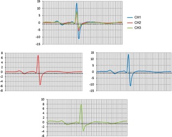
Foetal electrocardiographs were obtained at the following three gestational ages: 20–24 weeks (F1), 28–32 weeks (F2), and 34–38 weeks (F3). The study duration was 45 minutes. Foetal R waves were identified, and beat-to-beat foetal heart range in a range of 60–200 beats per minute (bpm) was calculated via automated analyses. Any value out of this heart rate range was excluded from further analysis. The signal-averaged waveform was calculated from tracings where at least 10% of foetal beats were detected. At least two physicians – I.W. and either B.Y. or H.N. – reviewed each sample independently to rate tracing quality as poor, moderate, or good and to calculate PR, QRS, and QT interval length. Only foetal electrocardiogram tracings of moderate to good quality were used in the final analysis (Fig 1). Therefore, success was defined first by obtaining a good quality foetal electrocardiogram recording with a detectable foetal electrocardiographic signal, and second by obtaining a high-quality signal-averaged waveform with readily identifiable P, QRS, and T waves.
An example of a signal-averaged waveform obtained from each recording channel, where 1000 foetal QRS complexes were averaged. Tracings obtained from channels (CH) 1, 2, and 3 were analysed independently to calculate PR, QRS, and QT interval lengths. Note that the T wave detection was poor in this participant.

Statistical analysis
Gestational ages for each group (F1 versus F2 versus F3) are reported as means (standard deviation). Pearson’s correlation coefficient was used to analyse the relationship between gestational age and foetal electrocardiographic interval length. Intervals from controls were compared with gestational-normed published values.Reference Taylor, Smith and Thomas 1 Comparisons between gestational age periods were assessed using analysis of variance and paired t-test. Comparisons between congenital heart disease and non-congenital heart disease foetuses were carried out using Student’s t-test. Inter-observer variability was calculated using the intraclass correlation coefficient, and the intraclass correlation coefficient for PR and QRS were 0.85 (p<0.001) and 0.78 (p<0.001), respectively. Alpha level was set at p≤0.05.
Results
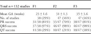
We enrolled 92 participants, 41 with normal cardiac anatomy and 51 with congenital heart disease. A total of 177 foetal electrocardiograms were obtained across gestation, with a mean gestational age of 23±1.6 weeks at F1, 31±1.3 weeks at F2, and 35±3.6 weeks at F3. Sufficient data quality to create a signal-averaged waveform was seen in 132 (75%) foetal electrocardiograms from 74 (80%) participants. Of these 74 participants, 33 (45%) were controls and 41 (55%) had congenital heart disease. Of the congenital heart disease foetuses, 15 had the diagnosis of hypoplastic left heart syndrome, 12 had transposition of the great arteries, and 14 had tetralogy of Fallot. Of the 132 foetal electrocardiograms, tracing quality was adequate to calculate PR in 102 (77%), QRS in 129 (98%), and QT in 39 (30%). Signal quality for interval analysis was highest at 20–24 weeks and at 34–38 weeks of gestation. Although waveform success rate did not change statistically across gestation for calculation of PR and QRS intervals, the least success was seen in F2. T waves appeared to become more detectable with increasing gestational age, although even late in pregnancy T wave identification was limited (Table 1).
Foetal electrocardiographic (ECG) interval calculation success at different gestational age (GA) periods.

Foetal electrocardiographic intervals of control foetuses and gestational age
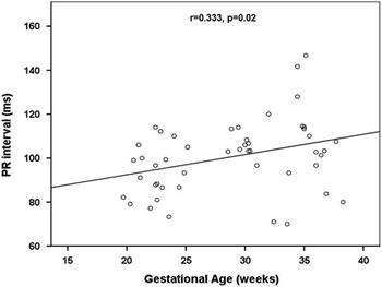
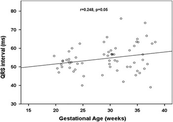
Among foetuses without structural heart disease, 88% of PR intervals, 87% of QRS intervals, and 86% of QT intervals fell within 2 standard deviations of previously reported norms. Among these control foetuses, PR (r=0.333, p=0.02) and QRS (r=0.248, p=0.05) intervals showed a mild- to-modest correlation with gestational age (Figs 2 and 3). Using paired t-test, differences in PR interval were observed between F1 and F2 as well as between F1 and F3 (Table 2); however, unlike in the correlation analysis, in the paired analyses, no significant differences were observed in QRS across gestational age periods. Owing to the low success of QT measurement, we did not compare QT intervals between groups or across gestation.
PR interval correlates positively with gestational age among non-congenital heart disease (CHD) foetuses, indicating that PR interval lengthens as pregnancy progresses.

QRS interval correlates positively with gestational age among non- congenital heart disease (CHD) foetuses, indicating that QRS interval lengthens as pregnancy progresses.

PR, QRS, and QT intervals among non-CHD foetuses during the different GA periods with ANOVA, p-values.

ANOVA=analysis of variance; CHD=congenital heart disease; GA=gestational age
Using the paired t-test, differences in PR interval between F1 and F2 (n=11, p=0.007) and F1 and F3 (n=8, p=0.02) were seen. Although QRS intervals appeared to lengthen, differences between GA periods were not statistically significant in paired analyses
Foetal electrocardiographic intervals of congenital heart disease foetuses and gestational age
Among foetuses with structural heart defects, there was no correlation between PR, QRS, and QT intervals and gestational age. Using paired analyses, we were not able to identify any significant differences in PR, QRS, and QT intervals among congenital heart disease foetuses at F1, F2, and F3.
Foetal electrocardiogram comparisons between control and congenital heart disease foetuses
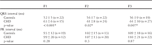
Foetuses with congenital heart disease demonstrated longer QRS intervals at F1, F2, and F3 compared with foetuses with normal cardiac anatomy (Table 3). Although PR intervals were longer in congenital heart disease foetuses compared with foetuses without structural heart defects, these differences were not statistically significant (Table 3).
CHD versus non-CHD QRS and PR intervals in different GA periods.

CHD=congenital heart disease; GA=gestational age
Foetuses with CHD consistently demonstrated longer QRS intervals compared with controls. Foetuses with CHD also demonstrated longer PR intervals compared with controls; however, these differences were not statistically significant
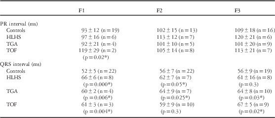
In the subgroup analyses among foetuses with congenital heart disease, foetuses with tetralogy of Fallot demonstrated the longest PR interval at F1 (Table 4). There was no significant difference in QRS interval found at any gestational age between subgroups, but each congenital heart disease subtype had longer QRS intervals than foetuses without structural heart disease (Table 4).
CHD subtypes (hypoplastic left heart syndrome (HLHS) versus transposition of the great arteries (TGA) versus tetralogy of fallot (TOF)) versus non-CHD PR and QRS intervals at different gestational age (GA) periods using the t-test.

Although the sample size within the individual CHD diagnostic groups was limited, foetuses with TOF had the longest PR interval at F1. There was no significant difference in QRS interval found across GA; however, within CHDs, each subtype had longer QRS than foetuses without structural heart disease
Discussion
Heart rate and electrocardiographic intervals are known to change over time from foetal life to adulthood,Reference Mehta and Dhillon 11 although there are limited data on the electrocardiographic intervals of foetuses and newborns with congenital heart disease. In a previously reported small series, PR and QRS intervals were shown to be prolonged in newborns with hypoplastic left heart syndrome compared with age-matched controls. It was proposed that these changes might be due to changes in the conduction system, including focal degeneration and increased connective tissue in the His bundle and other bundle branches.Reference Lev and Killian 12 Our study is the first prospective investigation carried out to assess electrocardiographic intervals of congenital heart disease foetuses across gestation and to compare these intervals with those of foetuses with normal cardiac anatomy. Similar to previous reports, we found that healthy foetuses demonstrated lengthening of PR and QRS intervals across gestational ages. In both correlation and paired t-test analyses, PR was shown to lengthen. Although QRS demonstrated a significant positive correlation with gestational age, we did not find a significant difference in the QRS interval in paired analyses, likely due to the limited sample sizeReference Taylor, Smith and Thomas 1 – Reference Pardi, Ferrazzi and Cetin 5 ; however, among foetuses with congenital heart disease, there was no correlation between foetal electrocardiographic intervals and gestational age, suggesting an abnormal developmental trajectory for these intervals. Furthermore, we observed as early as 20–24 weeks of gestation that foetuses with congenital heart disease demonstrated longer QRS intervals compared with controls. Although our sample size within individual diagnostic groups was limited, there was no significant difference in QRS interval found at any gestational age between congenital heart disease subgroups.
As opposed to previously proposed hypotheses,Reference Brambati and Pardi 2 – Reference Pardi, Ferrazzi and Cetin 5 , Reference Lev and Killian 12 it is unclear whether the increased interval length in the congenital heart disease group resulted from ventricular hypertrophy, increased ventricular mass, or conduction abnormalities, given that congenital heart disease subgroups had different anatomical and physiological characteristics. Differences in foetal electrocardiographic intervals may be explained by the underlying differences in RR interval or baseline heart rate. In a separate analysis carried out to investigate autonomical function utilising heart rate variability measures, we found that congenital heart disease foetuses demonstrated longer RR intervals compared with controls from 20 to 34 weeks of gestation.Reference Siddiqui, Wilpers and Weiss 13 Reasons for differences in autonomical variability remain speculative and include perturbation of brain stem development due to alterations in foetal blood flow secondary to structural cardiac anomalies. Although the exact underlying mechanism is unknown, these findings suggest that structural heart defects impact the normal progression of electrocardiographic development of the foetus.
Our rate of signal separation success was slightly lower compared with the success rates of 82–85% found in the literatureReference Taylor, Smith and Thomas 1 , Reference Graatsma, Jacod, van Egmond, Mulder and Visser 6 ; however, our rate of foetal electrocardiograph measurement was comparable with previous reports.Reference Taylor, Smith and Thomas 1 , Reference Chia, Ho, Rauff and Yic 3 Taylor et al, in a study using a different foetal electrocardiogram monitor, reported PR and QRS interval measurement success in 80% of the 250 foetal electrocardiogram recordings. Of the 250 recordings, 15% were deemed failures due to insufficient separation of the maternal and foetal signal, 84% of which occurred between 27 and 36 weeks of gestationa.Reference Taylor, Smith and Thomas 1 Chia et al, in a study using another foetal electrocardiogram monitor, reported success rates for detecting the P and QRS waves of 74 and 91%, respectively. Similar to these reports, we observed increased identification of T waves after 34 weeks of gestation, which was likely due to advancing foetal maturity; however, although both these reports identified T waves in 78–79% of tracings, we experienced a lower success rate for QT interval calculation. Differences between the foetal electrocardiogram systems likely explain the disparate results. Taylor et al used a foetal electrocardiogram monitor with 12–16 electrodes, whereas the Monica AN24 has five electrodes including the grounding electrode. It is important to note that the monitor cited in Taylor’s paper was developed for research purposes only and is not widely available for clinical use.Reference Taylor, Smith and Thomas 1
Graatsma et al used the same monitor, the Monica AN24, only for heart rate monitoring and not for electrocardiographic interval analysis. Good quality signal, defined as >60% of foetal electrocardiogram detection over a 15-hour study, was observed in 123 out of 150 (82%) women between 20 and 40 weeks of gestation.Reference Graatsma, Jacod, van Egmond, Mulder and Visser 6 The signal quality of the foetal electrocardiogram was reported to be the highest between 20 and 26 weeks of gestation, whereas the lowest signal quality was found between 26 and 34 weeks of gestation, similar to the previous reports, and was presumed to be due to interference of the vernix caseosa.Reference Taylor, Smith and Thomas 1 , Reference Chia, Ho, Rauff and Yic 3 , Reference Graatsma, Jacod, van Egmond, Mulder and Visser 6 In our study, the recording time was 45 minutes or less, shorter than that of Graatsma’s group, which likely explains the difference in tracing quality. Since the completion of our study, we have altered our protocol to include overnight studies and have noticed a pronounced improvement in foetal electrocardiogram signal quality.
Limitations
Our study was from a single centre. Although we attempted to record foetal electrocardiogram at the same gestational age periods in each individual, availability of participants varied. For these reasons, sample the size was limited, and subgroup analyses may have lacked sufficient power to discern statistical differences. In addition, a percentage of foetal electrocardiogram recordings demonstrated suboptimal quality. Changes to the protocol, including lengthening recording duration and performing overnight studies, as well as improvements in filtering and signal processing, are ongoing and have significantly increased foetal electrocardiogram signal detection.
Conclusion
PR and QRS intervals in foetuses with congenital heart disease do not lengthen across gestation as they do in normal foetuses. Differences between congenital heart disease and normal foetuses were detected as early as 20–24 weeks of pregnancy, with congenital heart disease foetuses demonstrating longer QRS intervals. Additional studies are warranted to investigate differences between specific congenital heart disease subtypes and test mechanistic hypotheses as to why these differences exist. Advances in foetal electrocardiogram monitor use, including automated interval calculation, may widen the applicability of this monitor and will ultimately determine the role of foetal electrocardiogram in routine care and obstetrical screening, especially in areas of the world where access to ultrasound techniques is limited.
Acknowledgement
This publication was supported by the National Center for Advancing Translational Sciences, National Institutes of Health, through Grant Number UL1 TR000040.
Financial Support
I.A.Williams also received support from Grant No. 1K23HD061601 from the National Institute of Child Health & Human Development of the National Institutes of Health. The contents of this paper are solely the responsibility of the authors and do not necessarily represent the official views of the NIH. Clinical Trial Registration NCT00115934.
Conflicts of Interest
None.
Ethical Standards
The Columbia University Medical Center Institutional Review Board approved this study. Reference number: IRB-AAAD3454. Date of approval: 9/24/2008.