Introduction
In the last decades, there has been an increase in the number of couples facing infertility problems, with reports indicating that c. 14% of couples in industrialized countries experience difficulties conceiving (Wilkes et al., Reference Wilkes, Chinn, Murdoch and Rubin2009), several of whom will ultimately need medically assisted reproduction (MAR) treatments, which are complex and expensive procedures (Thoma et al., Reference Thoma, McLain, Louis, King, Trumble, Sundaram and Buck Louis2013). In vitro fertilization (IVF) has markedly evolved over the past 40 years, resulting in the birth of more than 8 million babies globally (De Geyter et al., Reference De Geyter, Calhaz-Jorge, Kupka, Wyns, Mocanu, Motrenko, Scaravelli, Smeenk, Vidakovic and Goossens2018). In addition, in recent times, the world has noticed a marked evolution in communication technology. At this time, cell phones have become an integral part of everyday life. Their use is likely to continue to grow as the services offered by providers expand and go far beyond communication. As a result, radiofrequency electromagnetic radiation (RF-EMR) has increased in public areas and at home, raising concerns about possible adverse effects on human health.
The human body can absorb localized RF-EMR, which might cause harm (Grell et al., Reference Grell, Frederiksen, Schüz, Cardis, Armstrong and Siemiatycki2016). Indeed, associations between exposure to RF-EMR and adverse health outcomes have been reported. The IARC Working Group on the Evaluation of Carcinogenic Risks to Humans (IARC, 2013) has classified RF-EMR as a possible human carcinogen based on evidence for an association with brain tumours (Baan et al., Reference Baan, Grosse, Lauby-Secretan, El Ghissassi, Bouvard, Benbrahim-Tallaa, Guha, Islami, Galichet and Straif2011). Also, other health impairments might be related to RF-EMR, such as lymphoma (Hardell et al., Reference Hardell, Eriksson, Carlberg, Sundström and Mild2005) and childhood leukaemia (Ha et al., Reference Ha, Im, Lee, Kim, Kim, Gimm and Pack2007; Teepen and van Dijck, Reference Teepen and van Dijck2012; Kesari et al., Reference Kesari, Siddiqui, Meena, Verma and Kumar2013; Eghlidospour et al., Reference Eghlidospour, Ghanbari, Mortazavi and Azari2017). The potential adverse effects of RF-EMR on male reproductive health have also been investigated (Agarwal et al., Reference Agarwal, Singh, Hamada and Kesari2011; La Vignera et al., Reference La Vignera, Condorelli, Vicari, D’Agata and Calogero2012; Adams et al., Reference Adams, Galloway, Mondal, Esteves and Mathews2014). It has been suggested that the adverse effects of RF-EMR on biological tissues and organs are attributable to heat (Coulton et al., Reference Coulton, Harris, Barker and Pockley2004; Foster and Colombi, Reference Foster and Colombi2017). The fluctuating current and transfer of energy generated by the RF-EMR can increase testicular temperature, with consequential effects on sperm quality (Challis, Reference Challis2005). However, it has also been suggested that the damage could be non-thermal, involving excessive oxidative stress due to increased production of reactive oxygen species (ROS), leading to changes in protein conformation and binding properties as well as sperm DNA damage (La Vignera et al., Reference La Vignera, Condorelli, Vicari, D’Agata and Calogero2012; Adams et al., Reference Adams, Galloway, Mondal, Esteves and Mathews2014; Sciorio and Smith, Reference Sciorio and Smith2019).
Although RF-EMR is classified as non-ionizing radiation and in theory unable to induce ionization of atoms or molecules (Erogul et al., Reference Erogul, Oztas, Yildirim, Kir, Aydur, Komesli, Irkilata, Irmak and Peker2006), some evidence has indicated a potential adverse effect of non-ionizing radiation on human health, including headaches (Oftedal et al., Reference Oftedal, Wilen, Sandstrom and Mild2000), hypertension (Braune et al., Reference Braune, Wrocklage, Raczek, Gailus and Lücking1998), abnormal electroencephalogram patterns during sleep (Huber et al., Reference Huber, Graf, Cote, Wittmann, Gallmann, Matter, Schuderer, Kuster, Borbély and Achermann2000), and infertility (La Vignera et al., Reference La Vignera, Condorelli, Vicari, D’Agata and Calogero2012). Infertility due to male factors is commonplace, with estimates indicating that c. 40% of the treatments provided by Fertility Centre worldwide are male infertility related (Bechoua et al., Reference Bechoua, Hamamah and Scalici2016; Gatimel et al., Reference Gatimel, Moreau, Parinaud and Léandri2017), many of which are of unknown aetiology. At this time, most men of reproductive age in high-income or middle-income countries have at least one mobile phone, which emits RF-EMR (Agarwal et al., Reference Agarwal, Singh, Hamada and Kesari2011).
Human testicles seem to be particularly sensitive to RF-EMR (Dasdag et al., Reference Dasdag, Zulkuf Akdag, Aksen, Yilmaz, Bashan, Mutlu Dasdag and Salih Celik2003; Othman et al., Reference Othman, Ammari, Sakly and Abdelmelek2017). However, the published literature remains equivocal concerning the effect of mobile phone use and sperm parameters, with studies reporting decreased sperm motility, sperm concentration, and poor morphology (Agarwal et al., Reference Agarwal, Desai, Makker, Varghese, Mouradi, Sabanegh and Sharma2009, Reference Agarwal, Singh, Hamada and Kesari2011; Adams et al., Reference Adams, Galloway, Mondal, Esteves and Mathews2014), whereas others have shown no apparent effect on sperm quality and quantity (Liu et al., Reference Liu, Zhang, Liu, Cao, Ao and Zhang2014). Therefore, we sought to summarize the effects of RF-EMR on human male reproductive health, particularly on sperm quality. An extensive search was performed by retrieving relevant literature from PubMed up to June 2020. The searching strategies and keywords used were ‘cell phone and sperm damage’ or ‘radiofrequency radiation and sperm quality’ or ‘cell phone radiofrequency radiation and human spermatozoa’ and ‘spermatogenesis’ and returned 136 papers. In total, 32 original articles were used for data extraction, and their major findings are summarized in this manuscript.
Current status of RF-EMR on human health: sources of radiofrequency radiation
Mobile phones emit low-level RF-EMR (Agarwal et al., Reference Agarwal, Singh, Hamada and Kesari2011). This radiation includes electromagnetic microwaves that do not generate enough energy to completely remove an electron from atoms or molecules, therefore it is defined as a ‘non-ionizing radiation’. Instead, radiation such as X-rays, γ-rays, and α-particles is termed ‘ionizing radiation’, which brings sufficient energy to remove electrons from atoms or molecules, thereby ionizing an atom or a molecule (Erogul et al., Reference Erogul, Oztas, Yildirim, Kir, Aydur, Komesli, Irkilata, Irmak and Peker2006).
Significant sources of ionizing γ-rays include natural sources such as the decay of uranium in the earth, whereas artificial sources include radioactive waste, X-rays from medical procedures, and other devices. Such radiation might induce cancer by triggering chromosomal damage and instability (Baverstock, Reference Baverstock2000). Martin and collaborators (1986) showed that an increase in chromosomal abnormalities could result from radiation exposure. The widespread use of RF-EMR radiation on biological systems started during the Second World War, with radar development. Since the 1960s, several devices generating RF-EMR radiation have been manufactured and widely used, including AM and FM radio and television transmissions, and mobile phones. Moreover, major sources of these waves are airport security scanners and anti-theft devices operating at banks and shops.
Specific absorption rate
The rate of energy absorption in the presence of an electromagnetic field over a volume of tissue is called ‘specific absorption rate’ (SAR value). The SAR is used to measure radiofrequency energy absorption in the body, expressed in watts per kilogram (W/kg). The safe exposure in humans depends upon the frequency of the electromagnetic field (IEEE, 1992; International Commission on Non-Ionizing Radiation Protection, 2010; Teixeira and Hasan, Reference Teixeira and Hasan2016). The body SAR reaches maximal values when the human body’s long axis is parallel to the radiofrequency field vector.
The rate of energy absorbed depends on several factors, including the size of the exposed body. For instance, the exposure standards for the head and torso differ from those for the limbs. For heavy and tall individuals, the resonant absorption frequency is somewhat lower. For short adults, children or babies, and seated individuals, it may be increased. Furthermore, the energy absorbed by human tissue depends on the intensity, the duration of exposure, and the device’s position when used.
The exact path and magnitude of the resulting current induced in any part of the body will also depend on the tissue’s electrical conductivity as not all the body is electrically homogeneous (International Commission on Non-Ionizing Radiation Protection, 2010). It has been reported that a higher radiation absorption rate occurs when an individual is talking on the phone, keeping the phone near the head, in the pants’ pocket, or nearby the testicles (Agarwal et al., Reference Agarwal, Singh, Hamada and Kesari2011).
The testicles are indeed one of the most sensitive organs to this type of radiation; spermatogonia (the precursors of spermatozoa), can be damaged by radiation exposure (Xu et al., Reference Xu, Intano, McCarrey, Walter, McMahan and Walter2008). Several decades ago, Prausnitz and Susskind (Reference Prausnitz and Susskind1962) illustrated for the first time the effects of microwave radiation on the testes. Agarwal et al. (Reference Agarwal, Deepinder, Sharma, Ranga and Li2008) examined the effect of cell phone usage on semen parameters. The authors found a correlation between cell phone usage and decreased sperm quality in terms of number, motility, morphology, and consequently infertility. The drop was associated with the duration of daily exposure to cell phones and was found to be independent of the initial semen quality. This conclusion has been reinforced by several authors (Agarwal et al., Reference Agarwal, Desai, Makker, Varghese, Mouradi, Sabanegh and Sharma2009, Reference Agarwal, Singh, Hamada and Kesari2011; Avendaño et al., Reference Avendaño, Mata, Sanchez Sarmiento and Doncel2012; Shokri et al., Reference Shokri, Soltani, Kazemi, Sardari and Mofrad2015; Kesari et al., Reference Kesari, Agarwal and Henkel2018).
Thermal and non-thermal effects
Although the specific biological mechanism between cell phone use and its effect on human health is not fully understood, there is evidence suggesting that the toxic effect of radiation relates to thermal action given the capacity of RF-EMR to increase heat in tissues (Deepinder et al., Reference Deepinder, Makker and Agarwal2007; Kesari et al., Reference Kesari, Agarwal and Henkel2018). Exposure to electromagnetic fields at frequencies above 100 kHz can induce a significant absorption of energy and, consequently, temperature may rise.
Several studies have analyzed the tissue heating mechanism and reported that oscillating current and transfer of energy generated by the RF-EMR could induce rapid heating, with possible damage to body tissues and organs (Challis, Reference Challis2005; Avendaño et al., Reference Avendaño, Mata, Sanchez Sarmiento and Doncel2012; Kesari et al., Reference Kesari, Agarwal and Henkel2018). The so-called ‘thermal effect’ occurs when RF-EMR exposure is at high levels, and the absorption of energy by a biological system overcomes its ability to regulate the body temperature.
Thermal effects may disrupt cell function and development (Deepinder et al., Reference Deepinder, Makker and Agarwal2007). Radiation exposure may prompt biological tissue heating, and damage may occur if the body cannot dissipate the excessive heat. The human testicle is particularly vulnerable to heat; a thermoregulatory system exists involving countercurrent heat exchange in the pampiniform plexus to dissipate heat stress (National Radiological Protection Board, 2004). A boost in tissue or body temperature in response to radiation exposure has been shown to cause reversible spermatogenesis damage (Kandeel and Swerdloff, Reference Kandeel and Swerdloff1988; Jung and Schill, Reference Jung and Schill2000). Moreover, RF-EMR exposure can induce minimal temperature changes in localized areas of body tissues, leading to conformational changes in temperature-sensitive proteins and inducing the expression of heat-shock or stress response proteins (IARC Working Group on the Evaluation of Carcinogenic Risks to Humans, 2013).
Increasing scrotal temperature is one of the primary mechanisms by which the damage is caused (Deepinder et al., Reference Deepinder, Makker and Agarwal2007). As the human testis is an external organ, it represents an easy target as it can absorb more RF-EMR compared with internal organs. It is well established that the testicular temperature is 2°C or 3°C lower than body temperature, which is the optimal temperature for spermatogenesis (Kandeel and Swerdloff, Reference Kandeel and Swerdloff1988; Durairajanayagam et al., Reference Durairajanayagam, Agarwal and Ong2015).
A concern is that the radiation might have an adverse effect on the reproductive system by altering testicular function through thermal and oxidative stress damage (Deepinder et al., Reference Deepinder, Makker and Agarwal2007). On this basis, the habit of keeping a mobile phone in the trouser pocket and the prolonged use might influence the effect of hyperthermia on adjacent biological tissues and organs (Deepinder et al., Reference Deepinder, Makker and Agarwal2007; Kesari et al., Reference Kesari, Agarwal and Henkel2018).
A study published in 2005 found that carrying cell phones close to the waist decrease sperm concentration compared with men not using phones or keeping in another place (Kilgallon and Simmons, Reference Kilgallon and Simmons2005). However, to consider a ‘thermal noise”, the temperature should increase at least by 1°C (IARC Working Group on the Evaluation of Carcinogenic Risks to Humans, 2013), which is quite unusual, particularly if the device is used according to the manufacturers’ recommendations. However, it has been reported that elevated scrotal temperature might promote testicular germinative tissue loss and spermatogenic arrest in humans (Munkelwitz and Gilbert, Reference Munkelwitz and Gilbert1998; Durairajanayagam et al., Reference Durairajanayagam, Agarwal and Ong2015). Furthermore, a Danish study found that decreased inhibin B levels (a biochemical marker of spermatogenesis), might lead to lower sperm counts (Jensen et al., Reference Jensen, Andersson, Hjollund, Scheike, Kolstad, Giwercman, Henriksen, Ernst, Bonde, Olsen, McNeilly, Groome and Skakkebaek1997; Hjollund et al., Reference Hjollund, Storgaard, Ernst, Bonde and Olsen2002).
The effects of increased temperature and heating stress have also been investigated on germ cells, which are very sensitive as they have high mitotic activity (Shiraishi et al., Reference Shiraishi, Matsuyama and Takihara2012). Among them, human spermatocytes and early round spermatids are the most vulnerable to heating stress (Carlsen et al., Reference Carlsen, Andersson, Petersen and Skakkebaek2003). In animal models, the evidence is more supportive, as several studies have demonstrated that exposure to RF-EMR directly affects the seminiferous tubular epithelium by increasing testicular temperature. These studies show histological alteration of the seminiferous epithelium and semen abnormalities, including decreased sperm count and morphology (Varma and Traboulay, Reference Varma and Traboulay1975; Kowalczuk et al., Reference Kowalczuk, Saunders and Stapleton1983; Aitken et al., Reference Aitken, De Iuliis and McLachlan2009).
Adverse effects have also been described on the germ cells; the main mechanisms of damage include germ cell apoptosis (Yin et al., Reference Yin, Hawkins, DeWolf and Morgentaler1997; Lue et al., Reference Lue, Hikim, Swerdloff, Im, Taing, Bui, Leung and Wang1999), autophagy (Zhang et al., Reference Zhang, Jiang, Bi, Zhu, Zhou and Sha2012), and formation of ROS (Ikeda et al., Reference Ikeda, Kodama, Fukuda, Shimizu, Murata, Kumagai and Tanaka1999), which will be discussed later.
Additionally, there is an ongoing debate concerning non-thermal biological effects as a result of exposure to RF-EMR. Recent studies seem to indicate that testicular damage related to non-thermal effects involves the oxidative stress (OS) pathway (Adams et al., Reference Adams, Galloway, Mondal, Esteves and Mathews2014; Tök et al., Reference Tök, Nazıroğlu, Doğan, Kahya and Tök2014).
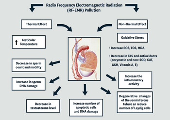
Non-thermal effects of mobile phones comprise raised generation of seminal ROS and reduction in antioxidant enzymes, chromosomal abnormalities, micronuclei formation, and change the spermatozoa membrane potential, as well as increased apoptosis (Agarwal et al., Reference Agarwal, Singh, Hamada and Kesari2011; Merhi, Reference Merhi2012). Leydig cells, seminiferous tubules, and spermatozoa are the main targets of the damage caused by the OS-mediated mobile phones’ adverse effect on the male reproductive tract. In particular, the injury might supress testicular steroidogenesis and reduce testosterone levels, with a consequent adverse effect on spermatogenesis (La Vignera et al., Reference La Vignera, Condorelli, Vicari, D’Agata and Calogero2012; McGill and Agarwal, Reference McGill, Agarwal, du Plessis, Agarwal and Sabanegh2014). Furthermore, Desai and co-workers (Reference Desai, Sharma, Makker, Sabanegh and Agarwal2009) reported that RF-EMR exposure might induce DNA sperm damage due to increased OS and accelerate spermatozoal cell death. Figure 1 shows the thermal and non-thermal effects following the exposure to RF-EMR on male reproductive organs.
Schematic representation of the mechanisms by which RF-EMR exposure might damage the testis. CAT, catalase; GSH, glutathione; MDA, malondialdehyde; ROS, reactive oxygen species; SOD, superoxide dismutase; TAC, total antioxidant capacity; TOS, total oxidant status.

Oxidative stress and ROS formation
The non-thermal effect represents a critical factor in creating damage to the male reproductive organ, through excessive ROS production that results in increased OS. Oxidative stress is a condition in which the natural balance between oxidants and antioxidants is lost. Free radicals are oxygen-containing molecules with an uneven number of electrons, which react with other molecules to cause a series of chemical reactions that might be toxic to cells, tissues, and organs (Elsayed, Reference Elsayed2001), as well as gametes and embryos (Sciorio and Smith, Reference Sciorio and Smith2019). It has been suggested that OS is a central element causing sperm chromatin and DNA damage (De Iuliis et al., Reference De Iuliis, Newey, King and Aitken2009; Roque and Esteves, Reference Roque and Esteves2018; Esteves et al., Reference Esteves, Santi and Simoni2020).
Exposure to cell phone radiation may induce OS leading to enhanced lipid peroxidation and changes in the body’s antioxidant activities. The increase in lipid peroxidation (Oksay et al., Reference Oksay, Naziroğlu, Doğan, Güzel, Gümral and Koşar2014; Shahin et al., Reference Shahin, Mishra, Singh and Chaturvedi2014) and 8-hydroxy-2′-deoxyguanosine activity, a marker of nucleic acid damage, may harm the sperm fertilizing potential (Atasoy et al., Reference Atasoy, Gunal, Atasoy, Elgun and Bugdayci2013). Although seminal plasma has a high endogenous antioxidant ability to protect spermatozoa from oxidative damage (Fraga et al., Reference Fraga, Motchnik, Shigenaga, Helbock, Jacob and Ames1991), cell phone exposure leads to the induction of OS through the generation of ROS in the sperm plasma membrane by activation of NADH oxidase (Aitken and Curry, Reference Aitken and Curry2011; Shi et al., Reference Shi, Chen, Huang, Yuan, Wu, Wu, Li, Shun, Chen and Shi2012; Jamaludin et al., Reference Jamaludin, Abdul Razak, Jaffar, Osman and Ibrahim2017; Ding et al., Reference Ding, Ping and Hong2018).
It has been found that OS, nitric oxide levels, and total oxidant status were increased following exposure to RF-EMR (Wiesner et al., Reference Wiesner, Nabha, Dos Santos, Yamamoto, Meng, Melchior, Bittinger, Thüroff, Vessella, Cher and Bonfil2008). These observations are consistent with a decrease in the spermatogonia and Leydig cell enzymatic antioxidant capacity (Shahin et al., Reference Shahin, Mishra, Singh and Chaturvedi2014; Almášiová et al., Reference Almášiová, Holovská, Šimaiová, Beňová, Raček, Račeková, Martončíková, Mihálik, Horváthová, Tarabová, Slanina and Cigánková2018). Spermatozoa seem to be particularly vulnerable to RF-EMR induced OS. Under physiological conditions, ROS play a crucial role in sperm capacitation, acrosome reaction, and sperm–oocyte binding (Garrido et al., Reference Garrido, Meseguer, Alvarez, Simón, Pellicer and Remohí2004). However, a contrary effect occurs in the presence of excessive ROS (Aitken and Curry, Reference Aitken and Curry2011), which is also seen in animal models when semen specimens are exposed to mobile phone radiation (Kumar et al., Reference Kumar, Kesari and Behari2010; Kesari et al., Reference Kesari, Kumar and Behari2011a).
Along these lines, it is well known that mitochondrial dysfunctions negatively effect sperm motility (Pelliccione et al., Reference Pelliccione, Micillo, Cordeschi, D’Angeli, Necozione, Gandini, Lenzi, Francavilla and Francavilla2011). Mitochondria regularly supply the energy for sperm motility; any metabolic disruption in the electron transport chain can significantly increase the mitochondrial ROS production, affecting sperm motility (Aitken et al., Reference Aitken, Jones and Robertson2012; Agarwal et al., Reference Agarwal, Virk, Ong and du Plessis2014). Mobile phone exposure can increase mitochondrial ROS production, induce sperm DNA fragmentation, and decrease sperm motility and viability (De Iuliis et al., Reference De Iuliis, Newey, King and Aitken2009; Awanti et al., Reference Awanti, Ingin, Jeevangi, Patil and Awanti2010).
The association between OS and male infertility has been well documented (Desai et al., Reference Desai, Sharma, Makker, Sabanegh and Agarwal2009; Aitken et al., Reference Aitken, Jones and Robertson2012; Guz et al., Reference Guz, Gackowski, Foksinski, Rozalski, Zarakowska, Siomek, Szpila, Kotzbach, Kotzbach and Olinski2013). Excessive ROS can alter the function of several seminal enzymes, including superoxide dismutase (SOD), catalase (CAT), and glutathione peroxidase (GPx), which play a crucial sperm protective role from ROS attack. It has been reported that a reduction in glutathione and superoxide production after exposure to mobile phone radiation relates to the disruption in sperm membranes as a consequence of induced OS and ROS formation (Moein et al., Reference Moein, Dehghani, Tabibnejad and Vahidi2007; Kesari and Behari, Reference Kesari and Behari2012). Accordingly, a few studies have investigated the protective role of antioxidants. A well studied antioxidant is melatonin (Reiter et al., Reference Reiter, Tan and Acuna-Castroviejo2000; Meena et al., Reference Meena, Kumari, Kumar, Rajamani, Verma and Kesari2014; Reiter et al., Reference Reiter, Mayo, Tan, Sainz, Alatorre-Jimenez and Qin2016), which can decrease OS and protect membrane lipids, proteins, nuclear and mitochondrial DNA from oxidative damage (Reiter et al., Reference Reiter, Tang, Garcia and Muñoz-Hoyos1997).
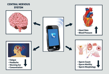
In addition, melatonin stimulates antioxidative enzymes (Ozguner et al., Reference Ozguner, Bardak and Comlekci2006) and prevents the decline in the mitochondrial membrane potential; the latter effect helps prevent the apoptotic cascade trigger (Martín et al., Reference Martín, Macías, Escames, León and Acuña-Castroviejo2000; Rodriguez et al., Reference Rodriguez, Mayo, Sainz, Antolín, Herrera, Martín and Reiter2004; Zhang et al., Reference Zhang, Cui, Wang, Chen, Chen, Lan, Wei, Pan and Lan2020). Figure 2 summarizes the effect of cell phone exposure on the human body (Figure 2).
Effects of cell phone exposure to human body. Changes might include the central nervous system, and cardiovascular and reproductive systems.

Summary evidence
Sperm DNA damage
Cell phones have been recognized as a lifestyle factor inducing DNA damage as a result of an overproduction of ROS. In one study, Kumar and colleagues (Reference Kumar, Nirala, Behari and Paulraj2014) have shown that testis exposure to 3G mobile phone radiation for 2 h per day for 2 months induced DNA strand break damage in rat sperm. Using the Comet assay, the authors noticed a significant increase in sperm DNA damage in the exposed group compared with the control group. Interestingly, they also observed that the duration of exposure and power density was directly correlated with the magnitude of the effect size.
Other investigators have also found that exposure to RF-EMR could increase sperm DNA fragmentation (Sheikh et al., Reference Sheikh, Amiri, Farimani, Najafi and Hadeie2008; Avendaño et al., Reference Avendaño, Mata, Sanchez Sarmiento and Doncel2012), and induce DNA breakage in epididymal spermatozoa (Baverstock, Reference Baverstock2000). However, it has also been shown that short-term exposure might not be strong enough to cause sperm genomic modification (Agarwal et al., Reference Agarwal, Singh, Hamada and Kesari2011).
In another study, Nikolova and co-workers (2005) reported that RF-EMR could induce DNA damage and double-stranded DNA breaks in neural progenitor cells. The authors also reported a significant increase in transcript levels of apoptosis-related genes. Apoptosis regulates the number of proliferating germ cells during spermatogenesis (Blanco-Rodríguez, Reference Blanco-Rodríguez1998; Berensztein et al., Reference Berensztein, Sciara, Rivarola and Belgorosky2002).
In another study, apoptosis was tested using terminal deoxynucleotide transferase dUTP nick end labelling assay, and it was found that the frequency of sperm showing apoptosis increased as a function of mobile phone radiation exposure (14.30% ± 1.92%, P < 0.005) compared with the control group (7.43% ± 1.30%, P < 0.005), respectively. In addition, a significant decrease in the levels of glutathione peroxidase (GPx) and SOD activities, as well as an increase in catalase (CAT) activity, was observed in the exposed group, indicating that radiation exposure may cause apoptotic in the testis and exert a negative effect on male reproductive function (Kesari and Behari, Reference Kesari and Behari2010).
Semen parameters
Spermatogenesis is a complex and delicate process that might be disrupted by radiation exposure (Kumar et al., Reference Kumar, Nirala, Behari and Paulraj2014; Meena et al., Reference Meena, Kumari, Kumar, Rajamani, Verma and Kesari2014). Although male infertility causes are multifactorial (Wilkes et al., Reference Wilkes, Chinn, Murdoch and Rubin2009; Bechoua et al., Reference Bechoua, Hamamah and Scalici2016; Gatimel et al., Reference Gatimel, Moreau, Parinaud and Léandri2017), the increasing usage of cell phones with RF-EMR could contribute to infertility by decreasing semen parameters. The relationship between cell phone radiation and semen quality has been reported in many animal models and human trials (Oni et al., Reference Oni, Amuda and Gilbert2011).
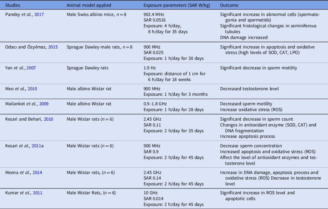
Overall, these studies indicate a link between cell phone usage and changes in sperm count, motility, normal morphology, and viability (Tables 1 and 2). Nevertheless, the role of confounders is difficult to ascertain, as numerous other factors might alter semen quality, including co-morbidities, advanced paternal age, lifestyle factors (e.g. obesity, alcohol consumption, recreational drugs consumption or cigarette smoking).
Summary evidence of human studies evaluating sperm parameters after exposure to cell phone radiation

ROS, reactive oxygen species.
Summary evidence of animal trials evaluating sperm parameters after exposure to cell phone radiation

CAT, catalase; LPO, lipid peroxidation; ROS, reactive oxygen species; SOD, superoxide dismutase.
Sperm count
Cell phone usage has been associated with a reduction in the epididymis and seminal vesicle weight in rats, as well as in sperm count (Meo et al., Reference Meo, Arif, Rashied, Khan, Vohra, Usmani, Imran and Al-Drees2011) and sperm maturation (Sullivan and Mieusset, Reference Sullivan and Mieusset2016). Using flow cytometry, Kesari and collaborators (Reference Kesari, Kumar and Behari2011b) investigated the sperm count in the rat model after cell phone exposure for 2 h per day for 1 month. The authors showed a significant reduction in sperm count in the exposed group compared with the sham group (31.14 ± 13.6% vs. 61.33 ± 3.68%, P < 0.0001).
Several other authors have confirmed the association between decreased epididymal weight and reduced sperm count in an animal model (Kesari and Behari, Reference Kesari and Behari2010; Saygin et al., Reference Saygin, Caliskan, Karahan, Koyu, Gumral and Uguz2011; Shahin et al., Reference Shahin, Mishra, Singh and Chaturvedi2014). In humans, the few studies available have yielded mixed results. A 1997 study investigated whether increased scrotal temperature was associated with decreased sperm production in 21 healthy male volunteers. The authors were unable to demonstrate a relationship between the increased scrotal temperature and reduction in sperm number.
Additionally, in a 2014 systematic review and meta-analysis, including 1376 participants, a clear reduction in sperm concentration during cell phone radiation exposure was not obtained (Adams et al., Reference Adams, Galloway, Mondal, Esteves and Mathews2014). Similar results were also noted by another meta-analysis published in the same year (Liu et al., Reference Liu, Zhang, Liu, Cao, Ao and Zhang2014). Evidence from current studies suggests potential harmful effects of mobile phone use on semen parameters. However, overall, the literature remains equivocal, and additional standardized studies are needed to assess the risk of mobile phone use on the reproductive system.
Motility, morphology, and viability
Fejes and colleagues (Reference Fejes, Závaczki, Szöllosi, Koloszár, Daru, Kovács and Pál2005) found that the prolonged use of cell phones might have adverse effects on sperm motility. The authors analyzed three main aspects in 371 White men of reproductive age (mean age 30.8 ± 4.4 years, range 17–41): including duration of phone possession (in months), duration of standby position closer than 50 cm to the patient (in hours) and duration of daily transmission (in minutes), which was divided into low transmitters (less than 15 min per day) and high transmitters (over 60 min per day). The authors showed that the duration of cell phone possession correlated negatively with the proportion of rapid progressive motile sperm and positively with the proportion of slow sperm motility. The low and high transmitter groups diverged significantly in the proportion of rapid sperm motility (48.7% vs. 40.6% P = 0.01).
Similar results were found by Erogul and co-workers (Reference Erogul, Oztas, Yildirim, Kir, Aydur, Komesli, Irkilata, Irmak and Peker2006), who exposed unprocessed raw spermatozoa to the cell phone radiation. In total, 27 males were included in the study. The semen sample obtained from each man was divided into two parts. One part was exposed to the radiation emitted by an activated 900 MHz cellular phone (10 cm for 5 min), whereas the other was not. The authors found a significant differences in percentage of sperm with rapid progressive motility (13.6% ±10.2% vs 9.1% ± 7.9%, P = 0.0007), and slow progressive motility (43.7% ± 19.4% vs 33.9% ± 20.6%, P = 0.0007) in the exposed group compared with the control group.
In another retrospective study involving 304 men of reproductive age, Wdowiak and colleagues (Reference Wdowiak, Wdowiak and Wiktor2007) reported a significant decrease in the percentage of sperm with normal forward progressive motility under the exposure of cell phone radiation. In this trial, 65.7% of patients who did not use cell phones had over 50% of sperm with forward progressive motility compared with only 17% of patients who frequently used cell phones and had been using them for more than 2 years.
Another observational study investigated the effect of cell phone use on semen quality (Agarwal et al., Reference Agarwal, Desai, Makker, Varghese, Mouradi, Sabanegh and Sharma2009). The study included more than 300 men undergoing infertility treatments, divided into four groups according to their cell phone usage (from 0 to >4 h per day). The authors showed a significant reduction in sperm motility (42.0% ± 17.2% vs. 58.9% ± 12.3%, P < 0.05), viability (44.6% ± 17.5% vs. 62.4% ± 12.8%, P < 0.05), and normal morphology (14.9% ± 9.1% vs. 27.7% ± 13.1%, P < 0.05) in men using cellular phones for variable lengths of time compared with men who did not use them.
Gorpinchenko and collaborators (2014) selected 32 healthy men (age 27.5 ± 3.5 years) with normal semen parameters to study the influence of mobile phone radiation on sperm quality. Semen samples were split into two parts: one was placed in a thermostat, and the second was placed into another thermostat for the same period of time, and at the same temperature of 27°C and exposure to mobile phone radiation (range 900/1800 MHz). After 5 h of incubation, an operator – blinded to which specimen had been exposed to cell phone radiation – analyzed both samples. Results showed that the number of spermatozoa with progressive movement was statistically lower in subjects exposed to mobile phone radiation than the control group (66.5% ± 6.3% vs. 81.3% ± 7.2%, P < 0.05). The same trend was also seen in the number of sperm with non-progressive motility (25.3% ± 4.7% vs. 12.8% ± 5.8%, P < 0.05).
Adams and colleagues (Reference Adams, Galloway, Mondal, Esteves and Mathews2014) outlined the effects of mobile phones on sperm motility in their meta-analysis, which combined 1448 samples from 1353 men. The mean total motility ranged from 36.6% to 86.8%. Six studies found a significant negative effect of mobile phone exposure on human sperm motility. The authors also investigated sperm viability in five studies, including 816 samples. The mean viability ranged from 52.3% to 89.0%, and results revealed a significant negative association between mobile phone exposure and sperm viability.
Lastly, Falzone and colleagues (Reference Falzone, Huyser, Becker, Leszczynski and Franken2011) have analyzed the sperm competence to fertilize the oocyte after exposure to RF-EMR. They used motile human spermatozoa collected from healthy donors, exposed for 1 h to mobile phone radiation to evaluate the sperm’s ability to undergo acrosome reaction and bind to the zona pellucida. They found that although RF-EMR exposure did not negatively affect the acrosome reaction rate, it reduced the sperm ability to bind the zona pellucida compared with unexposed sperm. The authors of the above study also performed a computerized morphometric sperm assessment, and a significant reduction in sperm head area (9.2 ± 0.7 μm2 vs. 18.8 ± 1.4 μm2) and acrosome percentage of the head area (21.5 ± 4% vs. 35.5 ± 11.4%) was reported among exposed sperm compared with controls.
These findings suggest an adverse effect of cell phone radiation on sperm fertilization potential. Overall, the existing evidence from human studies seems to indicate that exposure to mobile phone radiation decreases sperm motility, particularly rapid progressive motility, normal morphology, and viability. These abnormalities seem to be directly related to the duration of mobile phone use, with longer duration to lower sperm quality.
Limitations of existing studies
Despite several studies investigating the association between mobile phone radiation exposure and semen quality, the literature remains inconclusive. The reasons relate to the challenges in designing such studies and controlling for the many possible confounders. Factors such as inclusion of an adequate control group, cell phone type, testing procedure, device use duration, presence of other lifestyle confounders and co-morbidities, to cite only a few, account for these difficulties.
Naturally, it is difficult to find a control group of reproductive age individuals who have not been exposed to any form of mobile phone radiation. If such individuals exist, they most probably comprise older men. Furthermore, there is no standardized method to analyze the effects of cell phone exposure; no consensus has been reached on the accepted SAR value for different cell types, including sperm. Added to this, the distance between the cell phone device and the exposed tissue/organ, length of exposure concerning transmission model, type of mobile, usage mode (e.g. stationary at one point or in movement as when travelling in a car, bus, or train, and use in standby or not) are confounders that might impact the study results.
The SAR effect in a biological system depends on many factors, such as frequency, intensity, polarization, the distance from the source. Also, the SAR is related to the size, shape, and electrical properties of the investigated biological system. Unlike other animal species, humans have particular reproductive tract anatomical and physiological characteristics, therefore precluding the translation of animal trial findings to humans.
In addition, mobile phone technology continues to develop and increase in its complexity. At this time, multi-use smartphones with constant internet connection became the norm, and is associated with higher SAR compared with older mobile phones. On both an individual and societal level, little effort has been made to regulate the RF-EMR environmental pollution properly.
Future perspectives and conclusions
The rapid technological advances in personal computers and communication devices might pose a risk for human health. Cell phone devices emit radiofrequency electromagnetic waves that seem to affect male reproductive health and other body functions (McClelland 3rd and Jaboin, Reference McClelland and Jaboin2018; Sage and Burgio, Reference Sage and Burgio2018; Wall et al., Reference Wall, Wang, Kendig, Dobraca and Lipsett2019). Although the current data are not unequivocal, it seems sound to speculate that mobile phone exposure might be contributing to subfertility. However, the existing evidence primarily relates to adverse effects on sperm motility and morphology, which are limited endpoints for evaluating the male fertility potential.
The exact mechanisms of how RF-EMR might affect the testis, epididymis, and sperm have not yet been fully understood. Additional studies are warranted, particularly prospective studies assessing sperm functional markers, such as sperm DNA integrity and OS, in fertile and subfertile men. Equally important will be to analyze whether the decreased sperm quality associated with mobile phone exposure translates into impaired pregnancy chances. The effects of short-term and long-term exposure and energy intensity should be also investigated in more detail, taking into account relevant confounders. Only then will scientific societies and regulatory bodies be able to provide users with transparent information concerning the risks and guidance for proper use.
Financial support
None.
Conflicts of interest
No potential conflict of interest was reported by the authors.
Data availability statement
All data are included in the study.
Author contributions
RS, LT, and SCE contributed to the conception and designed the manuscript. RS and LT wrote the first draft of the manuscript with tables and figures. SCE wrote sections of the manuscript and revised it for content. All authors contributed to manuscript revision, read and approved the submitted version.
Ethical standards
Not applicable