Ventilator-associated pneumonia (VAP) is one of the most common infections encountered in intensive care units (ICUs) and is associated with prolonged durations of mechanical ventilation (MV), increased length of stay (LOS) in ICUs, and higher hospital costs.Reference Kollef 1 The VAP rate has been widely used as a key indicator of the quality of care for patients on MV for both internal assessment and external benchmarking. However, surveillance of VAP is labor intensive and is prone to biases and errors because VAP definitions are complicated, highly subjective, and nonspecific.Reference Klompas 2 – Reference Fabregas, Ewig and Torres 5 VAP surveillance is particularly problematic in hospitals in China because no uniformed or widely accepted definitions are available and because many different VAP definitions are used.
To overcome the shortcomings in VAP surveillance, the Centers for Disease Control and Prevention (CDC) developed a new approach to the surveillance of ventilator-associated events (VAEs).Reference Magill, Klompas and Balk 6 VAEs occur in 3 tiers: tier 1, ventilator-associated conditions (VACs); tier 2, infection-related ventilator-associated complications (IVACs); and tier 3, possible VAP or probable VAP.Reference Magill, Klompas and Balk 6 The National Healthcare Safety Network of the CDC plans to replace VAP surveillance by VAE surveillance for adult patients in the United States. However, only a few studies, most of which were retrospective, have demonstrated the validity of VAE surveillance. In this study, we investigated VAE incidence rates and sought to determine whether VAE is associated with poorer outcomes in China.
METHODS
A prospective multicenter surveillance study of VAEs was conducted in 15 ICUs in Sichuan, southwestern China. These ICUs (1 ICU per hospital) were located in 15 different hospitals including 3 university and 12 municipal hospitals. The bed capacity of the hospitals ranged from 800 to 4,100 beds (median, 1,708 beds). All but 1 ICU were medical-surgical units; the remaining ICU was a medical unit. In total, this study included 300 beds in the 15 ICUs with 12–50 beds in each unit (median, 18 beds). This study was conducted in accordance with the amended Declaration of Helsinki. The Ethics Committee of West China Hospital, Sichuan University, approved this study (No. WCH2013-13). The need for patient consent was waived by the ethics committee. Patient data were anonymized prior to analysis and investigators had no access to identifying patient information.
All ICU patients aged >18 years admitted to any of the study hospitals from April 1 to July 31, 2013, who received MV after admission were included in the surveillance study. Patients on MV on admission to the study hospitals or those who were hospitalized in the study hospitals during the study period but were admitted prior to April 1, 2013 were not included. Patients who were eligible for surveillance but were on MV ≤2 days were excluded for determining VAEs, but their ventilator days were counted in the denominator for calculating the VAE rate. Patients on high-frequency oscillary ventilation were also excluded. Extracorporeal membrane oxygenation was not available in any unit during the study period. Definitions of VACs, IVACs, possible VAP, and probable VAP developed by the CDC were applied.Reference Magill, Klompas and Balk 6 VAEs consisted of all events meeting at least the VAC definition. Sputa or endotracheal aspirates were collected for qualitative culture for patients with IVACs. Species identification and in vitro susceptibility testing were performed using Vitek II (bioMérieux SA, Marcy l’Etaile, France) or Microscan Walkaway 96 SI (Siemens, Erlangen, Germany) automatic microbiological systems. Surveillance was performed by infection control practitioners and ICU nurses by reviewing patient records daily to collect data such as demographic data, underlying diseases, MV days, daily minimum fraction of inspiration oxygen (FiO2), positive end-expiratory pressure (PEEP), and body temperature. Microbiological tests were conducted following the instructions of the CDC.Reference Magill, Klompas and Balk 6 The VAE incidence rate was also calculated according to the CDC protocol.Reference Magill, Klompas and Balk 6
Patients who had VAEs in this study (group A) were compared with patients who were on MV >2 days but had no VAE (group B) for features and outcomes. In previous studies,Reference Hayashi, Morisawa and Klompas 7 – Reference Muscedere, Sinuff and Heyland 9 patients who were on MV ≥2 days or >48 hours but had no VAE were typically regarded as the control group. Notably, ≥4 days on MV is required for determining a VAE; thus, a control group of MV ≥2 days might have shorter duration of MV and ICU LOS. To exclude the impact of this confounding factor, patients who had VAEs were also compared with an additional control group (group C) consisting of patients who were on MV with stable or decreasing daily minimum FiO2 or PEEP values ≥2 days followed by ≥2 more days on MV (total MV duration ≥4 d) but had no VAE during this study. The outcomes that were compared included mortality in hospital, mortality in ICU, mortality within 30 days after admission to ICU, duration of MV, LOS in ICU, overall hospital LOS, and days on antibiotics.
The potential etiology of VACs and whether VAE was an attributable cause were judged by 2 physicians independently for each case by reviewing the medical record. The determination of the attributable cause of death was based on their conclusion regarding whether the patients would have died if they had not had a VAE. In cases of disagreement, a consensus was reached via discussion.
Statistical analysis was performed using the SPSS program (version 18.0; SPSS, Chicago, IL). Means and standard deviations were used to describe the numerical variables that fit a normal distribution; otherwise, medians and interquartile ranges were used. Independent sample tests were used to compare differences in means. Wilcoxon signed rank tests were used to determine differences in median values and Fisher’s exact tests were used to determine differences in sex and mortality rates. To compare differences, statistical significance of P<.05 and 95% power were applied. A multivariate analysis was performed using a logistic regression model to compare the preexisting conditions (ie, age, sex, and APACHE II score on ICU admission) and underlying diseases between patients with VAEs (group A in Table 1) and without a VAE (group B). To verify the impact of VAE as an independent factor on the outcomes, multivariate analyses incorporating the preexisting conditions (listed above) and a VAE were performed using linear or logistic regression models when appropriate. The unstandardized coefficients (β) and the adjusted odds ratio (OR) were used for numerical and categorical variables, respectively. All tests were 2-sided, and P<.05 was considered statistically significant.
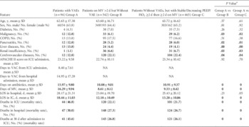
Features and Outcomes of Patients With or Without Ventilator-Associated Events (VAEs)Footnote a

NOTE. MV, mechanical ventilation; PEEP, positive end-expiratory pressure; FiO2, fraction of inspiration oxygen; SD, standard deviation; NA, not applicable; COPD; chronic obstructive pulmonary disease; ICU, intensive care unit; LOS, length of stay.
a Parameters with statistical significance (P<.05) are shown in bold.
RESULTS
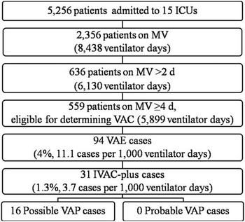
During the 4-month study period, a total of 5,256 patients were admitted to the ICUs for 28,248 ICU days; 2,356 of these patients (44.8%) received MV for a total of 8,438 ventilator days (Figure 1). However, only 636 patients (12.1% of all ICU patients and 27.0% of those on MV) were on MV >2 days (range, 3–63 days), including 409 (64.3%) males and 227 (35.7%) females. These 636 patients had 6,130 ventilator days, which accounted for 72.6% of total ventilator days. A total of 94 VAE cases (4.0% of all patients on MV and 14.8% of those on MV >2 d) were identified (Figure 1). The rate of VAE was 11.1 cases per 1,000 ventilator days (94 of 8,438). Among the 15 ICUs, VAE rates varied significantly from 3.1 to 26.6 cases per 1,000 ventilator days. The VAE rate in ICUs of the 3 university hospitals (23.0 cases per 1,000 ventilator days) was much higher than that (6.9 cases per 1,000 ventilator days) in ICUs of non-teaching municipal hospitals.
Summary of results in this study.

Patients with VAEs were 26 to 99 years old (mean, 62.65-year old), were mostly male, (63.8%) and had a mean APACHE II score of 23.22, which was measured on admission to ICU. VAEs developed 3–103 days after admission to hospitals (mean, 15.95 days) and 3–39 days after admission to ICUs (mean, 9.4 days). Our multivariate analysis revealed that patients with VAEs had greater incidence of pancreatitis (multivariate OR, 3.43; 95% CI, 1.51–7.78) and liver disease (multivariate OR, 2.68; 95% CI, 1.18–6.12) than those without VAEs (Table S1 in the online supplementary material). However, there were no significant differences in age, sex, APACHE II scores measured on admission to ICU, or other underlying diseases between patients who had VAEs and those who were on MV >2 days but had no VAEs (Table 1). Patients who had VAEs had more days on antibiotics (mean, 16.0 vs 10.1 days; P<.05), longer duration on MV (mean, 16.3 vs 8.6 days; P<.05), and increased ICU LOS (mean, 18.4 vs 12.2 days; P<.05) than those who had no VAEs (Table 1). Patients who had VAEs also had longer overall hospital LOS (mean, 28.2 vs 23.8 days), but this difference was not statistically significant (P>.05). The mortality rates in ICUs, in hospital wards, and at 30 days after admission to ICU of patients who had VAEs were 46.8%, 50.0%, and 43.6%, respectively. All of these rates were significantly higher than those of patients who had no VAEs (Table 1). The greater incidence of pancreatitis and liver disease among patients with VAEs could serve as confounding factors for evaluating the impact of VAEs on outcomes. Therefore, for patients who were on MV >2 days, those with and without pancreatitis or liver diseases were also compared regarding outcomes, and pancreatitis and liver disease as underlying diseases had no significant impact on outcomes (Table S2 in the online supplementary material). The impact of VAEs on outcomes was also challenged using multivariate analyses that included age, sex, APACHE II scores, and underlying diseases. These analyses also confirmed the significant impact of VAEs alone on outcomes (Table S3 in the online supplementary material).
Patients who had VAEs and those on MV with stable or decreasing daily minimum FiO2 or PEEP values ≥2 days followed by ≥2 more days on MV (total MV duration ≥4 d) but had no VAEs were also compared: the 2 groups shared similar compositions of age and sex and similar APACHE II scores (Table 1). The comparison of these 2 groups demonstrated that patients who had VAEs had more days on antibiotics (mean, 16.0 vs 10.9 days; P<.05), longer duration on MV (mean, 16.3 vs 9.5 days; P<.05), andincreased ICU LOS (mean, 18.4 vs 13.3 days; P<.05). Mortality rates were higher in ICUs, in hospital wards, and within 30 days after admission to an ICU (P<.05) than those who had no VAEs (Table 1).
The etiologies of VAEs included IVAC (n=31; 33.0%), mucous plugging (n=22; 23.4%), poor pulmonary hygiene (n=15; 16.0%), acute respiratory distress syndrome (n=13; 13.8%), pleural effusion (n=10; 10.6%), pulmonary edema (n=5; 5.3%), atelectasis (n=3; 3.2%), abdominal compartment syndrome (n=2; 2.1%), pneumothorax (n=2; 2.1%), and aspiration of the gastric content (n=1; 1.1%). VAEs were considered the cause of death for 11 cases; the mortality rate attributable to VAEs in these hospitals was 11.7%.
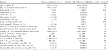
The rate of patients with IVACs was 3.7 per 1,000 ventilator days. Compared with those who had VACs alone (no IVACs, n=63), patients who had IVACs (n=31) had more days on antibiotics (median, 16 to 12 days; P<.05), longer duration on MV (median, 17 vs 12 days; P<.05), and longer ICU LOS (median, 19 vs 14 days; P<.05) (Table 2). However, no significant differences in overall hospital LOS or mortality rates were observed between the 2 groups (Table 2).
Comparisons of Patients with Ventilator-Associated-Events (VAEs) With or Without Infection-Related Ventilator-Associated Conditions (IVACs)Footnote a
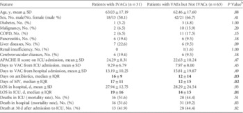
NOTE. SD, standard deviation; COPD; chronic obstructive pulmonary disease; ICU, intensive care unit; VAC; ventilator-associated condition; MV, mechanical ventilation; LOS, length of stay; IQR, interquartile range.
a Interquartile range was used for data that did not fit a normal distribution.
b Parameters with statistical significance (P<.05) are shown in bold.
Among the 31 patients with IVACs, 28 had no obvious extrapulmonary source of infection. However, only 17 patients had a respiratory sample (sputum or endotracheal aspirate) collected during the VAE window period (ie, 2 calendar days before or after the onset of worsening oxygenation). In total, 16 cases of possible VAP were detected; these cases had a positive culture of sputum or endotracheal aspirate. The microorganisms recovered were Acinetobacter baumannii (all were carbapenem-resistant isolates) from 10 patients, Staphylococcus aureus from 2 patients (1 methicillin-resistant S. aureus), and Acinetobacter lwoffii, Pseudomonas aeruginosa, Stenotrophomonas maltophilia, and Enterobacter aerogenes from 1 patient each. Probable VAP was not identified in this study.
DISCUSSION
To our knowledge, this is the first VAE surveillance study conducted in a developing country. Notable findings of this study include the following: (1) VAEs were frequently observed in patients on MV ≥4 days (16.8%, 94 of 559) but were uncommon among all MV patients (4%). (2) VAE rates varied significantly among different ICUs. (3) Only <25% of all MV patients were eligible for determining a VAE. The determination of a VAE requires that the patient be on MV for ≥4 days. Most ventilator days of all MV patients were still captured for determining VAEs; those of patients on MV ≥4 days accounted for more than half of all ventilator days. (4) VAEs were associated with prolonged duration of ventilator days, more days on antibiotics, prolonged ICU LOS, and higher mortality rates. (5) IVACs were associated with prolonged duration of ventilator days, days on antibiotics, and prolonged ICU LOS but not with higher mortality rates in patients with VAEs.
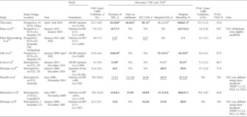
VAE rates varied significantly in previous studies, from 4.6% to 28.2% (Table 3). These statistics should be interpreted with caution because different patient populations were used as the denominator for the calculation of the VAE rate, and different FiO2 or PEEP values were used to define VACs in these studies (Table 3). The VAE rate in this study is consistent with those in other studies using the same VAE definitions and using all MV patients or episodes as the denominator (ranging from 4.6% to 5.8% and 12.4 to 13.8 cases per 1,000 ventilator days).Reference Klompas, Kleinman and Murphy 10 – Reference Lewis, Li, Murphy and Klompas 12 In contrast, much higher VAE rates (>20%) were reported in 2 previous studies that used lower values of PEEP or FiO2 to define VACs (Table 3).Reference Hayashi, Morisawa and Klompas 7 , Reference Klompas, Khan and Kleinman 8
Summary of Findings in Studies on Ventilator-Associated Events (VAEs)

NOTE. MV, mechanical ventilation; ICU, intensive care unit; LOS, length of stay; IVAC, infection-related ventilator-associated condition; US, United States; NA, not available; VAC, ventilator-associated condition; PEEP, positive end-expiratory pressure; FiO2, fraction of inspiration oxygen.
a Median days of duration, number of days, and LOS. Parameters with statistical significance (P<.05) are shown in bold. Parameters without P values are underlined.
b Compared with all MV patients >2 d rather than non-VAE patients using mean values.
c Compared with all MV patients ≥2 d rather than non-VAE patients.
d Compared with matched cases (matching the patient’s time from intubation to VAC onset).
e Compared with all MV patients rather than non-VAE patients.
A potential shortcoming of VAE surveillance is that most patients on MV are excluded for surveillanceReference Lilly and Ellison 13 when the prerequisite for determining a VAE is ≥2 days of stable or improving ventilation followed by ≥2 more days on MV. In this study, only 23.7% of patients on MV met the prerequisite. These patients had 5,899 ventilator days, corresponding to 69.9% (5,899 of 8,438 days) of the total ventilator days. Therefore, although most MV patients were not eligible for determining VAEs, most ventilator days were still captured.
Previous studies have demonstrated that VAEs are associated with prolonged duration of MV,Reference Hayashi, Morisawa and Klompas 7 – Reference Lewis, Li, Murphy and Klompas 12 , Reference Boyer, Schoenberg, Babcock, McMullen, Micek and Kollef 14 increased LOS in ICUReference Hayashi, Morisawa and Klompas 7 – Reference Muscedere, Sinuff and Heyland 9 and more days on antibiotics.Reference Hayashi, Morisawa and Klompas 7 , Reference Muscedere, Sinuff and Heyland 9 Notably, the control group in these studies included patients on MV <4 days, whereas a duration ≥4 days on MV is required for determining VAC. Therefore, the impact of a VAE might have been overestimated previously. Nonetheless, the comparison between patients who had VAEs and those who had no VAE but were on MV ≥4 days in this study revealed that VAEs were indeed associated with prolonged duration of MV, increased ICU LOS, and more days on antibiotics. However, it remains unclear whether VAEs cause longer duration of MV and increased ICU LOS or vice versa.
Most previous studiesReference Hayashi, Morisawa and Klompas 7 – Reference Lilly, Landry and Sood 11 also demonstrated that VAEs are associated with increased hospital LOS, but this finding was not supported by this study and a previous report.Reference Lewis, Li, Murphy and Klompas 12 The reason for the discrepancy remains unclear. In addition, all but 1 of the previous studies found that VAEs are associated with a higher mortality rate (Table 3).Reference Klompas, Khan and Kleinman 8 – Reference Lewis, Li, Murphy and Klompas 12 , Reference Boyer, Schoenberg, Babcock, McMullen, Micek and Kollef 14 The only study that found no difference in mortality rates between patients with and without VAEs used lower values of PEEP or FiO2 to define VACs and therefore might not be reliable for evaluating VAE outcomes.Reference Hayashi, Morisawa and Klompas 7 Our study results indicate that VAEs are indeed associated with significantly greater mortality, suggesting that VAE surveillance is likely to identify MV patients with poorer outcomes. Please note that the VAE definition was designed for surveillance and should not be used to direct clinical management.
Among VAE types, the data required to determine VACs and IVACs can be easily obtained and do not rely on microbiological results, making surveillance of VACs and IVACs practical in developing countries. In contrast, the determination of possible or probable VAP requires submission of ≥1 sample for microbiological examination. Either purulent respiratory secretions (determined using microscopy examination) or positive culture (ie, qualitative, semi-quantitative, or quantitative) of a sample from lower respiratory tract or lung is required to meet the definition of possible VAP. Microscopic examination and qualitative culture of sputum or endotracheal aspirate samples are relatively simple procedures, and in theory they can be performed in developing countries in microbiological laboratories of hospitals with the capacity to provide MV. However, in practice in China, sputum or extracheal aspirate samples are commonly not collected within the VAE window period or are not examined by microscopy before being processed for culture due to the shortage of personnel or lack of desire to do so. Therefore, the incidence of possible VAP might have been underestimated in this study. Furthermore, the determination of probable VAP requires quantitative culture of a non-sputum sample of the lower respiratory tract or lung or tests for Legionella and viruses causing pneumonia. The absence of probable VAP in this study was due to the fact that such quantified cultures and tests are not performed in the study hospitals. In developing countries, due to the lack of laboratory capacity and the absence of good practices for collecting and processing samples for microbiological examinations, surveillance of possible VAP and probable VAP can be problematic; thus, these rates may be underestimated.
Notably, although pulmonary embolism might be a cause of VAEs,Reference Hayashi, Morisawa and Klompas 7 , Reference Klompas, Khan and Kleinman 8 it was not identified as such in this surveillance study. Clinicians might be prone to under-recognize pulmonary embolism in patients on MV due to lack of awareness. However, it appears that pulmonary embolism is not a common cause of VAEs; it accounted for only 2% of all VAEs in previous studies.Reference Hayashi, Morisawa and Klompas 7 , Reference Klompas, Khan and Kleinman 8
Although VAE surveillance is meaningful, 2 major questions remain largely unanswered: (1) What proportion of VAEs is preventable? And (2) how can we reduce the occurrence of VAEs?Reference Lilly and Ellison 13 A recent study found that only 37.3% of VAEs were potentially preventable.Reference Boyer, Schoenberg, Babcock, McMullen, Micek and Kollef 14 However, in this study, ~50% of VAEs were attributable to preventable causes such as mucous plugging, poor pulmonary hygiene, pulmonary edema, pneumothorax, and aspiration of gastric content. A previous study demonstrated that the VAC rate had dropped ~30% along with the modest elevation of compliance with VAP bundle measures.Reference Muscedere, Sinuff and Heyland 9 Therefore, a significant proportion of VAEs appears to be preventable, and the current VAP prevention bundle might also work for preventing VAEs in certain patients. Nonetheless, more studies are warranted regarding the preventability of VAEs.
We are aware of several limitations of this study. First, this study was performed in a relatively short time period and the sample number was relatively limited. Second, we did not compare patients who had VAEs with those who had VAP as defined using traditional VAP standards. Therefore, we were unable to assure consistency between the 2 surveillance systems used in our local settings. Third, we did not investigate the potential risk factors for the development of VAEs and did not specifically look at the preventability of VAEs. In addition, this study was performed in a developing country, and the interpretation of this study might not be applicable to other settings due to the great divergence of patient populations and huge variations in patient treatment. Fourth, the approach of ‘attributable mortality’ used here may be subjective and lack sensitivity; it is possible that VAEs do not cause immediate mortality but rather extend the duration of MV and thus the time at risk to develop complications (with an associated increased mortality) or that VAEs cause excessive weakness that may lead to increased mortality following ICU discharge. However, reliable estimation of attributable mortality is difficult (if not impossible) to accurately assess in observational studies.Reference Melsen, Rovers and Groenwold 15
In conclusion, in this study setting, our surveillance of VACs and IVACs captured most ventilator days and identified MV patients with poorer outcomes. Surveillance of possible VAP and probable VAP might be problematic partially due to insufficient lab capability. However, before VAE surveillance can be widely adopted in developing countries, more studies are required to determine the preventability of VAEs.
ACKNOWLEDGMENTS
We are grateful to many other investigators who participated in this project: Weijia Yin and Jiancheng Ye from West China Hospital, Jia Wen and Yue Zhao from Mianyang Central Hospital, Lin Li and Jian Rao from The People’s Hospital of Dujiangyang City, Yin Liu and Chuan Zhang from The Third People’s Hospital of Chengdu, Chunlian Tang and Chaoqun Yang from Nanchong Central Hospital, Xuemei Li and Chunlin Zhang from The Second People’s Hospital of Yibin City, Zonglin Wang and Mina Zou from The First People’s Hospital of Neijing City, Neijiang; Li Yin and Lingxu Lin from The People's Hospital of Deyang City, Xin Hu and Hongchuan Zhang from The Affiliated Hospital of North Sichuan Medical College, Xiaoyan Leng and Xiaoyi Liu from Dazhou Central Hospital, Shicheng Xu and Huiping Yan from Bazhong Central Hospital, Chunrong Su and Fuxiang Li from Chengdu Military General Hospital, Lihua Wang and Qin Nie from The People's Hospital of Jiangyou City, Shenyan Gu from The First Affiliated Hospital of Chengdu Medical College, and Wei Huang and Yuanhua Zhang from The First People’s Hospital of Yibin City.
Financial support. This study was supported by a grant from the Sichuan Society for Infection Control, China (grant no. 2013-05).
Potential conflicts of interest. All authors report no conflicts of interest relevant to this article.
SUPPLEMENTARY MATERIAL
To view supplementary material for this article, please visit http://dx.doi.org/10.1017/ice.2015.200