Introduction
Avian tuberculosis (TB) is a chronic wasting disease caused by Mycobacterium avium belonging to serotype 1, 2, 3 and 6 and M. genavense. Other species of Mycobacterium that cause avian tuberculosis are M. intracellulare, M. scrofulaceum, M. fortuitum, M. tuberculosis, and M. bovis. According to the current taxonomy, M. avium contains four subspecies, namely, M. avium subsp. avium; M. avium hominissuis; M. avium paratuberculosis and M. avium silvaticum (Mijs et al., Reference MIJS, DE HAAS, ROSSAU, VAN DER LAAN, RIGOUTS, PORTAELS and VAN SOOLINGEN2002; OIE Manual, 2014). These mycobacterial species affect a vast range of bird species characterised by weight loss and deficient egg production. Generally, this infection is fatal for birds (Dhama et al., Reference DHAMA, MAHENDRAN, TIWARI, SINGH, KUMAR, SINGH and SAWANT2011). This affects production and causes financial loss. The disease is more rapidly onset by M. genavense than with M. avium. These bacteria can reach the flock from infected birds if they are allowed roaming freely. Domestic and pet birds can acquire the disease and become infected. When infected chickens come into contact with healthy birds, the disease is transmitted (Witte et al., Reference WITTE, HUNGERFORD, PAPENDICK, STALIS and RIDEOUT2010). If birds are kept for a longer time with infected birds, then incidences of avian TB may increase (Witte et al., Reference WITTE, HUNGERFORD, PAPENDICK, STALIS and RIDEOUT2010). Baled grass silage can transmit the disease to the chickens (Khol et al., Reference KHOL, BERAN, KRALIK, TRCKOVA, PAVLIK and BAUMGARTNER2010). Infected or deceased birds and the visceral organs and entrails of a butchered animal may infect pen-mates, rodents, and predators, if eaten.
The other sources of infection are contaminated water and soil; plants and bedding material. The susceptibility of birds to mycobacterial diseases varies from species to species. Mycobacterium avium may infect a number of mammalian species. It causes non-progressive lesions, especially in lymph nodes of mammals, specifically pigs (OIE Manual, 2014). Individuals with impaired immunity, such as HIV infected individuals, individuals who are on chemotherapy, the elderly and children are at high risk due to susceptibility to M. avium infection (Prince et al., Reference PRINCE, PETERSON, STEINER, GOTTLIEB, SCOTT, ISRAEL, FIGUEROA and FISH1989; Morris et al., Reference MORRIS, BERMUDEZ and CHAPARAS1991; Pesek, Reference PESEK1998).
Mycobacterium avium subsp. avium and M. avium subsp. hominissuis can cause opportunistic infections in animals and human beings (Thorel et al., Reference THOREL, HUCHZERMEYER, WEISS and FONTAINE1997, Ashford et al., Reference ASHFORD, WHITNEY, RAGHUNATHAN and COSIVI2001). All over the world, a number of studies revealed the transmission of infection from infected bird to human beings (Kincade, Reference KINCADE1959; Cheung and Konst, Reference CHEUNG and KONST1963; Bisseru, Reference BISSERU1967). The most usual mode of transmission of TB is from contaminated droplet nuclei. In some parts of the world bovine tuberculosis is carried by unpasteurised milk and other dairy products from affected cattle (O'Toole, Reference O'TOOLE2005). M. avium is the second most common reason for pulmonary infections in the United States and England (Johnson and Odell Reference JOHNSON and ODELL2014). Tuberculosis caused by avian mycobacterium may present a serious threat to man, especially in those places where the population is no longer exposed to pervasive infection from mammalian Mycobacterium spp. (Bisseru, Reference BISSERU1967).
It is utmost important to control the spread of infection in the human beings, animals, and poultry and pet birds. For this, timely diagnosis of disease is an important condition. This can be done by acquiring the knowledge of diagnostic techniques and treatment strategies in order to prevent the spread of disease in a flock and humans. Therefore, in this review, we focused on the diagnostic techniques currently available for avian tuberculosis disease.
Diagnostic techniques
pathological observation
In chronic diseases like avian TB, external symptoms are neither pathognomonic nor seen in the initial stages (pre-clinical or sub-clinical). However, in clinical and later stages, despite having a normal appetite, birds are apparently dull, depressed, weak, fatigue easily, have progressive and striking loss of weight, atrophy of breast muscles with prominent keel, body fat disappears, the head appears smaller than normal and feathers are dull and ruffled. Comb, wattle and earlobes look pale and thinner than normal and have a dry epidermis. Bluish discoloration of comb and wattles is infrequent, but the icterus condition indicates advanced hepatic damage. Affected bird may not have fever. Few birds develop unilateral lameness and walk with jerky hopping gait. Tuberculous arthritis may lead to paralysis. In cases of advanced emaciation, intestinal nodules may become ulcerative, resulting in severe diarrhoea. In some cases, hepatomegaly and splenomegaly can be seen (Mendenhall et al., Reference MENDENHALL, FORD, EMERSON, WELLS, GINES and ERIKS2000). Haemorrhage from rupture of liver or spleen may cause sudden death.
Infected birds in the same flock may exhibit variable clinical signs. Occasionally, birds may have good body condition, continue to lay eggs despite nodular masses in the infra-orbital sinuses, liver and intestines. While others may be emaciated, do not lay eggs, lack sinus lesions but possess numerous nodular masses in intestines (Gonzalez et al., Reference GONZALEZ, RODRIGUEZ-BERTOS, GIMENO, FLORES and PIZARRO2002). Life of affected birds depend on severity and extent of damage by disease.
gross lesions
Most frequently, liver, spleen, intestines and bone marrow are involved while heart, ovaries, testes, lungs and skin are involved infrequently (Francis, Reference FRANCIS1958). The size of lesions range from a pinpoint to several centimetres. More often, irregular grayish yellow or white nodules can be seen in spleen, liver and intestines which can result in fatal haemorrhage on rupture. Large nodules have an irregular knobby contour, with smaller nodules are present over the capsular surface of affected organs. Microscopically, a nodule may contain variable number of small yellowish foci or a single soft yellowish caseous centre surrounded by a fibrous capsule which is of variable thickness and consistency depending on size and duration and is apparently absent in small lesions but can measure 1-2 mm in thickness in larger nodules. Continuity of the capsule may be interrupted by small circumscribed necrotic foci. Intestines may have white firm nodules that bulge from the serosal surface. Granuloma formation is frequent within bone marrow. Infection of bone marrow probably occurs at early onset of the disease which results from the bacteremia (Fulton and Sanchez, Reference FULTON and SANCHEZ2013).
microscopic lesions
Infection of M. avium leads to formation of multiple granulomas with central caseous necrosis. Populations of epithelioid macrophage increase within granuloma and fuse near the periphery to form multinucleate giant cells. In larger nodules, the central area of the granuloma may have caseous (coagulative) necrosis. In large nodules, only multinucleate giant cells may persist around a necrotic core and is surrounded by collection of epitheloid and histiocytic macrophages . A fibrous capsule consisting of fibrocytes and minute blood vessels may be seen near outer portion of peripheral area. Acid-resistant bacilli are numerous in the central or necrotic zone of the tubercle and can be found in large numbers in the epithelioid zone adjacent and distal to multinucleate giant cells. Outermost regions of the granulomas are encapsulated by fibrous connective tissue, macrophages, some lymphocytes, and the occasional granulocyte (Mendenhall et al., Reference MENDENHALL, FORD, EMERSON, WELLS, GINES and ERIKS2000). Calcification of the tubercle rarely occurs in fowl. Amyloid deposition in the surrounding parenchymal elements can also be observed in liver, spleen, and kidney (Fulton and Sanchez, Reference FULTON and SANCHEZ2013).
Pathological examination is not sufficient to diagnose TB positive cases correctly because these may appear due to some other reasons and, occasionally, specific signs and symptoms are not specific to the disease.
identification of causative agent by the ziehl-neelsen method
If typical lesions are found in birds at post-mortem, then Mycobacterium spp. can be detected in smears by Zeihl-Neelson method. This method is known as ‘acid-fast staining’ and bacilli stained by this method are known as acid fast bacilli (AFB). In this method, carbol fuschin stain is used which binds strongly with Mycobacterium spp. and is difficult to remove by acid-alcohol treatment.
This technique is useful for the detection of AFB in faeces and eggs. For AFB staining, 1 g of faeces is homogenised in 1 ml of sterile distilled water. This mixture is allowed to settle for 30 min and then 10 µl of the supernatant is used to prepare smear. Then, this smear is stained with AFB staining technique. Egg samples (yolk, egg white, and eggshell) can be used to observe Mycobacterium by AFB staining technique. Eggshell must be ground with the little amount of water and then the supernatant is used for slide preparation (Shitaye et al., Reference SHITAYE, MATLOVA, HORVATHOVA, MORAVKOVA, DVORSKA-BARTOSOVA, TRCKA, LAMKA, TREML, VRBAS and PAVLIK2008a).
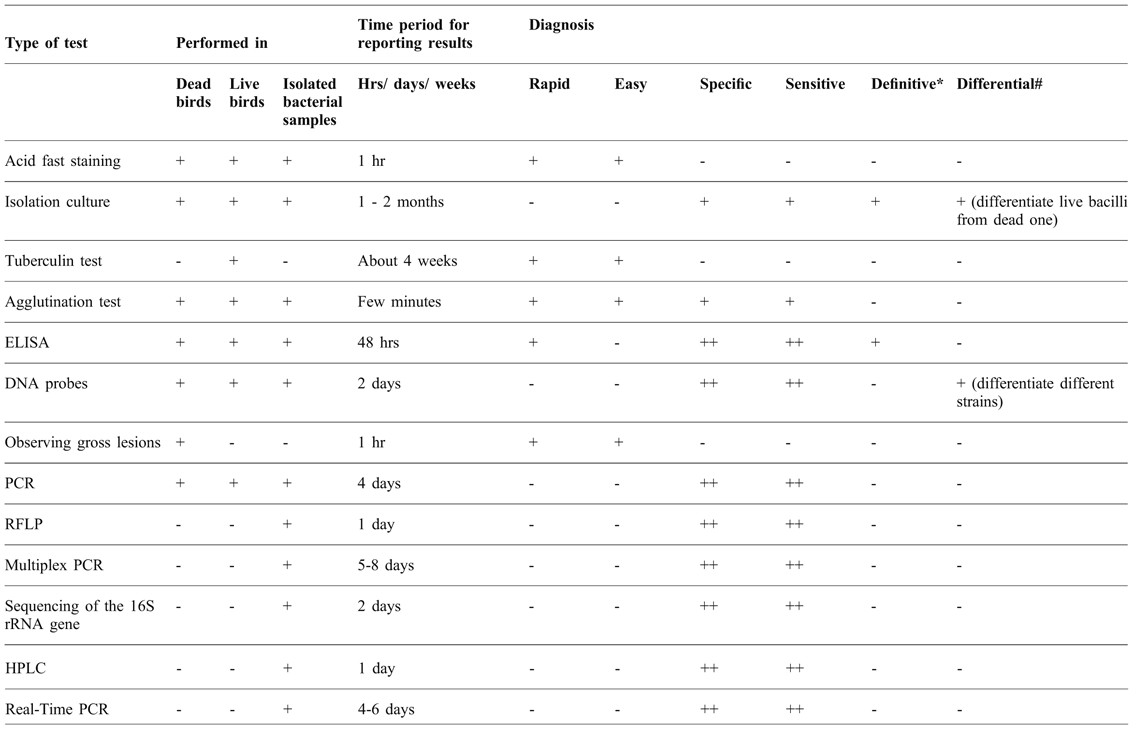
In many cases, smear microscopy fails to stain the bacteria by standard methods. This is the major limitation of this technique. Moreover, this technique depends on the microbial load. If the microbial load is low in the sample then it cannot be used successfully to observe the bacteria in the sample ( Table 1 ).
Comparative analysis of different techniques used for the diagnosis of avian tuberculosis.

(+): Present or effective; (++): more effective than other for particular parameter; (-): absent or ineffective
The definitive diagnosis means a final diagnosis which is made after getting all the results of tests
# Differential diagnosis helps in differentiating two or more strains/conditions
In one case of avian TB, high doses of Mycobacterium induced acute overwhelming disease which affected the liver and appeared as ‘morocco leather’ with fine grayish or yellow mottling. In such cases, AFB may not be observed easily. Instead, careful examination of the smear revealed the parallel bundles of brownish refractile bacilli. If the carbol fuschin stage of Zeihl-Neelson is performed for a longer time i.e. 10 min with heating then AFB can be observed. This demonstrates that AFB is highly resistant to stain penetration.
Direct microscopic observation of certain clinical samples for the presence of mycobacteria can be helpful. However, sensitivity is poor and many false negatives occur ( Table 1 ). Acid fast staining of the faecal sample shows poor sensitivity because few birds excrete tubercle bacilli in the faeces and is therefore difficult to diagnose.
histopathology
The AFB technique becomes more valuable when it is combined with histopathological studies. In birds, lesions developed from TB contain several AFB which can be observed by histopathological slide preparation of the lesions. This histopathological slide can be stained by the AFB technique which reveals the presence of mycobacteria in tissues. In some cases, congo red staining is used to observed amyloid deposits in liver kidney and spleen. These amyloid fibrils can be seen as green birefringence in a dark background under polarised light. These techniques are not widely accepted for the diagnosis of mycobacterial infections due to its non-applicability in living birds.
cytology
AFB staining technique is useful for detecting the presence of mycobacteria in cells. Granuloma (mass of macrophages) can be isolated from skin, conjunctiva, liver, spleen, intestine and other organs and stained with acid-fast staining technique. It shows the presence of large number of AFB in the cytoplasm of macrophages or multinucleated giant cells (Tell et al., Reference TELL, WOODS and CROMIE2001; Lennox, Reference LENNOX2007).
culture
In some cases, even after the prolonged stain application with extended heating time, AFB cannot be seen and therefore, smear microscopy fails to detect the presence of Mycobacterium spp. Special care must be taken in this circumstance and diagnosis of AFB can be done by culture method. Culturing is recommended where there is a characteristic flock history and indicative lesions are found at necropsy. Traditionally, M. avium subspecies are distinguished from common non-chromogenic slow-growing organisms by their ability to grow at 42°C (OIE Manual, 2014). Culture is used to isolate the causative organism from the necropsy material. Mostly, liver or spleen is the best organ to use, but if the carcass is decomposed, bone marrow may be proved more satisfactory as it may be less contaminated (Haque, Reference HAQUE2014).
It is necessary to take special care of culture methods, as rapidly growing bacteria may grow in the medium and contaminate the culture, producing false positive results and decreasing the sensitivity and specificity of the test. Therefore, samples must be pre-processed with detergent, alkali or acid in order to eliminate rapidly growing microorganisms before culture. The best media for the growth of Mycobacterium avium are Lowenstein–Jensen, Herrold's medium, Middlebrook 7H10 and 7H11 or Coletsos with 1% sodium pyruvate (Mayahi et al., Reference MAYAHI, MOSAVARI, ESMAEILZADEH and ASADOLLAHI2013). Occasionally, mycobactin J is supplemented in the media for the isolation of M. avium paratuberculosis and M. avium silvaticum. For M. genavense, the optimal solid medium is Middlebrook 7H11 medium which is supplemented with blood and charcoal and acidified to pH 6. This is incubated at 37°C and period should be extended for at least six months (Shitaye et al., Reference SHITAYE, HALOUZKA, SVOBODOVA, GRYMOVA, GRYM, SKORIC, FICTUM, BERAN, SLANY and PAVLIK2010).
The growth of these mycobacterial species may be confined to the edge of the condensation water. Typically, M. avium spp. produce smooth colonies within two to four weeks, however, rough variants do occur (Dhama et al., Reference DHAMA, MAHENDRAN, TIWARI, SINGH, KUMAR, SINGH and SAWANT2011). A number of species of M. avium require incubation for at least eight weeks. This is the main drawback of the culture based method. BACTEC or automated fluorescent MGIT 960 culture systems can be used for reducing the time of incubation (Dhama et al., Reference DHAMA, MAHENDRAN, TIWARI, SINGH, KUMAR, SINGH and SAWANT2011). Hence, BACTEC culture can provide growth in a shorter duration.
A fully automated BACTEC MGIT 960 system (A-MGIT) was compared with conventional media and the mycobacteria growth indicator tube (M-MGIT) method by Shitaye et al. (Reference SHITAYE, BERAN, SVOBODOVÁ, MORÁVKOVÁ, BABÁK and PAVLÍK2009). The conventional culture method is based on the use of media which support the growth of mycobacteria but takes a long duration. It includes media such as solid Herrold's and Stonebrink media and liquid Sula medium. This experiment has proven that detection rate of positive culture is highest, and detection time and contamination rate is lowest in the case of A-MGIT (Shitaye et al., Reference SHITAYE, BERAN, SVOBODOVÁ, MORÁVKOVÁ, BABÁK and PAVLÍK2009). BACTEC culture method is still considered as the gold standard for the definitive diagnosis of tuberculosis. It provides isolates for identification and drug susceptibility testing, although the time required for culture of Mycobacterium is still a problem ( Table 1 ).
haematology
This is a conventional method of diagnosis. Haematological changes are characterised by severe leukocytosis which comprises predominantly monocytosis, with a concurrent milder heterophilia, hyperfibrinogenaemia. It includes microcytic hypochromic anaemia, with reduced packed cell volume. The limitation of this technique is its sensitivity. Haematological analysis is reported to be less sensitive (Cromie et al., Reference CROMIE, BROWN, FORBES, MORGAN and STANFORD1993). In subclinical avian TB, birds show a number of haematological changes (Bush et al., Reference BUSH, MONTALI, SMITH and PERATINO1978; Hawkey et al., Reference HAWKEY, KOCK, HENDERSON and CINDERY1990; Forbes, Reference FORBES1992).
immunological tests
Immunological tests are based on the use of the specific antibodies for determining antigens on the bacterial cell surface in samples collected from the infected organs of birds. These tests are used for observing the health of chickens used for export (Duffus et al., 1968). The selection of particular antigen depends on the importing criteria of different countries.
tuberculin test
The most popular tests based on the use of antigen are the tuberculin test or haemagglutination (stained antigen) test. These are most frequently used for testing poultry for deciding the export quality. The tuberculin test is the most widely used test in domestic fowl and hens (Shitaye et al., Reference SHITAYE, MATLOVA, HORVATHOVA, MORAVKOVA, DVORSKA-BARTOSOVA, TRCKA, LAMKA, TREML, VRBAS and PAVLIK2008a). It is the only test for which reagents of international standards exists.
Tuberculin is the principle avian purified protein derivative (PPD). Birds are tested by intradermal inoculation in the wattle with 0.05 ml or 0.1 ml of tuberculin (containing approximately 2000 International Units [IU]), using a fine needle of approximately 10 mm × 0.5 mm (Shitaye et al., Reference SHITAYE, MATLOVA, HORVATHOVA, MORAVKOVA, DVORSKA-BARTOSOVA, TRCKA, LAMKA, TREML, VRBAS and PAVLIK2008a). The test is read after 48 hours and a positive reaction is by observing any swelling at the site and an increase in the diameter of a small firm nodule. On the day of inoculation, a small firm nodule is approximately 5 mm in diameter but after 48 h it produces oedema which extends into the other wattle and down the neck. This method can be successfully applied to immature birds possessing very small wattles. In immature birds, the comb may be used, although results are not reliable.
The most important factor that needs to be considered before performing a tuberculin test is the site of inoculation of the antigen in birds. In domestic fowl, tuberculin testing is a good technique, however; it is not reliable in turkeys (Pavlas et al., Reference PAVLAS, MICHALSKA and HUNADY1993). Therefore, inoculation of antigens in the wing web is recommended in the case of turkeys, while for other birds, inoculation in the wing web is not satisfactory (OIE Manual, 2014). The bare skin areas on Muscovy ducks and some species of pheasant can be used as inoculation site, however, reliability is doubtful and interpretation of results is difficult (OIE Manual, 2014). In waterfowl, the tuberculin antigen can be inoculated in the foot web but the test is not sensitive and is often complicated by infections at the inoculation site.
In pheasants, the tuberculin test can be performed in either of two ways. In the first method, 0.05 ml or 0.1 ml of tuberculin antigen is injected into the skin of the lower eyelid. If swelling occurs after 48 hrs, this indicates a positive result. The other method is based on the injection of 0.25 ml tuberculin antigen into thoracic muscles and the birds are observed for 6-10 hrs. Birds infected with Mycobacterium spp. show signs of depression and isolate themselves from the flock. Occasionally, sudden death of the infected birds occurs, or birds may show false negative test ( Table 1 ). The reason of this is the infection with another disease that interferes with the development of immune responses or improper administration of antigens.
Agglutination test
The Mycobacterium strain specific antigen is stained with 1% malachite green which is used for the rapid whole blood plate agglutination test (Rozanska, Reference ROZANSKA1965). The blood sample can be collected from the infected flock by venipuncture. A drop of antigen (0.05-0.1 ml) is mixed with the same volume of fresh whole blood on the slide or white tile. After 2 minutes it can be assessed for the agglutination reaction, which is seen as an accumulation of the malachite-green-stained antigen around the edge of the drop, leaving the centre a normal blood-red colour.
Besides blood, agglutination tests can be performed on serum samples. For this purpose, blood is collected from the infected birds and serum is isolated and mixed with antigen, and agglutination occurs if antibodies are present against the mycobacterial antigen. Shitaye et al. (Reference SHITAYE, MATLOVA, HORVATHOVA, MORAVKOVA, DVORSKA-BARTOSOVA, TRCKA, LAMKA, TREML, VRBAS and PAVLIK2008a) reported the use of two antigens MAA-b and MAA-p that are isolated from birds and pigs, respectively and possess the same genotype but a different serotype. MAA-b is bird isolate of serotype 1 and genotype IS901+ and IS1245+ while MAA-p is pig isolate of serotype and genotype IS901+ and IS1245+.
This test is suitable for immediate screening of an infected flock so that it can be culled, as is quick and easy to perform. It is used for screening domestic fowl as well as domestic and ornamental waterfowl. It has been reported that this technique is superior to the tuberculin skin test (Munang'andu et al., Reference MUNANG'ANDU, SIAMUDAALA, MATANDIKO, NAMBOTA, MUMA, MWEENE and MUNYEME2011). The main drawback of the technique is associated with the appearance of false positives and negatives (Cromie et al., Reference CROMIE, BROWN, FORBES, MORGAN and STANFORD1993).
elisa (advance laboratory technique)
Using the enzyme-linked immuno-sorbent assay (ELISA), antibodies present in the serum of birds are detected by using an enzyme linked conjugate which binds to the substrate and produces colour. This colour can be observed visually or more precisely by an ELISA reader. The kit is prepared by using antibodies against the mycobacterial antigens. These are isolated and purified by affinity chromatography and used to prepare secondary antibodies. These antibodies are inoculated in rabbits along with Freund's adjuvant. When secondary antibodies are produced, they are purified by affinity chromatography and then linked to an enzyme isolated from horseradish, known as the conjugate. When this conjugate reacts with a suitable substrate (2,2 azino-di-(3 ethyl benthiazoline sulphonic acid), change in colour occurs which can be visually identified or measured by using an ELISA reader. The intensity of colour reveals the presence of antibodies in the fowl serum.
The results of serum testing can be expressed as percentages of the differences between the positive and negative controls, after subtracting background responses (from wells containing coating buffer, but no antigen). The positive control used is serum from a duck with advanced avian TB, whereas the negative control was serum from a healthy ten-day-old duckling. According to Zsivanovits et al. (Reference ZSIVANOVITS, NEUMANN, BROWN and CROMIE2004), results were categorised as follows:
Avian TB negative: values B/20 of the adjusted positive control
‘Borderline’: values of 20 /50 of the adjusted positive control
Avian TB positive: values /50 of the adjusted positive control
Seroagglutination is based on sugar residue specificity of surface glycopeptidolipids. This allows identification and classification of M. avium complex for 28 serovars. More sophisticated typing methods directed at cell-wall-specific targets are currently available, such as enzyme-linked immunosorbent assays with monoclonal antibodies to major serovars.
The limitation of this technique is associated with delayed production of antibody after the infection. Serological identification of an antibody response to the Mycobacterium spp. can only be successful several weeks after an infection has occurred, because this is when antibodies are produced, and is a common technique for screening flocks. ELISA is a highly sensitive test to estimate mycobacterial infection. However, false-positives results may be seen (Zsivanovits et al., Reference ZSIVANOVITS, NEUMANN, BROWN and CROMIE2004) due to non-specificity of antibodies which leads to the cross reaction ( Table 1 ). Besides, the use of specific antigens produced from the local strain of M. avium is critical (Singh, personal communication).
Molecular biology techniques
polymerase chain reaction (pcr): rapid diagnosis without prior culture
Certain pathogens cannot be isolated in the laboratory due to their poor growth or fastidious nature regarding substrates. Successful isolation of mycobacteria can be slow and in some instances, impossible. Therefore, techniques are required that are based on the direct detection of bacteria without culturing. Such tests have been developed for the detection and identification of M. avium complex directly from tissue samples (Kaevska et al., Reference KAEVSKA, SLANA, KRALIK and PAVLIK2010). Bacterial DNA can be extracted in the laboratory from liver, spleen and intestine and amplified directly from the samples by PCR using specific primers. PCR products can be evaluated in an agarose gel and amplify the 192bp amplicon. Great success has been achieved in rapid diagnosis of avian tuberculosis by this technique.
Mycobacterium avium can be detected in massively infected tissues by conventional PCR. For identification purpose, nucleic acid hybridisation probes are available commercially. Nowadays, these probes are the ‘gold standard’ and are used to identify M. avium, M. genavense and M. intracellulare species. Multiplex PCR is an old method but remains suitable for differentiating M. avium from M. intracellulare and M. tuberculosis complexes. Multiplex PCR is rapid and simple to perform and is suitable for routine identification of M. avium and M. intracellulare as it is more economic than the commercial probe method (Cousins et al., Reference COUSINS, FRANCIS and DAWSON1996). At present, direct detection and quantification of M. avium using quantitative real-time PCR can be considered the best method.
The detailed conditions for performing PCR include initial denaturation: 94°C, 5 min; denaturation: 95°C, 1 min; annealing: 56°C, 1 min; elongation: 72°C, 1 min; number of cycles: 40; final elongation: 72°C, 10 min and expected product size: 193 bp M. avium. PCR can be carried out using the specific primers based (Forward: 5'-CGAGCGGATGACCACCCAGGACGTC-3' and reverse: 5'-biotin-CAGCGGGTTGTTCTGGTCCATGAAC-3') which are based on the RNA polymerase (rpoB) gene of M. avium subsp. avium (Hsieh et al., Reference HSIEH, TSAI, WANG, HUNG, TSAI and LIU2009).
Another method is based on the amplification of a portion of a 65 kDa heat shock protein gene. This gene possesses sequences that are conserved and unique to various species of mycobacteria. PCR based on hsp65 gene is used not only to diagnose or detect mycobacterial infection but also to differentiate M. avium from M. genavense. PCR is performed on analogous portions of hsp65 of both organisms. For this purpose, Mycobacterium spp. are cultured on media and cell and collected for preparing the cell lysate. DNA is extracted from cell lysate using zirconium beads and guanidine thiocyanate method. Primers possessing sequences 5’-AGG-CGA-TGG-ACA-AGG-T-3’, and 5’-CCT-CGA-TGC-GGT-GCT-TGC- 3’ are used to amplify a 693-bp fragment from hsp65 genes of M. avium and M. genavense. PCR products are cloned and sequenced and then, compared using the Genbank database. This helps in differentiating M. avium and M. genavense (Mendenhall et al., Reference MENDENHALL, FORD, EMERSON, WELLS, GINES and ERIKS2000).
The drawback of this technique is its high cost (Speer et al., Reference SPEER, SCHUMACHER, STETTER, GARNER and ISAZA1999). Another problem associated with the identification of Mycobacterium spp. is cross-reactivity of the pathogen, which may lead to the misidentification of M. avium (van Ingen et al., Reference VAN INGEN, AL-HAJOJ, BOEREE, AL-RABIAH, ENAIMI, DE ZWAAN, TORTOLI, DEKHUIJZEN and VAN SOOLINGEN2009).
Manarolla et al. (Reference MANAROLLA, LIANDRIS, PISONI, SASSERA, GRILLI, GALLAZZI, SIRONI, MORONI, PICCININI and RAMPIN2009) detected the presence of mycobacteria in pet birds using PCR and sequencing of an rRNA hypervariable region, and reported that avian mycobacteriosis can occur in very young birds ( Table 1 ). The most commonly affected species of birds are the canary (Serinus canaries), Eurasian goldfinch (Carduelis carduelis) and red siskin (Spinus cucullatus) and the main etiologic agent is M. genavense.
rapid-bap assay
RAPID-BAP assay stands for reliable assay protocol for identification of diseases (RAPID)-bioactive amplification with probing (BAP) assay. It has been used via a PCR and magnetic bead-based probe-based approach to detect M. avium in Swinehoe's pheasant. This hybridisation-based technique which is performed after PCR, whereby the rpoB gene-specific oligonucleotide is covalently coupled to unique sets of carboxylated-modified magnetic beads by using water-soluble carbodiimide (Yang et al., Reference YANG, TRAN and WANG2001; Hsieh et al., Reference HSIEH, TSAI, WANG, HUNG, TSAI and LIU2009). It is successful in diagnosing clinical cases of avian TB, although, unfortunately, probes used in this assay are not available for common use in clinical samples. The other limitation of this technique is the high cost.
restriction fragment length polymorphism (RFLP)
Mycobacterium avium subsp. avium has two genotypes based on two insertion sequences i.e. IS901 and IS1245. These were found to infect hens (Gallus domesticus) naturally (Shitaye et al., Reference SHITAYE, MATLOVA, HORVATHOVA, MORAVKOVA, DVORSKA-BARTOSOVA, TRCKA, LAMKA, TREML, VRBAS and PAVLIK2008a). These genotypes can be isolated from tissue, faecal and environmental samples. They are present in little egret (Egretta garzetta), buff-backed heron (Bubulcus ibis), great white egret (Egretta Alba), and bittern (Botaurus stellaris) and sacred ibis (Threskiornis aethiopicus) (Dvorska et al., Reference DVORSKA, MATLOVA, AYELE, FISCHER, AMEMORI, WESTON, ALVAREZ, BERAN, MORAVKOVA and PAVLIK2007). These isolates can be diagnosed and identified using RFLP methods. In this method, hybridisation probes for the identification of IS901 strain can be used. At first, DNA is isolated from Mycobacterium spp. and purified. This DNA is digested with restriction endonucleases PvuII and then these fragments are isolated in agarose gel. RFLP profiles can be analysed by two methods: (i) DNA fragments are transferred to a nylon membrane by vacuum blotting. DNA is fixed and hybridised with a labelled probe. This probe is used to diagnose RFLP profiles. (ii) After the gel run, DNA fingerprints can be scanned by CCD camera and analysed by the Gel Compare software using a standard isolate control profile. DNA can be digested in parallel with restriction endonucleases PvuII or PstI and hybridised to standard probes prepared by PCR. DNA fingerprints were scanned by CCD camera and analysed by the Gel Compare (Applied Maths, Version 4.1, Kortrijk, Belgium) software using a standard isolate control profile (Dvorska et al., Reference DVORSKA, BULL, BARTOS, MATLOVA, SVASTOVA, WESTON, KINTR, PARMOVA, SOOLINGEN and PAVLIK2003).
RFLP typing is useful for epidemiological studies. The main prerequisite for RFLP typing is the stability of IS901 profiles. IS901 was isolated from birds and used for generating RFLP profiles. It was observed that these profiles are stable in both in vivo and in vitro conditions. The identical type PvuII-IS1245 RFLP profiles was found in IS901 positive isolates which show their clonal nature and host specificity. However, these profiles are not associated with virulence for pullets (Dvorska et al., Reference DVORSKA, BULL, BARTOS, MATLOVA, SVASTOVA, WESTON, KINTR, PARMOVA, SOOLINGEN and PAVLIK2003).
Similar to IS1245, IS1311 can be used for discriminating M. avium subspecies. IS1311 has the ability to discriminate the strains. This is helpful in obtaining information about the type of infecting strain, and has been used to compare isolates from humans and animals in different regions of the world (Johansen et al., Reference JOHANSEN, OLSEN, JENSEN, DAHLE, HOLSTAD and DJØNNE2007). Later, Radomski et al. (Reference RADOMSKI, THIBAULT, KAROUI, DE CRUZ, COCHARD, GUTIÉRREZ, SUPPLY, BIET and BOSCHIROLI2010) reported the use of PCR-based mycobacterial interspersed repetitive-unit-variable-number tandem-repeat (MIRU-VNTR) typing for the molecular characterisation of M. avium subsp. paratuberculosis and M. avium strains. M. avium subspecies, including M. avium subsp. avium, M. avium subsp. hominissuis, and M. avium subsp. silvaticum, isolated from numerous animal species, HIV-positive and HIV-negative humans, and environmental sources, can be typed by IS1311 with discriminatory index 0.885 (Radomski et al., Reference RADOMSKI, THIBAULT, KAROUI, DE CRUZ, COCHARD, GUTIÉRREZ, SUPPLY, BIET and BOSCHIROLI2010). This showed that RFLP pattern is similar to that obtained by IS1311. The main advantage of MIRU-VNTR typing is associated with its ability to distinguish different subspecies of M. avium. It is convenient to use and provides useful information regarding epidemics caused by M. avium subspecies. Therefore, MIRU-VNTR typing is preferable to use as compared to IS1311 typing.
RFLP was used to detect pathogenic and conditionally pathogenic M. avium subsp. avium in tissues, swabs of cloacae and faecal samples. This technique confirms its presence in tissues and faecal samples, which are the main source of infection (Shitaye et al., Reference SHITAYE, MATLOVA, HORVATHOVA, MORAVKOVA, DVORSKA-BARTOSOVA, TREML, LAMKA and PAVLIK2008b). RFLP is used for the typing bacterial samples, and taxonomical characteristics can be identified by using RFLP which further helps in constituting a separate taxa (Ritacco et al., Reference RITACCO, KREMER, VAN DER LAAN, PIJNENBURG, DE HAAS and VAN SOOLINGEN1998).
RFLP profiles depend upon the restriction enzyme used for cutting DNA into fragments. Generated fragments help in the identification of Mycobacterium species. SalI and BsteII cut M. genavense PCR products but not the products of M. avium isolates. In contrast to this, BglI cuts the M. avium PCR products but not M. genavense. The restriction enzyme patterns generated by aforesaid restriction enzymes are consistent with culture results. This helps in correctly identifying and differentiating M. avium and M. genavense in all cases (Mendenhall et al., Reference MENDENHALL, FORD, EMERSON, WELLS, GINES and ERIKS2000).
high-performance liquid chromatography (HPLC)
High-performance liquid chromatography (HPLC) is based on the detection of mycolic acid, a constituent of the mycobacterial cell wall. HPLC is a time-consuming process and involves several steps such as saponification, extraction, derivatisation and chromatographic separation. It separates the compounds from the mixture by means of their different solubilities in the mobile phase and stationary phase. The mobile phase is the liquid which moves through the column and stationary phase is packed in the column. When a sample is injected into the port, it spreads over the stationary phase in the column. Thereafter, the mobile phase runs through the column and separates different compounds which are eluted out at different times. The HPLC chromatogram is a graphic output of UV absorption versus elution time. Absorption of the analytes is measured at 254 nm which is provided by UV lamp. Each separated compound forms a peak corresponds to a single compound. Each fraction is identified by the corresponding retention time. Retention time is defined as the time taken by analyte to travel from the injection port to the detector (Tortoli and Bartoloni, Reference TORTOLI and BARTOLONI1996).
The limitation of this technique is time requirement and cost ( Table 1 ). Minor changes have been made with time to overcome the limitations of the techniques. The first modification in the protocol is saponification, where the time is reduced from 18 hrs to 1 hr and the corresponding temperature raised from 85°C to 121°C, and the methanol-chloroform solvent is replaced with methanol-methylene chloride. The use of a shorter column reduces the elution period from 65 min to 10 min but increases the flow rate (Tortoli and Bartoloni, Reference TORTOLI and BARTOLONI1996).
Problems in diagnostics
The diagnosis of avian tuberculosis in the live bird can be difficult due to irregular faecal shedding and varying symptoms (Dhama et al., Reference DHAMA, MAHENDRAN, TIWARI, SINGH, KUMAR, SINGH and SAWANT2011). Single techniques provide inadequate information to give a confirmed diagnosis of avian tuberculosis cases. A combination of techniques, if applied correctly, may give more reliable results, but increases the cost of diagnosis. Due to these issues, diagnostic techniques cannot be incorporated in routine examination of birds. Other issues are related to the sensitivity and specificity of diagnostic techniques for identifying TB infection.
Conclusions
In the comparison of different diagnostic approaches for the diagnosis of avian TB, it has been seen that single technique is not effective in diagnosing the disease due to the lack of required sensitivity and specificity. Current efforts are underway to increase the sensitivity and specificity in order to diagnose the etiological agent at subspecies level. This is important to determine the cause of infection in the birds in order to prevent it at source. Multidisciplinary approaches can help us to fully understand the aetiology and epidemiology of the disease.