Tumours develop from normal cells when they sustain unrepaired mutations that directly or indirectly cause defects in the regulatory pathways that control cellular proliferation and homeostasis. These mutations can result in uncontrolled cell growth and genomic instability, which promote further mutations in larger populations as a cascading effect (Ref. Reference Hanahan and Weinberg1). Normal cells have a built-in mechanism to deal with such transformations via the activation of programmed cell death through apoptosis. Therefore, apoptosis represents a brake that must be disabled for any cancer to develop.
Apoptosis was first formally described and named in 1972 as a stereotypic morphological response to many different types of cellular insult, different from necrosis. However, despite the ultimate utility of the concept, little work was done over the ensuing decade and a half – largely because of the lack of experimental handles to manipulate the process. In the early 1990s two parallel developments changed that dramatically. First, as the ‘roadmap’ for the complete development of the nematode Caenorhabditis elegans from fertilised unicellular zygote to multicellular adult was elucidated, it became clear that certain ‘intermediate’ cells were destined to die, and that this process was both positively and negatively regulated by specific genes (i.e. depending on the gene mutated, the number of cells in the adult animal could be increased or decreased) (Ref. Reference Ellis and Horvitz2). Second, the gene called B cell chronic lymphocytic leukaemia (CLL)/lymphoma 2 (BCL2, encoding Bcl-2), which as a consequence of a reciprocal chromosomal translocation common in B cell follicular lymphoma is located in the immunoglobulin heavy-chain gene locus and hence is strongly upregulated (Refs Reference Bakhshi3, Reference Cleary, Smith and Sklar4, Reference Tsujimoto5), functioned not as a classic oncogene by increasing proliferation but rather by preventing cell death in normal lymphocytes (Ref. Reference McDonnell6). When it was shown that human BCL2 could substitute for one of the C. elegans genes inhibiting cell death, the generality of the process was recognised (Ref. Reference Hengartner and Horvitz7). An explosion occurred in the literature, with presently over 180 000 articles written on apoptosis (PubMed, http://www.ncbi.nlm.nih.gov/pubmed; accessed June 2010).
Apoptosis inhibition as essential to developing and maintaining cancer: from the orphan concept to clinical reality
The original observation that Bcl-2 was involved in the first stages of the development of a haematological malignancy has now been broadened into two important directions from a clinical viewpoint. First, Bcl-2 was shown to be transcriptionally upregulated by other mechanisms that did not involve chromosomal translocation (as is also the case for other antiapoptotic Bcl-2 family members, which will be discussed below). Thus, the impact of the original observations has been extended far beyond haematological malignancies to include many different types of cancers (Refs Reference Green and Evan8, Reference Look9). Indeed, a recent systematic survey and meta-analysis of the transcriptional profiles of many different types of cancer has indicated that the dysregulation of Bcl-2 and other antiapoptotic family members is one of the key distinguishing features between normal and cancer cells (Ref. Reference Slavov and Dawson10). Although the molecular mechanism mediating this deregulation is not known in detail in all cases, these observations are consistent with recent extensive analysis looking at copy number variations across the entire human genome in both cancer cell lines and primary samples from patients. These data indicate that increases in the gene dosage of two members of the Bcl-2 family, namely BCL2 and MCL1, are commonly involved in many different types of malignancy (Ref. Reference Beroukhim11).
This widespread involvement of the family of proteins in cancer development (Ref. Reference Brown and Attardi12) would be stimulus enough to look into its biochemistry, but a second feature makes it even more intriguing. It was shown that inhibition of apoptosis by Bcl-2 was involved not only in the development of cancer but also in the mechanism whereby cancers develop resistance to cancer therapeutics (including chemotherapy and radiotherapy) (Ref. Reference Kang and Reynolds13). Therefore, because most cancer therapies funnel through the induction of apoptosis, being able to ‘open this funnel wider’ with drugs that interfere with Bcl-2 was recognised early as an important general way of improving conventional cancer therapy. This concept has recently been extended even further with the idea that even in the absence of exposure to chemotherapy many cancer cells are addicted to the ongoing presence of Bcl-2 proteins. This novel concept is predicated upon the idea that the development of cancer itself causes the cell to generate apoptotic signals that must be neutralised. Being able to abolish Bcl-2 antiapoptotic signalling would therefore represent a form of synthetic lethality that would kill predominantly cancer cells rather than normal cells that do not automatically engage apoptotic machinery.
These suggestive findings have therefore motivated a widespread attempt to find drugs that would inhibit the mechanism whereby Bcl-2 prevents apoptosis. Many agents are in development, but we discuss here only the two that are currently in early-phase clinical trials as anticancer agents. Furthermore, even though Bcl-2 is widely involved in many cancers, because of historical precedent and ease of tissue sampling most of the early clinical work has been done in haematological malignancies. Therefore, we review these early results not only for their importance to these diseases in their own right, but also as sentinels to what might happen as the agents are applied to other types of cancers. To understand how these new agents work (and potentially how they can be improved for second-generation drugs), we need to look at the different members of the Bcl-2 family and how they interact to either promote or prevent apoptosis.
The dance between Bcl-2 family member proteins: the role of the players depends on the programme
To understand apoptosis at a general level, it is useful to remember that it is governed by two different clusters of threes. The temporal sequence of apoptosis can be divided into three phases. There is first an initiation phase whereby a cell has undergone some sort of insult and signalling molecules that indicate this are activated. In general, these signals can be initiated by processes that occur internally in the affected cell such as DNA damage (the intrinsic pathway), or after the binding of ligands to death receptors on the cell surface (the extrinsic pathway) (Ref. Reference Schmidt14). There is then a regulatory phase when the sum of all these signals is integrated to decide whether enough damage has occurred to warrant that the cell undergoes apoptosis. Once the decision has been made to undergo apoptosis, the third, and irreversible, execution phase is initiated, whereby various effectors such as activated proteases called caspases cleave specific substrates in the cell, resulting in the final phenotype of an apoptotic cell that can be engulfed by its neighbours. Elegant analytical studies using fluorescent readouts of these distinct phases in individual cells have demonstrated that after a uniform death signal, the length of the initiation phase is extremely variable even among a population of isogenic cells, probably due to stochastic differences in the levels of proteins that mediate these early changes. By contrast, once the ‘threshold’ to die has been reached at the regulatory phase, this is followed by an invariably swift execution stage. These data emphasise the importance of being able to manipulate the regulatory phase as the rate-limiting step in the whole process.
Division of the Bcl-2 family into three functional/structural classes
The second cluster of threes is that there are three separate classes of Bcl-2 family members (Ref. Reference Youle and Strasser15) (Box 1). They are distinguished structurally by the number of the four ‘Bcl-2 homology regions’ (BH regions of conserved sequences within the proteins) they share, and these structural classes can also be conveniently divided along functional lines that map onto the three different temporal phases of apoptosis. Proapoptotic family members are subdivided into two classes: Bax (Bcl-2-associated X protein; encoded by BAX) and Bak (Bcl-2-antagonist/killer; encoded by BAK), which contain BH regions 1–3; and another class designated as BH3-only proteins. The family members Bax and Bak are critically important in the regulation phase, because when they are activated they change from monomers to oligomers that disrupt the integrity of the outer mitochondrial membrane – a process called mitochondrial outer membrane permeabilisation (MOMP). MOMP causes the ‘leakage’ of components such as cytochrome c from the intermembrane space of mitochondria into the cytoplasm. Once released, cytochrome c interacts with the adaptor protein APAF1 to form a complex that binds to and activates the caspase proteases that digest intracellular substrates in the execution phase. Thus, MOMP mediated by activated Bax or Bak is the ultimate point of regulation for apoptosis and, consequently, the structural changes that occur in these proteins as they mediate MOMP is the subject of intense investigation, as discussed below.
Box 1 The three different classes of Bcl-2 family proteinsa
Proapoptotic
BH 1–3, pore-forming: Bax and Bak
BH3-only: Bad, Bid, BIK, Bim, Bmf, Noxa and PUMA
Antiapoptotic
BH1–4: Bcl-2, Bcl-XL, Bcl-w, Mcl-1
aThe proteins can be grouped functionally according to their pro- or antiapoptotic effects, and structurally according to the Bcl-2 homology (BH) regions they contain.
Abbreviations: Bad, Bcl-2-associated agonist of cell death; Bak, Bcl-2-antagonist/killer; Bax, Bcl-2-associated X protein; Bcl-2, B cell CLL/lymphoma 2; Bcl-XL, B cell lymphoma-extra large; Bcl-w (BCL2L2), Bcl-2-like protein 2; Bid, BH3-interacting domain death agonist; BIK, Bcl-2-interacting killer; Bim, Bcl-2-interacting mediator of cell death; Bmf, Bcl-2-modifying factor; Mcl-1, myeloid cell leukaemia sequence 1; PUMA, p53-upregulated modulator of apoptosis.
The BH3-only proteins are the factors that sense cell damage, and it is these proteins that directly or indirectly activate Bax/Bak, causing them to oligomerise. There are many different BH3-only family members, which correspond to different types of cellular stress that must be sensed. Therefore in normal cells not undergoing stress, these BH3-only proteins either are absent and must be transcriptionally upregulated, like PUMA (BBC3) and Noxa (PMAIP1), or exist in an inactive state and must be activated by various post-translational mechanisms, including phosphorylation [e.g. Bad (BAD)], cleavage [e.g. Bid (BID)] or relocalisation within the cell [e.g. Bim (BCL2L11) and Bmf (BMF)]. The BH3-only proteins are therefore involved in both the sensing phase of apoptosis and the initiation of the regulatory phase.
The third class of Bcl-2 proteins (although the first discovered) are antiapoptotic members such as Bcl-2, Bcl-XL (BCL2L1), Mcl-1 (MCL1) and Bcl-w (BCL2L2): they contain BH regions 1–4. As discussed below, these proteins inhibit the effects of the BH3-only proteins after apoptosis is initiated as well as the oligomerisation of activated Bax or Bak.
Models of the mechanism of apoptosis regulation by Bcl-2 family members
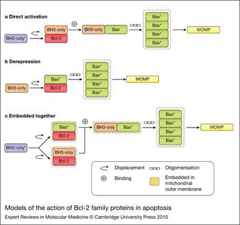
The different combinations of BH regions found in the three functional classes mediate the specific binding interactions between Bcl-2 family members. The BH3 region of one member, which forms an α-helix and has four spaced conserved hydrophobic residues (H1–4) with a charged residue at the C-terminal end, can fit into the hydrophobic cleft formed by BH regions 1–3 of another family member (Ref. Reference Giam, Huang and Bouillet16). However, an additional binding surface is required for oligomers larger than dimers to form. The recent discovery of an alternative BH3-binding pocket on the opposite side of the protein (the rear pocket, as discussed further below) probably fulfils this function (Ref. Reference Gavathiotis17). From this arrangement, it can be seen that many different combinations of hetero- and homodimers are possible (as well as oligomers with more than two subunits). To understand the principles governing the outcome of the interactions between different classes of Bcl-2 family members, three models have been proposed, which are shown in simplified form in Figure 1.
Models of the action of Bcl-2 family proteins in apoptosis. The three different models of apoptosis are shown in schematic form. (a) The direct activation model. A BH3-only activator (such as Bim or Bid, orange) directly binds to Bax (green) and activates it (denoted as Bax*). Activation of Bax permits integration into the mitochondrial outer membrane (MOM) and subsequent oligomerisation, resulting in Bax pore formation and MOM permeabilisation (MOMP). Antiapoptotic proteins (such as Bcl-2, red) can sequester the BH3-only activators and prevent Bax oligomerisation. BH3-only sensitisers (such as Bad or Noxa, purple denoted as BH3-only') act as ‘decoys’ and displace the BH3-only activators from the antiapoptotic proteins, which then bind to Bax, resulting in Bax oligomerisation, pore formation and MOMP. (b) The derepressor model. The sole function of the antiapoptotic proteins such as Bcl-2 in this model is to bind to and inhibit Bax, which is active (Bax*). The role of the BH3-only proteins is to bind to the antiapoptotic proteins, causing the displacement of Bax. The active Bax is then able to integrate into the MOM, oligomerise and form pores resulting in MOMP. The BH3-only proteins are distinguished by varying binding affinities to the different antiapoptotic Bcl-2 family proteins. (c) The embedded together model. Antiapoptotic proteins are able to sequester both active Bax and BH3-only activators. BH3-only sensitisers displace Bax and BH3-only activators from the antiapoptotic proteins. The ‘freed’ Bax can oligomerise whereas the BH3-only activators can bind and recruit additional Bax, which also oligomerises, resulting in pore formation and MOMP. These important interactions occur within the MOM where all three classes of Bcl-2 family members undergo specific conformational changes. Some of the binding interactions occur only in membranes (i.e. Bcl-2 or tBid binding to Bax).

The ‘direct activation’ model (Refs Reference Kim18, Reference Letai19) proposes that there are two subclasses of BH3-only proteins. The ‘activator’ subclass of proteins [including truncated Bid (tBid) and Bim)] directly bind to and activate Bax or Bak. In this case, the sole function of antiapoptotic proteins such as Bcl-2 is to sequester these Bax/Bak activators. The second subclass of ‘sensitiser’ BH3-only proteins (such as Bad or Noxa) do not directly activate Bax/Bak, but instead act as a ‘decoy’ and displace tBid or Bim from binding to antiapoptotic members; these ‘freed’ activator BH3-only proteins can then activate Bax/Bak. In this view, the outcome depends on which BH3-only subclass Bcl-2 binds to: if it binds to activator BH3-only proteins, apoptosis is shut down because Bax/Bak cannot be activated; however, if it binds to a ‘decoy’ sensitiser BH3-only protein, activators such as tBid and Bim become free, and then Bax/Bak can be activated.
The competing ‘derepression’ model (Refs Reference Chen20, Reference Willis21) postulates that Bax/Bak is always active and that the antiapoptotic Bcl-2 proteins must constitutively bind to them to prevent apoptosis. The model states that the only role of the BH3-only proteins is to displace activated Bax/Bak from antiapoptotic Bcl-2 proteins. According to this model, the different BH3-only proteins are distinguished by which antiapoptotic Bcl-2 family members they bind to: all of them (such as tBid, Bim or PUMA) or just a subset (e.g. Bad binds only to Bcl-2 and Bcl-XL and not to Mcl-1, whereas for Noxa it is the reverse). In this view, the critical question is whether Bcl-2 is bound to Bax (apoptosis shut down) or a BH3-only protein (constitutively active Bax released; apoptosis active).
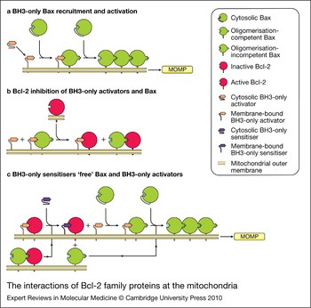
The ‘embedded together’ model proposed by our group combines features of both models, because from studies using an in vitro system with purified recombinant proteins it is clear that antiapoptotic Bcl-2 family member Bcl-XL binds to and sequesters both BH3-only activators and activated Bax/Bak (Ref. Reference Billen22). Furthermore, a major factor that regulates these binding interactions is the membrane because interaction with the membrane alters the binding surfaces between proteins for all three classes of Bcl-2 family members (Ref. Reference Leber, Lin and Andrews23). In this way the interactions with membranes change the relative affinities of specific interacting pairs. The critical conformational change for triggering apoptosis occurs when Bax and Bak change shape in the membrane such that they can bind to each other and form homo-oligomeric structures that permeabilise the outer mitochondrial membrane (Ref. Reference Lovell24). As shown in Figure 2, by binding to both the ‘trigger’ (BH3-only proteins) and ‘propagator’ (previously activated Bax molecules) without permitting further oligomerisation, Bcl-XL and the other antiapoptotic proteins act as ‘dominant negative’ versions of Bax, thereby preventing membrane permeabilisation.
Interactions of Bcl-2 family proteins at the mitochondria. (a) BH3-only Bax recruitment and activation. Various damage signals to the cell turn on BH3-only activators (orange), which undergo a conformational change as they embed into the mitochondrial outer membrane (MOM). By binding to cytosolic Bax (green), BH3 activators cause a conformational change in Bax, disengaging its C-terminal helix from the BH3 groove, allowing it to embed in the MOM, oligomerise and form pores resulting in MOM permeabilisation (MOMP). Furthermore, this activated Bax can recruit other cytosolic Bax molecules to the MOM, thereby allowing increased oligomerisation and MOMP. (b) Bcl-2 inhibition of BH3-only activators and Bax. In response to activated Bax and BH3-only activators in the MOM, antiapoptotic proteins such as Bcl-2 (red) change conformation and bind to both active Bax, preventing oligomerisation, and BH3-only activators, preventing Bax activation. (c) BH3-only sensitisers ‘free’ Bax and BH3-only activators. Cellular signals cause BH3-only sensitisers (purple) to change conformation and associate with antiapoptotic proteins at the MOM. This allows active Bax and BH3-only activators to be ‘freed’, resulting in Bax activation, oligomerisation and subsequent MOMP.

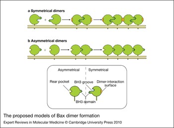
All models agree that the heterodimeric interactions between the different classes of the Bcl-2 family occur through the BH3 domain of one protein acting as a ‘ligand’ binding to a ‘receptor’ binding groove formed by BH1–3 regions on the other. Indeed, this is the structural rationale for the successful drug screen outlined below where small molecules acting as BH3-peptide mimetics fit into the ‘receptor’ binding groove of antiapoptotic Bcl-2 family members. However, as mentioned above, there must be other binding interactions that occur to permit Bax or Bak to form homo-oligomers of higher stoichiometry that create pores, as the binding groove formed by BH1–3 regions would only permit ‘face-to-face’ dimers that could not enlarge. Either the dimers themselves must join, or another ‘rear’ binding pocket must exist to allow a Bax to join with a partner both in front of it and behind (Fig. 3). This is an active area of investigation and there is conflicting evidence for both possibilities (Ref. Reference Gavathiotis17). Whichever proposal is correct, it is clear that there is another binding interface that is a potential, and currently unexplored, drug target.
Proposed models of Bax dimer formation. (a) Symmetrical dimers. The BH3 domain of one Bax interacts with the BH3-binding groove of another. Bax dimer formation causes a conformational change, opening up a dimer interaction surface and thereby permitting Bax dimers to join to each other, thus forming higher-order oligomers. (b) Asymmetrical dimers. The BH3 domain of an activated Bax can interact with the ‘rear pocket’ of another Bax. This interaction causes a conformational change in Bax, allowing the BH3 domain to be exposed so that it binds to the ‘rear pocket’ of an adjacent Bax. Through this mechanism Bax oligomerises, causing mitochondrial outer membrane permeabilisation.

Oncogene addiction to antiapoptotic Bcl-2 family proteins
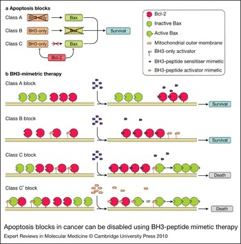
The concept of oncogene addiction implies that when a cancer develops as a consequence of the altered function of an oncogene, its ongoing survival is dependent on the continuous activity of this oncogene in a way that the normal cell is not dependent on the activity of the corresponding normal proto-oncogene (Ref. Reference Felsher25). Therefore, inhibiting the activity of the oncogene (such as catalytic activity or binding interactions) would differentially kill cancer cells. This is the basis of the clinical activity of tyrosine kinase inhibitors like imatinib against cancers bearing oncogenes such as BCR–ABL or activated KIT in chronic myeloid leukaemia and gastrointestinal stromal tumours, respectively (Ref. Reference Tolomeo26). Elegant work initiated in the laboratories of the late Stanley Korsmeyer and Anthony Letai has identified the presence of Bcl-2 on the mitochondria of lymphoid and other malignancies as another type of oncogene addiction: inhibiting Bcl-2 on mitochondria isolated from cell lines or patient samples with peptides from BH3-only proteins causes MOMP by releasing the ‘block to apoptosis’ (Refs Reference Letai27, Reference Del Gaizo Moore28). Based on the many steps required to initiate MOMP discussed above, this concept of a block to apoptosis has been further refined to define three different classes (Ref. Reference Letai29) (Fig. 4).
Apoptosis blocks in cancer can be disabled using BH3-peptide mimetic therapy. (a) Blocks in apoptosis. The three classes of blocks to apoptosis are outlined. Typically, BH3-only proteins (orange) respond to various cellular signals and activate Bax (green), leading to MOMP. Antiapoptotic proteins (such as Bcl-2, red) can bind to and inhibit both activated Bax and BH3-only activators, resulting in cellular survival. When the amount of proapoptotic proteins outweighs that of antiapoptotic proteins, apoptosis occurs. If cells have an insufficient amount of BH3-only proteins, apoptosis does not occur because the antiapoptotic proteins are able to bind to both activated Bax and the very low amount of BH3-only activators, resulting in cellular survival (Class A block). A Class B block occurs when Bax and Bak are inactive or at low levels such that the antiapoptotic proteins are able to prevent Bax/Bak oligomerisation and mitochondrial outer membrane permeabilisation (MOMP). Class C blocks occur when the amount of antiapoptotic proteins is increased to the point where they can bind to and sequester all of the activated Bax/Bak and BH3-only activators, thereby promoting cellular survival. These cells are addicted to the antiapoptotic proteins, and once the action of the antiapoptotic proteins has been prevented cells are able to die. (b) Treatment with BH3-peptide mimetic drugs. Mitochondria with a Class A block are resistant to an influx of BH3-peptide sensitiser mimetics (purple) and apoptosis due to an insufficient amount of BH3-only activators (orange) to activate Bax/Bak (green). Mitochondria with a Class B block have insufficient or absent levels of Bax/Bak so that BH3-only activators displaced by BH3-only sensitisers do not have a target to bind to; therefore, MOMP and apoptosis cannot occur. Mitochondria with a Class C block are addicted to the antiapoptotic proteins (red) when they are in excess. Class C mitochondria respond to BH3-peptide mimetic therapy because if the excess antiapoptotic proteins are inhibited, Bax/Bak oligomerisation and pore formation occur, resulting in MOMP and cell death. As an alternative scenario (class C′ block), antiapoptotic proteins also bind to activated Bax, preventing oligomerisation. BH3-peptide mimetic drugs may act as activators that, by displacing the antiapoptotic proteins from this complex, allow Bax to oligomerise by binding to and activating other Bax proteins, thereby causing oligomerisation and MOMP.

Mitochondria with a Class A block have insufficient BH3-only proteins, due to a number of reasons such as an altered stress pathway that otherwise turns them on, or BH3-only gene deletions or mutations. As a consequence, the amount of these proteins is reduced so that the antiapoptotic proteins are able to sequester all the BH3-only activator proteins. Mitochondria with a Class B block have inactive or low levels of Bax and Bak to mediate MOMP, even if sufficient BH3-only activators are present. A Class C block occurs when cells overexpress antiapoptotic proteins such as Bcl-2 or Mcl-1: as a result, all activator BH3-only proteins and activated Bax/Bak are sequestered (Refs Reference Certo30, Reference Brunelle and Letai31). Class C blocks can be further divided by which antiapoptotic protein is present, as they have different affinities for BH3-only proteins; for example, Bcl-2 and Bcl-XL bind to Bad but not Noxa, whereas Mcl-1 has the opposite binding pattern. Therefore, by using peptides derived from either Bad or Noxa on mitochondria derived from cell lines or clinical samples, the predominant antiapoptotic Bcl-2 family member can be identified (Ref. Reference Deng32).
Development of first-generation Bcl-2 antagonists for cancer: a new clinical paradigm
Accumulating evidence points to a crucial role for the Bcl-2 family in regulating apoptosis in cancer cells, and it is clear that exploiting this relationship is an attractive approach for novel anticancer agents. The first drug developed with a goal of inhibiting Bcl-2 function was oblimersen sodium, an antisense modified 18-mer oligonucleotide complementary to the BCL2 gene with a phosphothiorate backbone. This agent has had only moderate success against low-grade lymphoid malignancies, where it would be anticipated to be most active (Refs Reference Pro33, Reference O'Brien34), and did not obtain US Food and Drug Administration (FDA) approval after failing to result in survival differences in a pivotal melanoma trial (Ref. 35). Potential explanations for these disappointing outcomes include suboptimal penetration in cancer cells, a long half-life of the Bcl-2 protein and the narrow specificity of the target.
All these problems are potentially surmountable with small-molecule inhibitors that release proapoptotic family members from the binding pocket of several members of the antiapoptotic class. However, this poses new sets of problems, including the relatively large hydrophobic surfaces involved in the binding interaction compared with inhibiting the catalytic sites of other oncoproteins, and the possibility of off-target effects. Off-target toxicity can be very conveniently and precisely studied using mitochondria derived from mice or cell lines with knocked-out genes for both Bax and Bak – if any cell death occurs under these circumstances in response to candidate inhibitors, it is not apoptosis and presumably the agents would not have selectivity towards cancer cells. Many of the candidate Bcl-2-binding compounds have this unfortunate property (including gossypol, apogossypol, EM20-25 and chelerythrine) (Ref. Reference Vogler36), and one of the two compounds (obatoclax) currently in clinical trials has given conflicting results with this assay in different publications (Refs Reference Vogler36, Reference Nguyen37).
ABT-737/263 and obatoclax, the first two drugs in the clinic
The two Bcl-2 inhibitor drugs furthest in clinical development are the Abbott compound ABT-263 and obatoclax (GX-15-70) from Gemin X Biotechnologies. The former is based on a derivative of the widely used experimental compound ABT-737. Using nuclear magnetic resonance structure-based design with the BH3 region of Bad as a template, ABT-737 was developed as a rational Bcl-2 inhibitor (Ref. Reference Oltersdorf38). ABT-737 binds to and inhibits Bcl-2, Bcl-XL and Bcl-w with nanomolar affinities when truncated versions of these proteins are used as binding targets. This is the highest affinity of all the reported compounds currently in clinical or preclinical testing (Ref. Reference Vogler36). ABT-737 is also extremely effective at enhancing the cytotoxicity of a variety of chemotherapy agents in many different cancer cell lines in vitro (Ref. Reference Kang39), and notably causes significant apoptosis as monotherapy in cell lines derived from lymphoid malignancies such as acute lymphoblastic leukaemia, and small-cell lung cancer. However, drug delivery is problematic for ABT-737 and it does not bind to Mcl-1, with resistance observed in cells that express Mcl-1 (Refs Reference Konopleva40, Reference Van Delft41). If Mcl-1 is neutralised by silencing RNA, cells regain sensitivity to ABT-737 (Refs Reference Chen42, Reference Hauck43). More convincing, an unbiased screen with an RNA-interference library indicated that Mcl-1 expression was the only predictor of resistance to ABT-737 (Ref. Reference Lin44). Several strategies discussed below are being tested to complement the potent activity of ABT-737 by ‘attacking’ Mcl-1, either by making structural modifications in related compounds or by adding conventional or novel agents that will independently inhibit Mcl-1. By contrast, the drug disposition problem with ABT-737 does seem to be solved. Although ABT-737 has low oral bioavailability, high nonspecific binding to proteins and low solubility that would be difficult for intravenous delivery, Abbott has developed a related derivative, ABT-263, that is orally active and has a similar binding profile and affinities to purified Bcl-2, Bcl-XL and Bcl-w proteins as ABT-737 but with less nonspecific protein binding. Furthermore, ABT-263 has potent activity against cancer cell lines in vitro and against orthotopic tumour models in experimental animals (Ref. Reference Tse45).
Obatoclax was discovered using a high-throughput screen of natural compounds that disrupt protein–protein interactions in the Bcl-2 family (Ref. Reference Shore and Viallet46). In vitro, obatoclax binds to all antiapoptotic Bcl-2 family members and by fluorescence polarisation assays has IC50s in the 1–7 µm range for Bcl-2, Bcl-XL, Bcl-w and Mcl-1 (Ref. Reference Zhai47). Furthermore, obatoclax has been shown to overcome Mcl-1-mediated resistance to ABT-737 in cell lines (Ref. Reference Nguyen37).
Specific targeting of Mcl-1
Mcl-1 has therefore been recognised as an important determinant of chemoresistance. Moreover, it is a structural ‘outlier’ compared with the other antiapoptotic Bcl-2 family members, because there are important differences in the BH3-binding groove of Mcl-1 that determine ligand-binding capacity.
To rigorously test the binding requirements for Mcl-1, a structural analysis of native and mutated BH3 peptides in complex with different antiapoptotic proteins was carried out (Ref. Reference Lee48). After binding BH3 peptides or BH3-peptide mimetics such as ABT-737, the hydrophobic BH3-binding groove of Bcl-XL opens up and accommodates the binding partner. The plasticity of the binding groove allows Bcl-XL to create an induced fit of the BH3-peptide mimetic. However, the hydrophobic BH3-binding pocket in Mcl-1 is deeper, but also more rigid and it does not accommodate the binding of ABT-737 (Ref. Reference Lee48). The different shapes of the binding pockets might explain why obatoclax, a pan Bcl-2 inhibitor, binds with lower affinities to antiapoptotic proteins than does ABT-737, which only targets a subset of these proteins. Using phage-display peptide libraries, the same group examined in an unbiased fashion binding requirements for the Mcl-1 BH3-binding groove (Ref. Reference Lee49). The BH3-binding groove in Mcl-1 is more accepting of a bulkier residue at the H1 +1 position in the BH3 domain, and increasing the helical propensity of the peptide by introducing flanking regions to the core or using an amide lock improved the affinity of the peptide for Mcl-1. Thus, intentional refinements in the core structure of BH3-peptide mimetics may greatly improve activity against Mcl-1.
Mcl-1 has another feature not shared by other antiapoptotic family members that is exploitable therapeutically: it has an extremely short half-life because of ubiquitin-dependent proteasomal degradation, mediated by a BH3-domain-containing E3 ligase that is selective for Mcl-1 (Ref. Reference Zhong50). The physiological control of ubiquitination has long been known to be regulated by various upstream kinases, including Akt (AKT1), GSK3β (GSK3B) and others. These kinases are inhibited by drugs such as sorafenib as well as other anticancer drugs that are in development (Ref. Reference Yu51). Most recently, the pathway has been shown to converge on the USP9X deubiquitinase (USP9X): knocking down its expression causes rapid degradation of Mcl-1, and thus represents another potential drug target (Ref. Reference Schwickart52).
Other strategies to regulate Bcl-2 family member interactions
Both obatoclax and ABT-737 were designed using the soluble forms of the antiapoptotic proteins as targets; however, it is known that these proteins undergo substantial conformational changes in order to insert into the MOM. Most importantly, Bax and Bak must change shape within the MOM to form the oligomers that permeabilise the membrane, and the exact binding interface that allows this to occur is still under debate. However, it is clear that a binding interface other than that between the BH3 domain and the BH3-binding pocket (such as the BH3 domain and the rear pocket) must be involved. Designing a Bax/Bak activator that acts as an agonist for this interaction would be the most direct way to trigger apoptosis in a cell that was already primed by having inhibitory Bax–Bcl-2 dimers on its mitochondria, the way the derepression model and the oncogene addiction concept would predict.
Furthermore, the inhibitory interactions between antiapoptotic proteins and their targets (the BH3-only proteins and activated Bax/Bak) occur within the MOM. These interactions are known to be associated with conformational changes in all three classes of Bcl-2 family members (Ref. Reference Letai19). For example, Bcl-2 changes conformation and embeds α helices 5 and 6 into the MOM in response to apoptotic stimuli (Refs Reference Dlugosz53, Reference Kim54, Reference Peng55). Bcl-XL probably undergoes even more marked conformational changes that determine its function, based on the fact that it starts out as a cytoplasmic protein that must translocate to and insert into the MOM to be effective (Refs Reference Billen22, Reference Leber, Lin and Andrews23). It was these soluble forms of the antiapoptotic proteins that are not in their final functional conformation that were used for the screen for this first generation of Bcl-2 family inhibitors. Therefore, screening for compounds that disrupt (or enhance) specific interactions of these proteins in membranes might lead to cancer therapies that are more selective and potent.
Bcl-2 family members at the endoplasmic reticulum (ER): recognised but underexploited
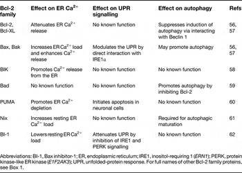
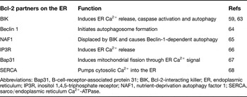
Because the Bcl-2 family proteins regulate MOMP, much research has been focused on the mitochondrial events and the downstream activation of caspases. However, there is growing realisation that some of the newer and investigational cancer drugs work by inflicting a different kind of damage to cancer cells as a way of killing them. Two of these processes – the ER stress response and macroautophagy – involve other membrane systems. Significantly, members of all three classes of the Bcl-2 family are associated with the ER, where they have been implicated in regulating these and other processes (Table 1). These functions are probably mediated by other proteins that Bcl-2 family members bind to at the ER (Table 2). Therefore, any pharmacological interference with Bcl-2 function will affect these processes as well, and we discuss here two that are relevant to cancer and its treatment.
Proposed functions of known ER-localised Bcl-2 family proteins

Abbreviations: BI-1, Bax inhibitor-1; ER, endoplasmic reticulum; IRE1, inositol-requiring 1 (ERN1); PERK, protein kinase-like ER kinase (E1F2AK3); UPR, unfolded-protein response. For full names of other Bcl-2 family proteins, see Box 1.
Bcl-2 binding partners on the ER and their corresponding function

Abbreviations: Bap31, B-cell-receptor-associated protein 31; BIK, Bcl-2-interacting killer; ER, endoplasmic reticulum; IP3R, inositol 1,4,5-triphosphate receptor; NAF1, nutrient-deprivation autophagy factor 1; SERCA, sarco/endoplasmic reticulum Ca2+-ATPase.
ER stress and Bcl-2
The ER is the main site for protein folding, lipid biosynthesis and calcium storage in the cell, and disturbances of any of these functions lead to ER stress (Ref. Reference Szegezdi56). The specific signalling pathway that senses excess unfolded-proteins in the ER is called the unfolded-protein response (UPR). Most normal cells are not subject to this form of stress and their UPR pathway is inactive, suggesting that selective inhibitors of the UPR may not affect normal cells. However, tumour cells are often under stress due to conditions of hypoxia, glucose deprivation and misfolded mutated proteins, and therefore these cells rely on the UPR for survival. ER stress is prone to occur in cancer tissues either directly or as a result of the tumour microenvironment (Ref. Reference Moenner69), and some cancers derived from secretory cells with high protein synthesis, such as multiple myeloma, are especially susceptible (Ref. Reference Carrasco70).
Once activated, there are three effector arms of the UPR that have the net effect of increasing chaperone proteins in the ER to help with folding, and globally decreasing translation to decrease the protein load entering the ER (Refs Reference Xu, Bailly-Maitre and Reed71, Reference Kim72, Reference Ron and Walter73). Bax and Bak located at the ER are required for the function of IRE1 (ERN1), one of the three effectors of the UPR. IRE1 is a transmembrane ER-resident protein that oligomerises upon release by ER chaperones when unfolded proteins accumulate, and its cytoplasmic domain binds to the same pocket formed by BH1–3 in Bax/Bak that mediates binding to BH3-only proteins. Sustained activation of IRE1 is required to keep the cell alive during the UPR (Ref. Reference Lin74); hence disrupting the interaction with Bax/Bak would have the net effect of cell death. It is unknown whether either endogenous BH3-only proteins or the BH3-peptide mimetic anticancer drugs in development will disrupt this interaction, but specific agents designed for this might be particularly useful for treating those cancers with ongoing UPR activation.
Autophagy: another stress response modulated by the Bcl-2 family
Autophagy is a major intracellular process for the degradation and recycling of proteins and cytoplasmic damaged organelles. Autophagy is essential for maintaining cell survival following a variety of extracellular and intracellular stimuli, including starvation. Autophagy has been reported to initiate cell death in response to intracellular damage caused by hypoxia, chemotherapeutic agents, virus infection or toxins. Here we give a brief overview of how autophagy is mechanistically related to apoptosis in general, and how it represents another pathway that can be modulated by Bcl-2-interacting drugs; for a detailed discussion of autophagy in tumour development and cancer therapy, see Ref. Reference Rosenfeldt and Ryan75.
Unlike apoptosis, which is characterised by nuclear condensation and fragmentation without major ultrastructural changes in cytoplasmic organelles, autophagy is a caspase-independent process characterised by the accumulation of autophagic vacuoles in the cytoplasm, with extensive degradation of the Golgi complex, the polyribosomes and the ER preceding destruction of the nucleus. The final effector phase involves the fusion of the autophagosome-enclosed cargo with lysosomes for catabolism by intralysosomal enzymes. Thus the induction of MOMP by activated Bax or Bak is not required for autophagic cell death.
Recent studies suggest pre-autophagosomal vesicles are initially generated at an ER-associated location, the omegasome, which represents a specialised, phosphatidylinositol 3-phosphate (PtdIns3P)-enriched ER membrane platform for assembly of a phagosomal initiation complex (Ref. Reference Axe76). This platform is created by recruitment to the ER of vesicles containing Vps34 (PIK3C3), a class III phosphoinositide 3-kinase (PI3K) that then binds to Beclin 1 (BECN1) and other accessory proteins to generate a functional PI3K complex (Ref. Reference Kihara77). Beclin 1 is therefore necessary to trigger autophagy and functions as a haploinsufficient tumour suppressor.
Beclin 1 was first identified as a binding partner for Bcl-2 located at the ER (Refs Reference Liang78, Reference Pattingre79), and indeed Beclin 1 has recently been identified as a BH3-only protein. Canonical BH3-only proteins such as Bad have been shown to displace Beclin 1 from Bcl-2 (Ref. Reference Maiuri59), leading to the proposal that a signalling cascade analogous to the release of BH3-only activator proteins by BH3-only sensitisers to initiate the apoptosis pathway by Bax/Bak activation also extends to autophagy (Ref. Reference Levine, Sinha and Kroemer80). Indeed, one of the consequences of using BH3-peptide mimetic drugs is to trigger autophagy in an appropriately primed cell (Ref. Reference Maiuri59). This might be particularly relevant for obatoclax, which enhances the steroid-induced autophagic cell death of acute lymphoblastic leukaemia cells by displacing Beclin 1 from Mcl-1 (Ref. Reference Bonapace81). Furthermore, the ability of obatoclax to elicit Bax/Bak-independent cell death by autophagy might explain the apparent nonselective cytotoxicity previously reported for this compound (Ref. Reference Vogler36).
Because autophagy can be cytoprotective in some cancer cells exposed to chemotherapy, joint inhibition of autophagy and promotion of apoptosis may be especially effective. Chloroquine is a currently available drug that inhibits that last step of autophagy, and many new trials are under way combining chloroquine with conventional cancer drugs (Ref. Reference White and DiPaola82), as well as newer targeted therapy (Ref. Reference Bellodi83) that elicits apoptosis indirectly. Extending this logic, therapy with the BH3-peptide mimetic agents and anti-autophagy drugs might be an especially interesting combination to test in the future.
Moving Bcl-2 antagonists into the clinic
After almost 20 years of basic biochemical and cell biology research into sorting out the details of apoptosis and its critical role in cancer, and identifying ways in which it can be manipulated, is it ready for prime time in the clinical arena? These are very early days, because with very few exceptions the published clinical material on the two agents currently being tested is still in abstract form.
Confirmation of activity of ABT-263 and obatoclax in experimental systems
The mechanism of action of ABT-263/737 and obatoclax as BH3-peptide mimetics against purified proteins has been confirmed in cell lines, as both drugs displace Bax and BH3-only proteins such as Bim from antiapoptotic proteins, as assessed by immunoprecipitation and western blotting, and cause the classical hallmarks of apoptosis in cells such as cytochrome c release from mitochondria and caspase activation. As a further test, the enantiomer of ABT-263 is much less active in binding and cell viability assays.
As a prelude to clinical studies, ABT-263 (or ABT-737) and obatoclax have been studied and are shown to be effective in physiologically relevant systems such as primary patient samples or mouse xenograft models, either as monotherapy or in combination with other drugs. Important clinical insights have been gained from ABT-737 treatment of a mouse model of lymphoma using overexpressed MYC either alone or co-expressed with Bcl-2, Mcl-1 or Bcl-w. As predicted from in vitro binding data, ABT-737 reduced tumour growth and killed lymphoma cells when Bcl-2, but not Mcl-1, was overexpressed (Ref. Reference Whitecross84). However, it was ineffective against tumours overexpressing Bcl-w despite the in vitro binding data. Because Bcl-w adopts an unusual conformation in its final membrane topology in cells (Ref. Reference Hinds85), this confirms the view stated above that some potential targets will be hidden (or exposed) by membrane binding of the functional forms of the protein. Furthermore, there was a distinct gradient of sensitivity of cells to the cytotoxic effect of ABT-737, depending on their Bcl-2 status. Tumour cells overexpressing MYC and Bcl-2 were ten times more sensitive than those that overexpressed MYC alone, even though the latter cells must have developed a secondary shut-off to apoptosis often involving Bcl-2. Thus, different sequences of development of the same tumour in vivo will lead to different levels of oncogene addiction and susceptibility to BH3-peptide mimetics, something that is likely to be borne out clinically.
Early clinical results with ABT-263 and obatoclax
The maximum tolerated doses for both obatoclax and ABT-263 have been determined in Phase I/II clinical trials based on dose-limiting toxicities that would be unusual for conventional chemotherapy agents. Euphoria and confusion have been observed with obatoclax, which is related to the delivery rate of infusion and has led to a suggested dose of 28 mg/m2 over 3 h every 3 weeks (Ref. Reference O'Brien86). It is unclear whether this represents an expected effect of pan Bcl-2 inhibition. By contrast, the dose-limiting effect for ABT-263 is entirely consistent with its mechanism of action. Bcl-XL antagonises Bak to extend platelet lifespan (Ref. Reference Mason87). When ABT-263 inhibits Bcl-XL in platelets, caspase activation with subsequent externalisation of platelet inner-membrane phosphatidyl serine occurs, which is the primary signal for clearance of aged platelets by the reticuloendothelial system (Refs Reference Tse88, Reference Elmore89). Thus, administration of ABT-263 to mice (Ref. Reference Shoemaker90) and to patients in the Phase I trials (Refs Reference Wilson91, Reference Roberts92, Reference Wilson93) caused dose-dependent thrombocytopaenia. However, by modifying the clinical schedule with a lower lead-in dose (7 days of 100 mg followed by 21 days of 250 mg), thrombopoietin-mediated upregulation of megakarypoiesis occurs and the thrombocytopaenia is decreased (Ref. Reference Roberts92).
In these heterogeneous groups of advanced-stage, mostly heavily pretreated patients with CLL, a respectable overall response rate was observed for both agents of 7/26 patients for obatoclax (Ref. Reference O'Brien86) and 36/85 patients for ABT-263 (Refs Reference Wilson91, Reference Khaw94). For both drugs, spectacular deep responses were noted in a few patients: a complete response (CR) in an acute myeloid leukaemia patient treated with obatoclax (Ref. Reference Schimmer95), and nodal CR in bulky CLL after ABT-263 (Ref. Reference Wilson93). A preliminary analysis of response determinants indicated that the classic poor-prognosis markers in CLL – 17p13 or 11q22.3 deletion – did not predict poor response, whereas baseline Mcl-1 expression was inversely correlated with decrease in lymphocyte count after treatment with ABT-263. The intriguing observation was made that CLL lymphocytes persistent in the patient after several cycles of ABT-263 retained in vitro sensitivity, suggesting the upregulation of Bcl-2-independent antiapoptotic pathways in vivo, possibly through adhesion-based or paracrine survival signals. It is hoped that this will be a rare finding in less heavily treated patients, or amenable to other drugs combined with Bcl-2 antagonists.
Bcl-2 antagonists as drugs: the next big thing, or much ado about nothing?
The preclinical data indicate that the use of BH3-peptide mimetics may introduce a novel paradigm to treat a broad range of haematological malignancies and selected solid tumours, especially considering that the toxicities observed to date seem manageable and largely do not overlap with those of both standard and newer targeted anticancer drugs for use in combination. However, high expectations have been thwarted frequently in the past – witness the relatively few cancers that are strongly addicted to single druggable tyrosine kinases (Ref. Reference Knight, Lin and Shokat96) and the less than spectacular early results with angiogenesis inhibition (Ref. Reference Schmidt14). Furthermore, there may be better ways to target the Bcl-2 family than those agents currently being tested, and toxicity and efficacy may be considerable with combination therapy. With the very high interest in this approach as indicated by the numerous clinical trials initiated (any many further no doubt planned), we should be able to determine whether these drugs represent a true breakthrough in cancer treatment.
Acknowledgements and funding
The work in the authors' laboratory was supported by grant FRN12517 from the Canadian Institute of Health Research (CIHR), a Tier I Canada Research Chair in Membrane Biogenesis to D.W.A., and with funds from the Ontario Institute for Cancer Research TFRI-OICR Selective Therapies Program-PRAR Award. We thank the peer reviewers for their helpful comments and constructive criticism, which assisted in the preparation of this review.