Transcatheter closure of patent ductus arteriosus is a well-established procedure and has become the standard of treatment with high success rate for the vast majority of patients. Reference Baruteau, Hascoet and Baruteau1 – Reference Santoro, Giordano and Gaio3 The Amplatzer duct occluder I device is the most widely used device with excellent results. Reference Pass, Hijazi, Hsu, Lewis and Hellenbrand4 – Reference Masura, Tittel, Gavora and Podnar6 Although it has a safe profile, protrusion of the device to the descending aorta, in various degrees that complicate with coarctation of the aorta, may occur especially in small infants. Reference Saliba, El-rassi and Helou7 – Reference Backes, Kennedy and Locke10 Thus, this situation may require further interventions. Reference Backes, Kennedy and Locke10 , Reference Choi, Song, Kim, Lee, Huh and Kang11 There are some techniques performed by institutes and operators individually to reduce the risk of device protrusion or to correct the protruded device, such as balloon repositioning of the device Reference Saliba, El-rassi and Helou7 , Reference Masri, El Rassi, Arabi, Tabbakh and Bitar8 or supporting the device by a pigtail catheter during release. Reference Buys, Greig and Brown12
To our knowledge, there has been no report about balloon-assisted device releasing technique in the literature. The aim of this study is to present our experience with this technique and its benefits on reducing the risk of device protrusion and complications related to protrusion.
Methods
Definitions
The term “balloon-assisted device releasing technique” was defined for the technique that the patent ductus arteriosus occluder device is released simultaneously with inflation of a low-profile balloon just adjacent to the device in descending aorta. Balloon repositioning was defined for pushing the device with an inflated low-profile balloon from the aortic side towards the pulmonary side. Device protrusion was defined for the condition in which the device was visibly protruding into the lumen of the descending aorta or pulmonary artery on angiography or echocardiography. Jumping of the device was defined for distinctive movement of the occluder device towards the aorta immediately after releasing of the device.
Study population and indications for patent ductus arteriosus closure
One hundred and fifty-five infants, whose type A duct, according to Krichenko classification, Reference Krichenko, Benson, Burrows, Moes, McLaughlin and Freedom13 had percutaneously closed by an Amplatzer duct occluder I device between January, 2012 and December, 2018, were retrospectively analysed. All patients were under 2 years of age and had a haemodynamically important duct. Anti-congestive treatment was started for symptomatic small infants (left heart chamber enlargement, pulmonary hypertension, failure to thrive, and clinical symptoms of congestive heart failure). In the situation where the symptoms could not be controlled by medication, the transcatheter closure was performed if the patient was >5.0 kg in weight, whereas a surgical ligation was offered to those under 5.0 kg. Percutaneous patent ductus arteriosus closure was performed beyond 1 year of age or 10 kg in weight in asymptomatic patients.
The balloon-assisted device releasing technique was used in 20 cases (group 1, 12.9%), between January, 2015 and December, 2018. Procedures in which the balloon-assisted device releasing technique had been used was compared with the remaining ones (group 2, 87.1%, n = 135) with regard to device stabilisation, device protrusion, iatrogenic coarctation, and device embolisation. The demographic, clinic, echocardiographic, and other angiographic data were also compared between the groups. As we use double disc devices such as Amplatzer duct occluder II or atrial septal occluder device for Krichenko classification type B ducts and Amplatzer vascular plugs or Amplatzer duct occluder II additional sizes for Krichenko classification type C ducts and use the balloon-assisted device releasing technique only for Krichenko classification type A ducts as an intuitional policy, we only included the procedures of Krichenko classification type A ducts that were closed with Amplatzer duct occluder I to obtain a more homogeneous study population.
Catheterisation procedure and indications for balloon-assisted device releasing technique
Cephazolin, at a dose of 50 mg/kg, was given 30 minutes prior to the catheterisation. All procedures were performed under deep sedation or general anaesthesia. Intravenous heparin, at a dose of 50 units per kg, was administered after femoral artery and vein cannulation. Activated clotting time level was kept above 200 seconds during the procedure. Descending aorta angiograms were made in lateral and right anterior oblique projections. The device size was selected as pulmonic end of the occluder shank to be at least 1.5–2.0 mm larger than the narrowest diameter of the duct.
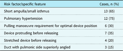
The balloon-assisted device releasing technique was used in infants with suspected increased risk for protrusion of the device to the aorta and complications related to that. The presence of pulmonary hypertension, short ampulla, small isthmus or descending aorta, or ducts with superiorly angled pulmonary side was considered as risky procedures for aortic protruding of the device. The stretched appearance of the device on fluoroscopy just before releasing was accepted as another risk factor of device protrusion to the aorta. In addition to the risk factors mentioned above, the balloon-assisted device releasing technique was used if the aortic retention disc did not fix on the rim of the ductal ampulla and not opposed to the ampulla properly or hanged on the lumen of the descending aorta before releasing. Protruding of the device into the aorta before releasing was also accepted as another indication for using the technique. Moreover, the technique was used if the occluder device was not able to be positioned precisely in the duct without giving an undue tug because in that situations, an attempt to pull the device into the ductal ampulla to avoid aortic obstruction may be needed and this manoeuvre could result with sudden displacement of the device into the aorta just after releasing.
A low-profile balloon (Tyshak II) was inflated in the descending aorta just adjacent to the device, while the device was opened but still screwed to the delivery cable. The size of the balloon was selected according to the isthmic and descending aorta diameter and the aortic disc diameter of the duct occluder device to just fit the aorta and hold the device at a stable position. The balloon was inflated using an indeflator device without exceeding the balloon’s nominal pressure. The duct occluder device was released after the balloon had been inflated, then the balloon was deflated. Final angiogram was performed 5–10 minutes after releasing the device to determine its position and residual shunting. Pressure measurements were done to determine the gradient in the aorta across the device in the case of device protrusion. Pulmonary angiogram was performed if device-related pulmonary artery stenosis was suspected.
Follow-up
Clinical examination and echocardiographic evaluation were made in all patients the day after the procedure and during the first, third, and sixth months after the procedure.
Statistical analysis
The statistical analyses were performed using Statistical Package for Social Sciences Software, version 21 (SPSS Inc., Chicago, IL, USA). Demographic and clinical variables were considered as descriptive statistics. Categorical variables were summarised as absolute frequency and percentage, whereas continuous variables were summarised as median, mean, and standard deviation. Paired t-tests were used to compare baseline and post-procedural numerical variables. Chi-squared test was used to compare ordinal and categorical variables.
Results
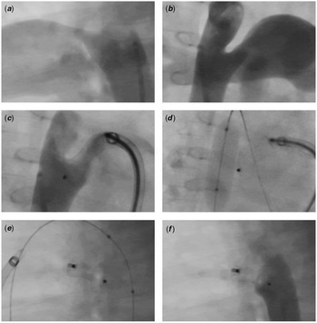
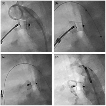
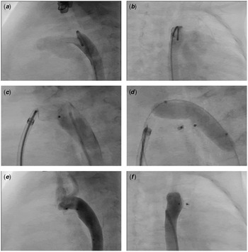
Group 1 consists of 20 patients, and 65.0% (n = 13) of them were females. Mean age at the time of catheterisation was 1.14 ± 0.43 years, and mean weight was 8.48 ± 1.47 kg. All cases had Krichenko classification type A duct and was closed by an Amplatzer duct occluder I device. The mean narrowest ductal diameter was 3.25 ± 1.15 mm, and the average mean pulmonary artery pressure was 29.30 ± 11.43 mmHg. The case-specific features that caused the concern for device protruding and related complications so the balloon-assisted technique was used are shown in Table 1. The sizes of the devices that were used for ductal closure were 06/08 mm (n = 12, 60%), 08/10 mm (n = 6, 30%), 04/06 mm (n = 1, 5%), and 10/12 mm (n = 1, 5%). Tyshak II balloon was used for all balloon assistance. The most used balloon size was 8 × 20 mm (58.3%) (Figs 1 and 2). The device was kept at a stable position in all, and there was no protrusion of the device to the descending aorta except one, in whom the device was already protruding to the aorta before releasing (Fig 3). In this particular patient with small isthmus, the aortic disc of the device was markedly protruding into the aortic isthmus and causing a gradient of 8 mmHg before device release. Therein, the balloon-assisted device releasing technique was used, and the device was kept in stable position and the mild protrusion regressed. There was a 5 mmHg pressure gradient in the aorta after releasing, and no further intervention was required. There was no device embolisation, procedure-related complication, or pulmonary artery stenosis due to the device in any patient of this group. The mean fluoroscopy time was 7.86 ± 4.78 minutes. Follow-up course was uneventful in all patients.
The specific features of patients during catheterisation in group 1.

Angiography and fluoroscopy images of a 1-year-old female child. Initial descending aorta angiograms in left lateral ( a ) and right anterior oblique ( b ) projections. ( c ) The device implanted into the duct protrudes to small-sized isthmus in right anterior oblique projection. Note the stretching of the 06/08 mm Amplatzer Duct Occluder I device before releasing. ( d ) A 8 × 20 mm Tyshak II balloon is inflated in descending aorta. ( e ) The device seems well fixed after releasing. ( f ): Final angiogram made in left lateral projection.

Angiography and fluoroscopy images of a 1.5-year-old female child. ( a ) Descending aorta angiogram made in left lateral projection shows protrusion of the aortic disc of the 06/08 mm before releasing. ( b ) A 8 × 20 mm Tyshak II balloon is inflated in descending aorta. ( c ) The device seems well fixed after releasing. ( d ) Final angiogram made in left lateral projection.

Angiography and fluoroscopy images of a 5-month-old female child. Initial descending aorta angiograms in left lateral ( a ) and right anterior oblique ( b ) projections. ( c ) Descending aorta angiogram made in left lateral projection shows protrusion of the aortic disc of the 06/08 mm before releasing. ( d ) A 8 × 30 mm Tyshak II balloon lying from transverse arcus to descending aorta is inflated. Final angiogram made in left lateral ( e ) and right anterior oblique ( f ) projections.

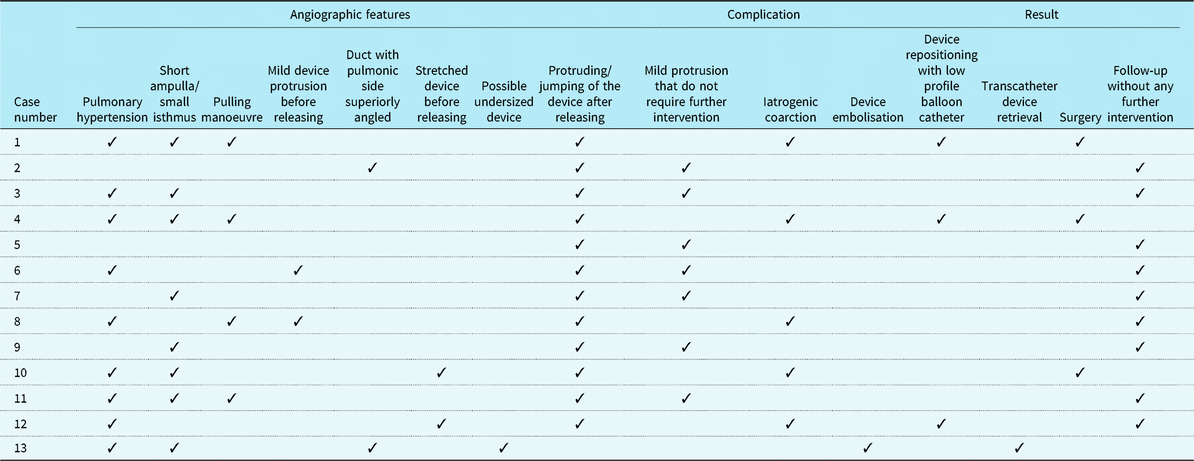
Group 2 consists of 135 patients, and 60.7% (n = 82) of them were females. Mean age at the time of catheterisation was 1.39 ± 0.87 years, and mean weight was 8.75 ± 1.97 kg. All had Krichenko classification type A duct and had been closed by Amplatzer duct occluder I device. The mean narrowest ductal diameter was 3.61 ± 1.08 mm, and the average mean pulmonary artery pressure was 23.51 ± 7.83 mmHg. The angiographic and procedural features of the all cases with complication (n = 13/135, 9.6%) in group 2 and final results of those procedures are detailed in Table 2. In this subgroup, the mean age at the time of catheterisation was 1.01 ± 0.57 years, mean weight was 7.91 ± 1.91 kg, and mean ductal diameter was 3.05 ± 1.37 mm. The sizes of the devices that were used for ductal closure were 06/08 mm (n = 7, 54%), 08/10 mm (n = 3, 23%), 04/06 mm (n = 2, 15%), and 10/12 mm (n = 1, 8%). The balloon-assisted device releasing technique was not applied in any of these cases. Jumping or protruding of the device towards aorta after releasing was observed in 12 of the patients (8.8%) and device embolisation in one (0.7%). In cases with device jumping or protrusion, seven patients (58.3%) had a pressure gradient of below 10 mmHg in aorta across the device, while five (41.7%) had a pressure gradient of equal or more than 10 mmHg. Balloon repositioning of the device was performed in three of these cases, and in one of them, repositioning was successful with decrease in pressure gradient, stabilisation of the device, and no requirement of further interventions. Two patients had surgical device removal and ductal ligation due to device-related aortic coarctation. The intra-operative and post-operative courses of both cases were uneventful. One patient with a residual pressure gradient of 10 mmHg was taken to close echocardiographic and clinical follow-up without any additional intervention. No evidence of coarctation was observed during the follow-up, and there was a decrease in systolic flow velocity obtained by echocardiography on the sixth month of outpatient visit (1.7 m/second) when compared with early post-procedural echocardiographic evaluation (2.5 m/second). Early device embolisation occurred in one patient. The device was snared via antegrade approach, and a bigger device was implanted following the same procedure. Protruding of the device into the pulmonary artery was observed in two patients, but both had no important pressure gradient obtained by catheterisation; thus, no additional interventions were required, and the echocardiographic follow-up was uneventful. The mean fluoroscopy time was 7.21 ± 6.84 minutes. Follow-up course was uneventful in all patients.
The angiographic and procedural features of the cases with any complication and final results, in group 2.

Comparing the two groups, no significant difference was observed with regard to gender, age, weight, and ductal diameter (p > 0.05), while mean pulmonary artery pressure was significantly higher in group 1 (p > 0.05). There was no significant difference with regard to age, weight, ductal diameter, and devices used between group 1 and the subgroup consisting of 13 cases with complication in group 2 (p > 0.05). There was a significant high ratio of device protrusion and iatrogenic coarctation in group 2 when compared with group 1 (p < 0.001). The mean fluoroscopy time did not differ significantly between the groups. However, it was significantly increased in patients with device protrusion in group 2, which had required additional interventions such as balloon repositioning or snaring of the device (mean = 12.36 ± 4.3 minutes), when compared with both groups (p < 0.001), as expected.
Discussion
Percutaneous transcatheter closure of patent ductus arteriosus is widely accepted as the gold standard treatment in infants, children, and adults. Reference Baruteau, Hascoet and Baruteau1 , Reference Pass, Hijazi, Hsu, Lewis and Hellenbrand4 , Reference Backes, Rivera and Bridge14 Despite increasing experience and technological advances, protrusion of the device to the aorta still remains an important issue. Reference Ewert9 , Reference Dimas, Takao and Ing15 Apart from premature babies, Amplatzer duct occluder I device is the most commonly used device for transcatheter ductal closure procedures Reference Pass, Hijazi, Hsu, Lewis and Hellenbrand4 , Reference Liddy, Oslizlok and Walsh16 and there are many reports evaluating the safety and efficacy of the device with high success rates. Reference Baruteau, Hascoet and Baruteau1 , Reference Pass, Hijazi, Hsu, Lewis and Hellenbrand4 , Reference Masura, Tittel, Gavora and Podnar6 , Reference Behjati-Ardakani, Behjati-Ardakani, Hosseini and Noori17
The large aortic disc of the Amplatzer duct occluder I device may sometimes slope over the aortic lumen despite appropriate size in accordance to ductal shape and diameter. Additionally, jumping of the device may be seen after release. This “jumping” is likely due to the rigid delivery system of the device which is screwed for the deployment. The delivery cable causes tension and retraction on the device and pulls it in the direction of the pulmonary artery before releasing. Reference Epcacan, Bulut, Yucel, Ceviz and Celebi18 This mechanism may act as a slingshot after releasing, resulting with jumping of the device into the aorta. In fact, this situation usually does not cause significant obstruction, but on the other hand, the device may dislodge, cause iatrogenic coarctation, or device embolisation into the aorta may occur, which requires additional interventions and disrupts patient’s condition. Based on our experience, this situation is likely to occur in small infants with pulmonary hypertension, long or large ducts with short ampulla, small isthmus and descending aorta, and ducts with pulmonic side angled superiorly.
In a meta-analysis Reference Backes, Rivera and Bridge14 investigating 635 infants (post-natal age < 12 months) who underwent percutaneous ductal closure, it is emphasised that percutaneous ductal closure interventions are more complex in infants than in children and adults. In this meta-analysis, the malposition of the device was found to be in 1.5% of cases. Reference Backes, Rivera and Bridge14 Liddy et al Reference Liddy, Oslizlok and Walsh16 have compared the results of percutaneous ductus closure between Amplatzer duct occluder devices and reported the frequency of device protrusion as 9.0% for Amplatzer duct occluder I. Saliba et al Reference Saliba, El-rassi and Helou7 determined that device-related minor complications had occurred only in the Amplatzer duct occluder I group and that 4.1% of the patients, all weighing less than 10 kg, showed a mild aortic narrowing due to device protrusion into the aortic isthmus. One of those patients had an initial gradient of 20 mmHg; thus, the device position was partially adjusted by pushing it with a balloon catheter from the aorta, reducing the gradient to 10 mmHg. Choi et al Reference Choi, Song, Kim, Lee, Huh and Kang11 reported three patients that had iatrogenic coarctation (9.0%), and the Amplatzer duct occluder I device was the device used in two of these and both patients had undergone surgical device removal and ductal ligation. The group also suggested that the absence of ampulla and underlying arch hypoplasia might increase the risk of device protrusion. Abadir et al Reference Abadir, Boudjemline and Rey19 have reported that other than the small size of the aortic isthmus, the acute angle between the duct and the aorta, and the absence of aortic ampulla in patent ductus arteriosus types B and C may also be potential factors of device protrusion. Masri et al Reference Masri, El Rassi, Arabi, Tabbakh and Bitar8 analysed the relationship between ductal type and risk of device protrusion into the descending aorta in a retrospective cohort study. Therein, they reported that non-conical types of ducts, lacking a sufficient ampulla, are more likely to have the risk of protrusion of the aortic retention disc into the descending aorta. However, they used only Amplatzer duct occluder II devices in all cases. They also experienced balloon repositioning of the protruding aortic retention disc and remarked that it has no significant effect on degree of the protrusion.
Although Krichenko classification type A duct, in which the ampulla provides space for the placement of the device and prevents aortic disc protrusion, and the narrowing part allows fixation of the device, is suggested to be favourable for device implantation, Reference Saliba, El-rassi and Helou7 , Reference Ewert9 our experience in this study showed that device protrusion into the aorta may still occur even in conical-shaped ducts. In our study, we included only the conical-shaped ducts closed with Amplatzer duct occluder I in patients under 2 years of age and determined a protrusion into the aorta with a percentage of 8.8% in the group in which the balloon-assisted device releasing technique was not used. Five (3.7%) of these patients had significant pressure gradient in aorta. Balloon repositioning of the device was successful in one of these, while two patients required surgical intervention. In contrast, we observed a good device stabilisation and did not observe jumping of the device into the aorta in the procedures in which the balloon-assisted device releasing technique was used. Furthermore, the device was well stabilised, and there was no significant protrusion after releasing, even in a case with visible protrusion before releasing. The mean fluoroscopy time, and thus the exposed radiation, increases with the occurrence of device protrusion and related complications. However, the balloon-assisted device release technique significantly decreases these parameters by preventing device protrusion. On the other hand, an important disadvantage of this technique is that it adds additional cost to the procedure. That is why it will be a reasonable cause to use this technique if the Amplatzer duct occluder I device is the only device available to close the duct. Additionally, if there is a concern about risk of device protrusion, an alternative device to Amplatzer duct occluder I may be advisable if the cost of the balloon-assisted technique exceeds the cost of the alternative device. In fact, it would not be difficult to apply this technique for procedures of young children that carries risks for device protrusion, we cannot strongly suggest to use the technique for those children as the complication rate is lower in that group. Although many young children would have small aortic dimensions, which we suggest as one of the risk factors for device protrusion, it would not be reasonable to use balloon-assisted technique for those having this situation as the only risk factor for that age group.
Conclusion
Protruding of the Amplatzer duct occluder I device into the aorta during percutaneous duct closure may occur in infants. Although this situation usually does not cause significant stenosis in the aorta, it may still result in iatrogenic coarctation, dislodging, or embolisation of the device. Balloon repositioning of the protruded device may be beneficial in some cases. The balloon-assisted device releasing technique is a very efficacious technique, which provides a good device stabilisation and prevents protrusion of the device and protrusion-related complications, thus increasing the success rate of the procedure. This technique can be safely applied in selected cases with Krichenko classification Type A duct where the Amplatzer duct occluder I is used and carries the risk factors of device protrusion and related complications.
Acknowledgements
None.
Financial Support
This research received no specific grant from any funding agency, commercial or not-for-profit sectors.
Conflict of Interest
All of the authors declare no conflict of interest in connection to the submitted article.
Ethical Standards
All procedures performed were in accordance with the ethical standards of the institutional research committee and with the 1964 Helsinki Declaration and its later amendments or comparable ethical standards.
Informed Consent
Informed written consent from parents was obtained from all individual participants included in the study.