Introduction
Pericardial effusion is an abnormal amount or quality of fluid accumulation in the pericardial space. Reference Adler, Charron and Imazio1 In children, inflammatory aetiologies (idiopathic, bacterial, and viral infections), connective tissue disease, malignancies, and post-cardiac surgery effusions are the leading causes for pericardial effusion. Reference Adler, Charron and Imazio1–Reference Shakti, Hehn and Gauvreau4 The true incidence and prevalence of pericardial effusion are unknown. In a 10-year study by Roodpyema et al., Reference Roodpeyma and Sadeghian5 it was reported that 0.12% of hospital admissions were related to pericardial diseases and pericardial effusion was detected in 0.02–0.23% of hospital admissions. Reference Peter, Asani and Aliyu6,Reference Mehdizadegan, Mohammadi and Amoozgar7 In most of the patients, pericardial effusion is mild and regress spontaneously or with medical treatment. However, in case of tamponade, life-threatening haemodynamic deterioration may occur and require urgent intervention such as pericardiocentesis or surgical drainage of the fluid. In our study, we aimed to evaluate the clinical characteristics and aetiological distribution in patients who have large pericardial effusion and are relieved by pericardiocentesis.
Material and method
We reviewed our hospital and echocardiography records in a 10-year period (2010–2020) for large pericardial effusion relieved by pericardiocentesis in a single tertiary care centre. We identified 59 paediatric cases (0–18 years old) who underwent pericardiocentesis for large pericardial effusion. Due to the differences in the treatment and clinical characteristics of the patients, cardiac surgery-related effusions are excluded and the remaining 38 patients were included in the study. The demographic (sex, age, and body weight), laboratory (complete blood count, erythrocytes sedimentation rate, and C-reactive protein), and clinical data (complaints, aetiologic factor, and management strategy) of the patients were obtained from hospital records. Cardio-thoracic ratio from the telecardiogram was calculated. Size of the effusion was recorded as maximum diameter during diastole in long axis, apical-4 chamber, and subcostal views. The presence of signs of tamponade was also determined (Figure 1). The amount, colour, and appearance of fluid obtained by pericardiocentesis were recorded. The results of microbiological (microscopic examination, culture) and biochemical analysis of the fluid were evaluated. Data were analysed with a statistical programme, and descriptive statistics were given as mean ± standard deviation, minimum and maximum, and percentages. Since it was a retrospective study, informed family consent form could not be obtained. The study was approved by local Ethical Committee for Clinical Investigation (Date:19/04/2019, Decision number:2019-092).
Echocardiographic images of a patient with large pericardial effusion and cardiac tamponade demonstrating right atrial wall collapse (left) and fibrinous network in effusion (RA = right atrium; LA = left atrium).

Results
Thirty-eight children with large effusions who underwent pericardiocentesis are included in the study. The demographic and clinical characteristics of the patients are given in Table 1.
The demographic and clinical characteristics of the non-surgery related pericardial effusion patients

Of the patients, 26 were male with a male-to-female ratio of 2.16. Mean age of the patients was 69.4 ± 74.9 months and the mean body weight was 22.5 ± 22.4 kg during pericardiocentesis. Patients were divided into different age groups as follows due to differences in aetiological factors. According to age, 11 (28.9%) were under 3 months, 10 (26.3%) were between 3 months and 3 years, and 17 (44.7%) were over 3 years of age. The male-to-female ratio varied by age group and was 1.75, 0.66 and 7.5, respectively. Dyspnoea (51.7%) was the most common complaint, followed by chest pain (37.9%), weakness (13.8%), and cough (6.9%). The mean cardio-thoracic ratio on telecardiogram was 0.62 ± 0.07 (range 0.48–0.78).
The largest diameter of effusion was 24.4 ± 10.4 mm and 2.8 ± 2.8 mm/kg when indexed to body weight. Echocardiographic signs of tamponade were present in nine (23.7%) of the patients. The amount of fluid drained with pericardiocentesis was 279.24 ± 279 ml and 12.63 ± 6.2 ml/kg when indexed to body weight. Macroscopic appearance of the fluid was serous in 12 (34.3%), haemorrhagic in 18 (51.4%), purulan in four (11.4%), and chylous in one (4.8%) patient.
Laboratory examination revealed erythrocyte sedimentation rate and C-reactive protein value of 50 mm/hr and 46.4 mg/L, elevated at 79% and 63.2%, respectively. Mild anaemia was present in 21.1% and white blood cell was increased in 23.7%. Mean haemoglobin, white blood cell, and thrombocyte counts were 11.8 ± 2.5 g/dL, 12.3 x10⁶/µl, and 315.8 x10⁶/µl, respectively. Pericardial fluid examination was positive for viral serology (Enterovirus) in one patient and mycobacterial culture and polymerase chain reaction (PCR) in one patient. Pericardial fluid bacterial culture was negative.
Aetiology for pericardial effusion was infectious in 26%, idiopathic in 18%, iatrogenic in 11%, rheumatological in 11%, malignancy in 8%, cardiomyopathy in 8%, and other factors related in 18% (Figure 2).
Aetiologic distrubition of the patients.

Infectious aetiology was present in 10 patients. Viral serology, culture, and PCR examination demonstrated Enterovirus, Mycobacterium, and Chlamidia in three patients. Pericardial fluid was purulan in three patients without bacterial culture growth. Sepsis and necrotising enterocolitis were present in two premature newborn. Two patients were also accepted as viral infection with clinical findings; however, viral examination was not studied. Rheumatological disease was present in four patients and three had neoplastic diseases. Pericardial effusion was due to iatrogenic causes in four patients; central venous catheter implantation-related pericardial effusion in three newborns and post-resuscitation pneumopericardium and pericardial effusion in one patient. Two patients had non-immune hydrops fetalis, and one patient each had congenital myopathy, unidentified neurometabolic disease, intestinal lymphangiectasia, Gorham-Stout syndrome, and congenital nephrotic syndrome. Seven (18.3%) patients were accepted as idiopathic after initial examination.
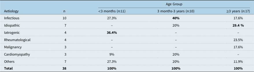
When aetiologic factor was evaluated according to age groups, iatrogenic causes were the most common in patients under 3 months, infectious causes in those between 3 months and 3 years, and idiopathic causes in those over 3 years (Table 2).
Aetiologic distribution of the patients according to age group

Appearance of the fluid in all of the patients with malignancy and iatrogenic effusion and 75% of the rheumatologic disease and 44% of idiopathic cases were haemorrhagic. Fluid was chylous in one patient with Gorham-Staut syndrome.
Of the 38 patients, 16 received nonsteroidal anti-inflammatory drugs (NSAID), and colchicine and steroids were added in nine and two patients, respectively. Empirical antibiotics were given to 32 patients. Surgical drainage of the fluid was performed in two of the patients. A total of eight (21%) patients died during follow-up: three due to sepsis, two due to cardiomyopathy and heart failure, two due to hydrops fetalis, and one due to severe lower respiratory tract infection. Six of these patients were born prematurely and were less than 3 months old.
Discussion
Pericardial effusion is asymptomatic in most patients, and regresses spontaneously; however, moderate and severe pericardial effusion can progress to cardiac tamponade, which causes life-threatening haemodynamic deterioration and may require urgent pericardiocentesis or surgical drainage. More urgent intervention is required in children due to the lower tolerance to ventricular stroke volume reduction compared to adults.
In a multicentre study by Bolin et al. including 9902 patients with pericardial effusion, pericardiocentesis was performed in 15% of the patients. Reference Bolin, Tang and Lang8 Mehdizadegan et al. reported that pericardial effusion was present in 0.23 % of all hospital paediatric admissions. Of these patients, 32.6% had moderate to large pericardial effusion having tamponade signs in 2% and pericardiocentesis were performed in 14.1%. Reference Mehdizadegan, Mohammadi and Amoozgar7 In our study, signs of tamponade were present in 23.7% of our patients.
Male-to-female ratio was found 2.16, which varied according to age group with a prominent male predominance (male to female:7.5) over 3 years of age. Shakti et al. reported a male-to-female ratio of 2.45 in patients who developed pericardial effusion due to idiopathic and viral pericarditis, while it was around one in the preadolescent age and approximately five in the adolescent age group. Reference Shakti, Hehn and Gauvreau4 In our study, in accordance with Shakti et al. it was 7.5 in patients over 3 years of age. In light of these findings, it was determined that the probability of its occurrence in the male gender increased with increasing age in the paediatric age group. Although the cause is not clear, it is thought that sex hormones may have an effect on viral concentration, immune response, and susceptibility to disease, as in adolescent men with myocarditis.
In our study, the mean age of the patients was 14 months and 21% of the patients were in the neonatal age group with a body weight varying between 0.8 and 80 kg. In the literature, the median age of the patients with pericardial effusion was reported as 10–11 years. Reference Bolin, Tang and Lang8,Reference Abdel-Haq, Moussa and Farhat9 Especially the presence of premature patients and the absence of any significant complications related to the technique showed that percutaneous pericardiocentesis can be performed safely in all age groups by providing a suitable environment.
Considering the complaints at the time of admission in our study, at least one complaint was reported in 89.7% of the patients. The most common complaints were respiratory distress (51.7%) and chest pain (37.9%). In the literature dyspnoea, chest pain and fever were reported as leading symptoms. Reference Mehdizadegan, Mohammadi and Amoozgar7,Reference Ozturk, Cansaran and Saygi10 It should be kept in mind that atypical findings such as fatigue and abdominal pain may be associated with pericardial effusion and tamponade.
The classical triad of tamponade is hypotension, jugular venous engorgement, and decreased heart sounds; however, very few patients have all three of these findings. Reference Stolz, Valenzuela and Situ-LaCasse11 In the study by Akthar et al. including 36 patients with moderate to severe pericardial effusion, it was reported that tamponade findings were present in 39%. Reference Akhtar and Malik12 Pulsus paradoxus increases the risk of tamponade 3.3 times in the presence of pericardial effusion; Reference Roy, Minor and Brookhart13 however, its evaluation may not be reliable, especially in a young and agitated child. Hearing deep heart sounds and jugular venous engorgement has 28% and 76% sensitivity, respectively. Reference Roy, Minor and Brookhart13–Reference Spodick15 One of the earliest signs of tamponade, tachycardia, was detected in 47% of our patients; it has been reported to have a sensitivity of 77%. Reference Roy, Minor and Brookhart13 Non-specific findings or unexplained haemodynamic disturbance may be observed in newborn patients. Reference Nowlen, Rosenthal and Johnson16 Considering that 21% of the patients were in the neonatal period, evaluation with echocardiography is recommended, especially in the presence of unexplained findings in this period.
The earliest echocardiographic sign of haemodynamic deterioration in cardiac tamponade is collapse of the right atrium in early systole and the right ventricle in early diastole (Figure 1). Collapse in any cardiac cavity has 90% sensitivity and 65% specificity for tamponade. Reference Mercé, Sagristà-Sauleda and PermanyerMiralda17 In our study, due to the different age groups of patients, these measurements were also evaluated by proportioning to body weight. Haponiuk et al. defined the “Pediatric tamponade index (PTI)” by proportioning the amount of fluid drained for 24 hours with the inserted chest tube to body weight. They suggested that the initial clinics of patients with paediatric tamponade index > 21 were more severe and may be associated with a longer hospital stay and higher inotropic requirement after pericardiocentesis. Reference Haponiuk, Kwasniak and Chojnicki18
The appearance of pericardial fluid was haemorrhagic in 51.4% of patients. Haemorrhagic effusion has been related to trauma, malignancy, vascular tumour, tuberculosis, bleeding diathesis, vasculitis, uraemia, aortic dissection, and iatrogenic causes. Reference Spodick19 Although rare, it may also develop after chlamydial infection as in our case . Reference Tenenbaum, Heusch and Henrich20
In children, large pericardial effusion can develop as a finding of many local or systemic diseases. While tuberculosis is still the most common aetiology in developing and underdeveloped countries, viral and idiopathic pericarditis are more common in developed countries. Reference Peter, Asani and Aliyu6,Reference Bagri, Yadav and Agarwal21,Reference Khanal, Gajurel and Sahi22 When our patients were examined according to aetiology, 44% were classified as inflammatory (idiopathic and infectious), 11% rheumatological disease, 11% iatrogenic causes, 8% malignancy, 8% cardiomyopathy, and 18% other. In retrospective study by Bolin et al. Reference Bolin, Tang and Lang8 and K FC;hn et al., Reference Kuhn, Peters and Marx3 idiopathic and neoplastic causes were leading aetiologies. Reference Bolin, Tang and Lang8 The reason for this difference is thought to be that our study only included large effusions and pericardiocentesis data.
In the study by Bolin et al., it had been suggested that undetermined viral infections may be responsible for idiopathic pericardial effusion due to the fact that mortality and re-admission rates were found to be similar in idiopathic and viral aetiologies. Reference Bolin, Tang and Lang8 When classified together, the viral and idiopathic groups constituted 26% of our patients. Patients with purulent effusions thought to be bacterial but no bacterial growth in blood or pericardial fluid cultures were observed to have had a history of antibiotic use before admission, and therefore the causative agent was not detected. In these patients, bacterial infection was diagnosed based on the appearance of the effusion and supporting clinical and laboratory tests. Mok et al. reported that 38% of patients with severe pericardial effusion had bacterial infection, and in 48% of these, no causative agent could be detected. Reference Mok and Menahem2 In an adolescent patient with pericardial effusion due to chlamydial infection, a simultaneous pleural effusion was observed, and improvement was achieved with antibiotic treatment. Two siblings in our study, whose aetiology could not be determined despite detailed history and examinations and one of whom had recurrent effusion, were accepted as having recurrent idiopathic pericarditis and were followed up with colchicine treatment.
Tuberculosis remains as one of the main causes of haemorrhagic pericardial effusion, especially in developing countries, accounting for approximately 1–2% of cases of acute pericarditis, with tamponade reported in 7% and mortality rate varying between 14 and 40%. Reference Peter, Asani and Aliyu6,Reference Khanal, Gajurel and Sahi22 G FC;ven et al. Reference Guven, Bakiler and Ulger23 from our country and Akhtar et al. Reference Akhtar and Malik12 reported that tuberculosis was shown as the causative agent in 30% and 11.1% of the patients with pericardial effusion, respectively. Diagnosis is made by direct detection of M. tuberculosis in tissue or fluid culture, histopathological observation of bacilli or granulomas, observation of granulomas in the pericardium in patients with a history of extracardiac tuberculosis, and response to tuberculosis treatment. Tuberculosis growth in pericardial fluid occurs in 1/3 of the cases. Reference Cherian24 In addition, studies have reported that elevated adenosine deaminase in pericardial fluid with a value of 63–117 U/L is 100% sensitive and 83.3% specific in the diagnosis. Reference Mathur, Tiwari and Trikha25 In our study, the M. tuberculosis was shown by both culture and PCR results in one patient and pericardial fluid adenosine deaminase level was found as 81.1 U/L. This patient died during follow-up due to disseminated involvement and sepsis.
Neoplastic diseases were detected in 8% of the patients diagnosed with carcinoid type neuroendocrine tumour, T-cell lymphoblastic lymphoma, and B-cell acute lymphoblastic lymphoma. Although the malignancy rate was found to be 24% in the multicentre study by Bolin et al., Reference Bolin, Tang and Lang8 in studies including only patients who underwent pericardiocentesis, it was reported as 7–20% . Reference Ozturk, Cansaran and Saygi10,Reference Akhtar and Malik12 Medary et al. reported that the most common diagnoses in patients with malignancy who developed pericardial tamponade during follow-up and underwent pericardiocentesis were acute leukaemia and lymphoma. Reference Medary, Steinherz and Aronson26
Mild fibrinous pericarditis or mild effusion is usually present in vasculitis or connective tissue diseases, but rarely massive effusion may be seen. Rheumatological diseases were detected in four (11%) patients, systemic lupus erythematosus and familial mediterranean fever (FMF) in two patients each. FMF, which is relatively common in our country, was detected at a rate of 15%. Reference Ozturk, Cansaran and Saygi10
The prognosis of pericardial tamponade in newborn patients is poor and is usually associated with heart failure, fetal restrictive cardiomyopathy, concomitant malformations, chromosomal abnormalities, fetal infections, intrauterine growth retardation, non-immune hydrops fetalis, erythroblastosis fetalis, and central and peripheral venous catheters. Reference Haponiuk, Kwasniak and Chojnicki18 In our study, 11 patients under 3 months of age, seven of whom were premature, underwent pericardiocentesis in three due to central catheter-related complications, two due to non-immune hydrops, two due to perforated NEC and sepsis, one due to respiratory distress syndrome and sepsis, one due to congenital myopathy-related heart failure, one due to hypertrophic cardiomyopathy, and one due to pneumopericardium and effusion after cardiopulmonary resuscitation. Pericardial effusion and tamponade constitute 0.7% of central catheter-related complications. It develops on average 3 days after the procedure and can develop on the same day as the procedure or even 112 days after the procedure . Reference Nowlen, Rosenthal and Johnson27 Although the aetiology of pericardial effusion in these patients is not clear, it has been suggested that damage to the endothelium may occur as a result of repeated traumas at the catheter tip, thrombus formation at the catheter tip as a result of adhesion and activation of platelets, and osmotic damage due to infusion may occur as a result of the tip of the catheter touching the heart and has also been reported in patients with a normal umbilical venous catheter position. Reference Onal, Saygili and Koc28,Reference Bentata, Hamdi and Chemlal29 Pericardiocentesis plays a role in these patients both as an emergency and definitive treatment. In the study by Novlen et al., mortality was reported as 8% in patients who underwent pericardiocentesis and 75% in those who did not. Reference Nowlen, Rosenthal and Johnson27
A patient who presented with tamponade and was found to have chylohemorrhagic pericardial effusion, multiple lytic lesions, and compression fractures in the vertebrae was diagnosed with Gorham-Stout syndrome with lymphatic drainage problems.
Pericardiocentesis was performed in one patient for pericardial effusion due to chronic renal failure. The aetiology of uraemic pericarditis is multifactorial. Albuminuria seen in these patients is associated with inflammatory abnormalities and pericarditis develops in the low inflammatory process as a result of increased endothelial permeability due to albuminuria. Reference Abdel-Haq, Moussa and Farhat9,Reference Bentata, Hamdi and Chemlal29
Treatment varied depending on the underlying disease. Although NSAIDs are primarily preferred in treatment, colchicine has recently become widely used in acute and recurrent pericarditis. Double-blind and placebo-controlled adult studies have shown that colchicine use reduces recurrences, duration of symptoms, and hospitalisation rates. Reference Imazio, Brucato and Cemin30 In our centre, colchicine use has increased in parallel with the trend in the literature. No significant complications were observed in our clinic due to colchicine use. It was observed that two patients (11.1%) with continuing pericardial effusion despite medical treatment underwent surgical pericardial tube insertion and pericardial window operation.
Of the 38 patients who underwent pericardiocentesis, eight (21%) died during follow-up. In the study by Bolin et al., mortality was reported as 8.2% in all pericardial effusion cases . Reference Bolin, Tang and Lang8 Six of our patients who died were under 3 months of age; two were diagnosed with hydrops fetalis, two with perforated NEC and sepsis, one with dilated cardiomyopathy, and one with hypertrophic cardiomyopathy. Although the mortality rate was high in our study, it was thought that this was due to the age group distribution of the patients and the inclusion of only patients with large pericardial effusion.
Although the most common approach for pericardial fluid drainage is subxiphoid percutaneous intervention, minimally invasive surgical intervention is also used in some centres. Reference Haponiuk, Kwasniak and Chojnicki18 In our centre, pericardiocentesis and/or pericardial catheter placement with a subxiphoid percutaneous approach is preferred. Although the number of newborns and low body weight patients who were more prone to complications in our study was high, we think that the percutaneous approach is more appropriate because no serious complication related to the procedure was observed and the success of the procedure was high. In case of surgical intervention, it seems to be a disadvantage to intervene in patients under operating room conditions and accompanied by general anaesthesia. We think that surgical intervention is more appropriate in patients who require a pericardial window due to the locality of the collection, the presence of widespread fibrous structures and the recurrent nature of the collection.
Limitations
The limitations of our study are that it is retrospective, that it includes only the group in which pericardiocentesis was performed among the pericardial effusion cases, and that some clinical and laboratory diagnostic tests cannot be accessed from the hospital medical records.
Conclusion
In this study, in which we evaluated the pericardiocentesis findings and demographic characteristics of patients with moderate and severe pericardial effusion, it was seen that percutaneous pericardiocentesis can be applied safely when necessary, and the underlying aetiology is decisive in the prognosis of the patient. Although pericardial effusion in children is often due to inflammation of the pericardium due to viral and bacterial factors, it can develop as a finding of many local or systemic diseases that should be kept in mind.
Author contributions
VD, FD: study concepts/study design, writing and data analysis, SKC, MB, ZD: data acquisition or data analysis/interpretation, IUS: literature research, All authors: manuscript drafting or manuscript revision for important intellectual content, approval of final version of manuscript.
Financial support
The authors have no relevant financial or non-financial interests to disclose.
Competing interests
The authors have no conflicts of interest to declare that are relevant to the content of this article.