Introduction
Since the introduction of the genus Minutoexcipula (Atienza & Hawksworth Reference Atienza and Hawksworth1994), three additional species have been described so that it now comprises: Minutoexcipula tuckerae (op. cit.), M. tuerkii (Hafellner, Reference Hafellner1994), M. mariana (Atienza Reference Atienza2002) and M. calatayudii (Atienza Reference Atienza2002).
Diederich (Reference Diederich2003) accepted nine Lichenodiplis species adding the species with simple conidia that had been previously placed in Laeviomyces by Hawksworth (Reference Hawksworth1981). Four of the species treated by Diederich (Reference Diederich2003) have 1-septate conidia and so conform to the original generic concept of Lichenodiplis (Hawksworth & Dyko Reference Hawksworth and Dyko1979): Lichenodiplis lecanorae, L. lichenicola (Hawksworth & Dyko, op. cit.), L. poeltii (Kondratyuk Reference Kondratyuk and Wasser1996) and L. hawksworthii (Berger & Diederich Reference Berger and Diederich1996).
There is a strong superficial similarity between Minutoexcipula and Lichenodiplis, and Diederich (Reference Diederich2003, Reference Diederich, Nash, Ryan, Diederich, Gries and Bungartz2004) pointed out the difficulties in distinguishing these two lichenicolous genera. However, there are fundamental differences in the complexity of the conidiogenous cells, presence of conidiophores, and further in the entirety of the exciple. The present paper describes and compares two new conidial lichenicolous fungi, one belonging to the genus Lichenodiplis, L. crespoae growing on Cyphelium notarisii, and the other to the genus Minutoexcipula, M. tephromelae growing on Tephromela atra, in order to underline their similar morphological characteristics and to emphasize their significant differences. As exemplified by these two new species, both merit retention as morphological genera, although molecular data could be valuable to resolve this conclusively.
Materials and Methods
The material was examined with standard microscopic techniques using a Leica DM 2500 microscope fitted with a drawing tube. Conidia measurements were made in water and their size ranges recorded as a range with the averages in italics and the number of spores measured indicated by ‘n’. Measurements outside the main ranges are given in parentheses. Specimens were photographed with the DFC320 Leica digital camera for both a Leica MZ 16 dissecting microscope and Leica DM 2500 microscope.
The following specimens were examined for comparison:
Lichenodiplis hawksworthii: Austria: Ober Österreich: Donautal, Schlögener Schlinge, Carpinus Unterhangwald kilometer 2185, on Pertusaria pustulata, alt. 340 m, 17 July 2002, F. Berger (Flechtenherbar Berger 16986).
Lichenodiplis lecanorae: Spain: Teruel: Cedrillas: Nacimiento del río Mijares, 30TXK9676, on Caloplaca hungarica on Pinus sylvestris, alt. 1500 m, 21 May 1993, V. Atienza & S. Fos, (VAL-Lich 26943).
The Species
Lichenodiplis crespoae Pérez-Ortega & V. Atienza sp. nov
MycoBank no.: MB 513062
Lichenodiplis lecanorae similis, sed differt praecipue conidiis minoribus et cellulis conidiogenis minoribus latioribus. Fungus in thallis lichenis Cyphellium notarisii vigens. Conidiomata globosa, pycnidiiformia, non ostiolata, atrobrunnea, 50–60(–100) μm diam. Conidiophora desunt. Cellulae conidiogenae cilindriformis vel ampuliiformis, 3–4·5–6 × (2·5–)3–4 μm diam. Conidia, singularia, holoblastica, pallida brunnea, ellipsoidea, basaliter truncata, 1-septata, 4–5·5(–6) × (1·5–)2–2·5(–3) μm.
Typus: Spain, Castilla y León, Valladolid, Pedradas de Santisteban, camino hacia la Virgen de Sacedón, 41° 19′ 12,7″N 4° 36′ 36,6″ W, on Cyphelium notarisii, on Pinus pinea, 1208 m alt., 18 September 2005, S. Pérez-Ortega 26942 (VAL-Lich.—holotypus).
(Figs 1 & 2)
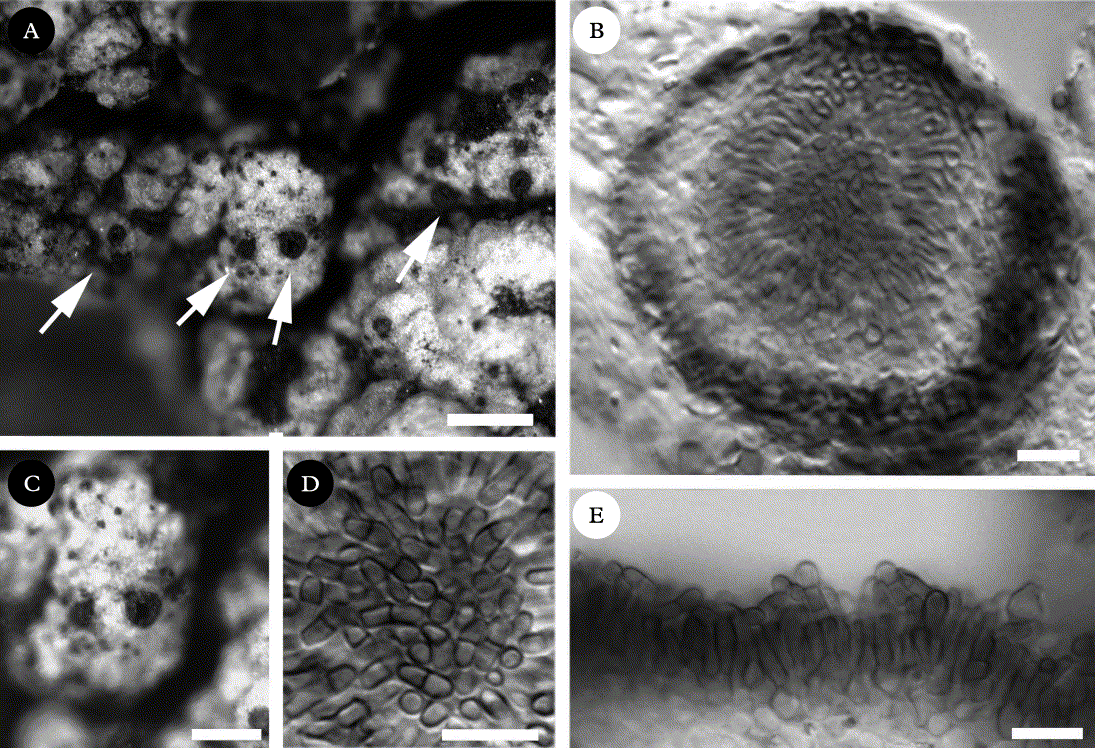
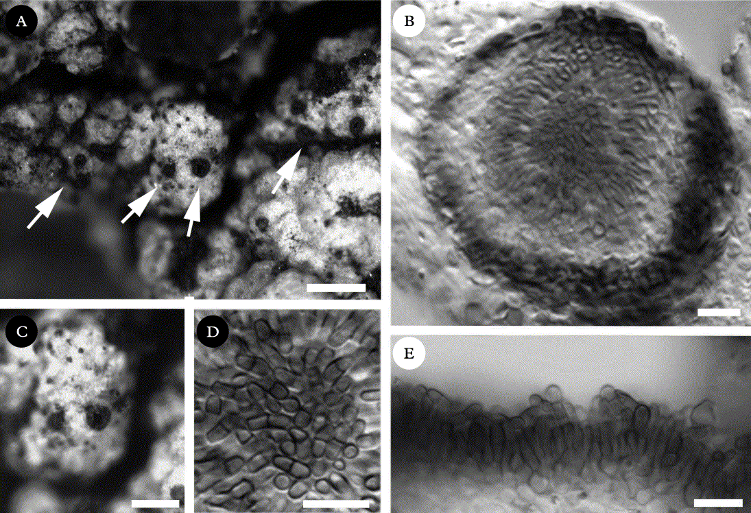
Lichenodiplis crespoae (holotype). A, habitus, arrows indicate conidiomata on Cyphelium notarisii thallus; B, cross section of a conidioma; C, detail of a conidioma in host thallus showing the wide and irregular ostiole of the conidiomata; D, conidia aggregation within the conidioma; E, detail of conidioma wall showing conidiogenous cells. B, D & E in lactophenol blue. Scale bars: A = 300 μm; C = 30 μm; B, D & E = 10 μm.
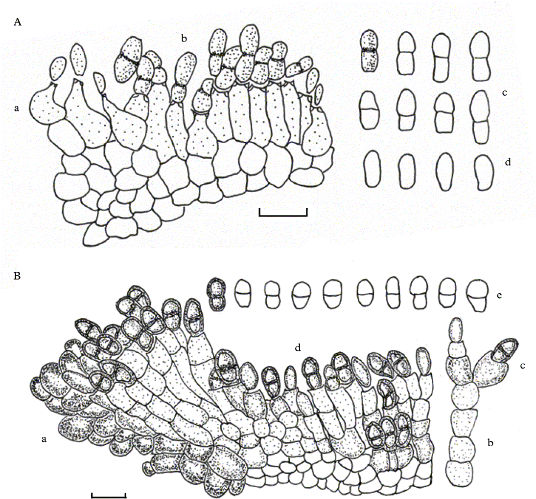
Vertical sections of conidiomata. A, Lichenodiplis crespoae (holotype) showing conidiogenous cells (a), young and mature conidia (b), eight conidia outlines with surface pigmentation in one (c) and four young conidia (d); B Minutoexcipula tephromelae (holotype) showing detail of exciple-like structure (a), conidiophores (b), conidiogenous cells, some bearing 1–2 extensions (c), young pale and mature dark conidia (d) and detail of one mature conidium and outlines of several conidia (e). Scales A & B = 10 μm.
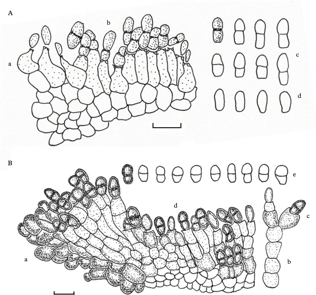
Conidiomata pycnidial, arising singly, semi-immersed mainly in the upper cortex of the thallus of the host, Cyphelium notarisii, but sometimes also in the host ascomata, black, 50–60(–100) μm diam., globose at first, not ostiolate, with an irregular opening becoming semiglobose (cup-shaped) because of the disintegration of the upper half of the conidiomatal wall. Conidial wall 10–20 μm thick, in vertical section composed of 4–5 layers of pseudoparenchymatous cells, cells isodiametric to irregularly polyhedral, the outermost layer brown with thick walls, the inner layers hyaline with thinner walls, cells (2·5–)3–4(–4.5) μm diam., around the conidiomata opening the cell walls being elongated and swollen with dark brown and unevenly thickened and more intensely pigmented tips. Conidiophores absent. Conidiogenous cells arising from the inner wall layer of the conidioma, lining the conidioma cavity, proliferation percurrent and enteroblastic, with 0(–1?) annellations, pale brown, smooth, subcylindrical to shortly ampulliform, mainly 3–4·5–6 × (2·5–)3–4 μm diam. Conidia arising holoblastically and singly, acrogenous, dry, abundant, the youngest aseptate, pale brown, smooth, mostly ellipsoid, with the apex rounded and the base truncated, brown, 1-euseptate when mature, sometimes constricted at the septa 4–4·7–5·5(–6) × (1·5–)2–2·2–2·5(–3) μm, (n=12), length/breadth ratio 2–2·7, without a gelatinous sheath.
Etymology. The epithet ‘crespoae’ honours Professor Ana Crespo de las Casas as a 60th birthday tribute for her continued support of young researchers in lichenology in Spain.
Distribution and ecology. So far, Lichenodiplis crespoae is known only from the original collection from Valladolid in central north-west Spain. It grows on Cyphelium notarisii, a rare lichen species which grows on dry wood, and occasionally on the bark of coniferous and deciduous trees in the Northern Hemisphere (Areskoug & Thor Reference Areskoug and Thor2005), where it behaves as a cool-temperate to southern boreal-montane species (Nimis & Martellos Reference Nimis and Martellos2008; http://dbiodbs.univ.trieste.it/). Apart from Valladolid, C. notarisii is known from only seven different localities in the Iberian Peninsula (Sarrion et al. Reference Sarrión, Aragón and Burgaz1999; Hawksworth Reference Hawksworth2004; Fos & Atienza Reference Fos and Atienza2007; Pérez-Ortega Reference Pérez-Ortega2007; J. Etayo unpublished). Cyphelium notarisii collections from four of these localities, Sra de El Toro in Castellón, Sierra de Guadarrama, and Sa de Albarracín in Madrid, Soria and Teruel, have been checked, albeit without success, for the new Lichenodiplis species.
Notes. Lichenodiplis crespoae is distinguished by its most characteristic conidiomata (Fig. 1), which open irregularly at first, becoming cupulate at maturity by disintegration of the upper half. Lichenodiplis crespoae differs from the other 1-septate spored Lichenodiplis species mainly in the conidiomata wall being composed of brown cells with thick walls in the outermost layers, and the innermost layers of hyaline cells with thinner walls. The new species has shorter and narrower conidia, 4–4·7–5·5(–6) × (1·5–)2–2·2–2·5(–3) μm, length/breadth ratio 2–2·7, without an obvious basal frill, than the other Lichenodiplis species, whereas in the closest species, L. lecanorae they are 4–7·5 × 2–3 μm, length/breadth ratio 2–2·5, and sometimes have an evident marginal frill. However, examination by scanning electron microscopy would be necessary to determine whether even a rudimentary frill was present in L. crespoae. In addition, the conidiogenous cells, which are 3–4·5–6 × (2·5–)3–4 μm in L. crespoae, are shorter than those of L. lecanorae which measure 5·5–12 × 2–3 μm. Furthermore, in L. crespoae, the enteroblastic conidial proliferation develops with or without visible annellations, probably due to environment or age, which are more visible in other Lichenodiplis species. This is also the first Lichenodiplis species reported on Cyphelium notarisii; it mainly infects the cortex, but also occurs in the apothecia.
Minutoexcipula tephromelae V. Atienza, Etayo & Pérez-Ortega, sp. nov
MycoBank no.: MB 513064
Minutoexcipula mariana similis, sed differt praecipue cellulis conidiogenis sacciformis et conidiis brevioribus. Fungus in thallis lichenis Tephromela atra vigens. Conidiomata sporodochioidea superficialia, concava vel, atrobrunnea, 30–100(–150) μm diam, Conidiophora generaliter absentia, cylindrica, simplicia vel rarissimo ramosa, septata, (6–)10–11(–15) × (2·5–)3–4(–4·5) μm. Cellulae conidiogenae sacciformis, enteroblasticae, 3–4·5 × 3–5 μm. Conidia singularia, holoblastica, fusca, ellipsoidea basaliter truncata, 1-septata, 5–5·5(–6) × (2·5–)3(–4) μm.
Typus: Spain, La Rioja, Logroño, sierra de San Lorenzo, subida al pico de San Lorenzo, UTM 30TWM0276, on Tephromela atra on sandstones, 1950 m alt., 7 September 2004, S. Pérez-Ortega 26941 (VAL-Lich.— holotypus).
(Figs 2 & 3)
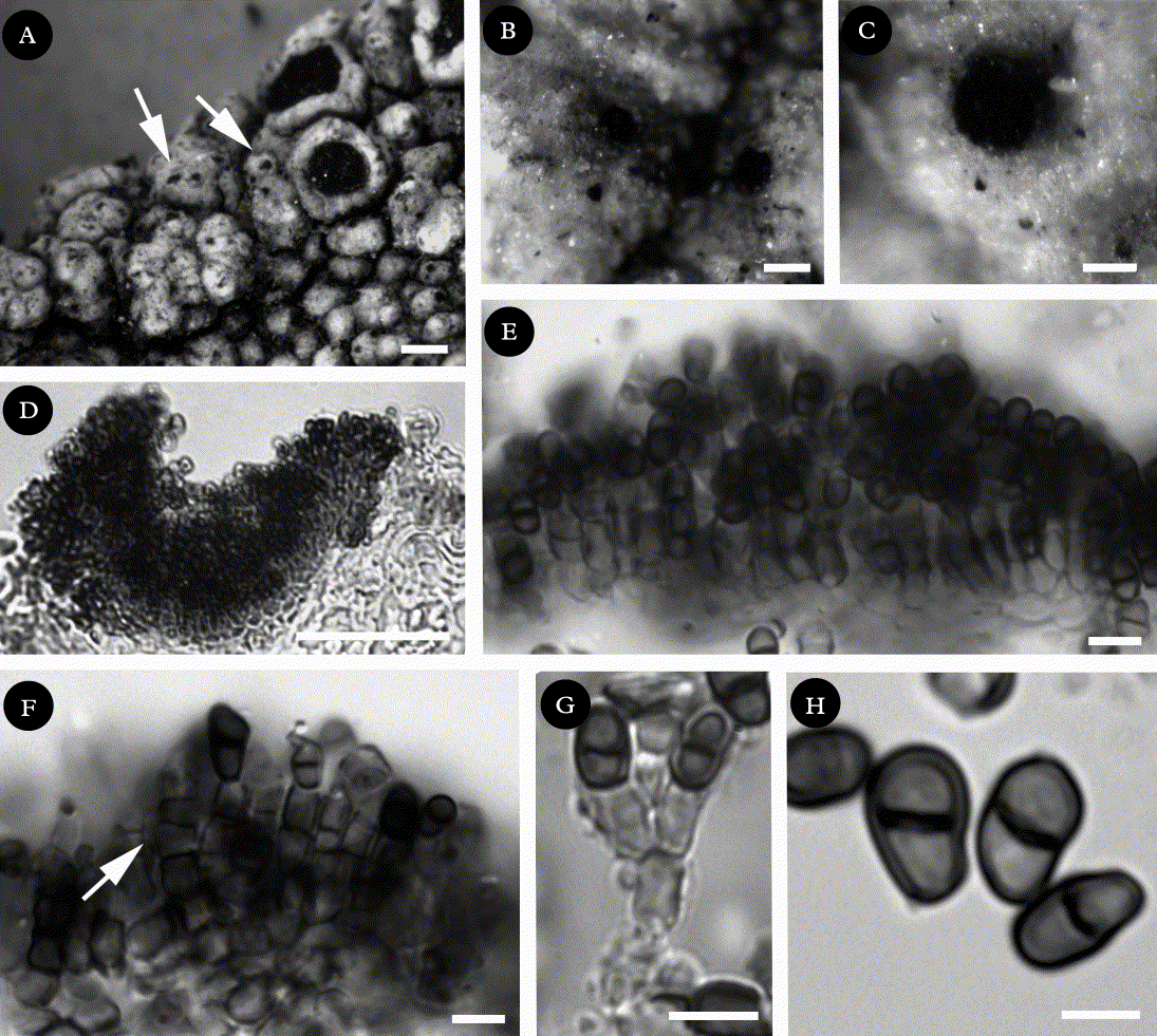
Minutoexcipula tephromelae (holotype). A, habitus, arrows indicating conidiomata on Tephromela atra thallus; B & C, detailed views of conidiomata habitus; D, cross-section of a conidioma in Tephromela thallus; E, conidioma cross-section showing conidiophores and cells at the base of conidioma; F, detail of conidiophores and conidiogenous cells, arrow indicates proliferations; G, detail of a branched conidiophore; H, conidia. D–G in lactophenol blue. Scale bars: A = 500 μm; B = 100 μm; C = 50 μm; D = 25 μm; E–G = 10 μm; H = 2·5 μm.
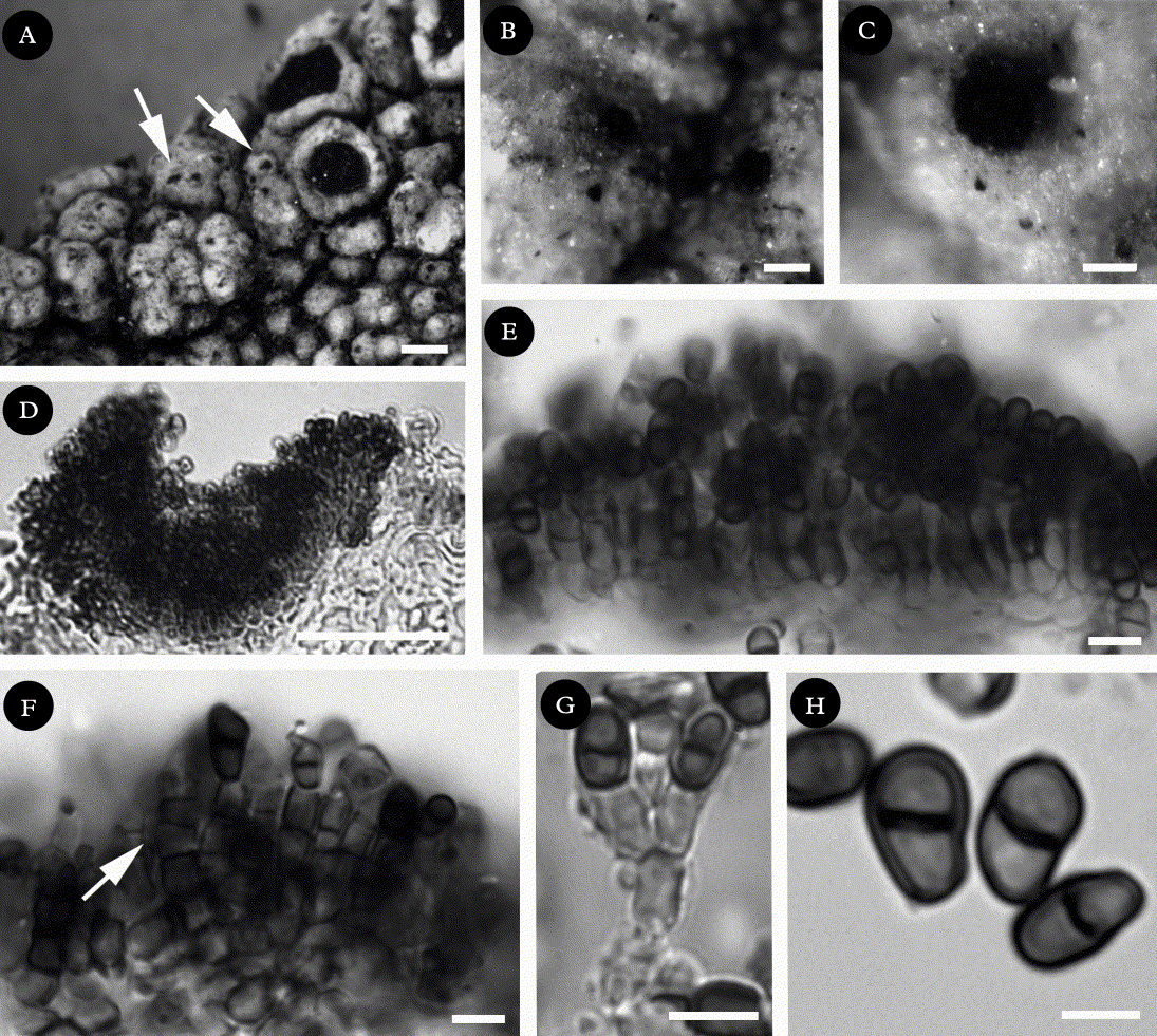
Conidiomata sporodochia-like, superficial, rounded to irregular shaped, plane to slightly concave, convex when mature and wart-like, arising from the upper cortex of the host lichen singly, scattered, rarely confluent, dark brown to black, granular, 30–100(–150) μm diam.; in section composed of a basal stromatic tissue formed by 3–4 irregular layers of pseudoparenchymatous cells; cells isodiametric to irregularly polyhedral, hyaline 10–20 μm diam., forming an exciple-like structure at the margins, with elongated, swollen dark brown, unevenly thickened and more intensely pigmented tips. Mycelium hyaline with poorly visible cells. Conidiophores mainly absent (not developed), in the central part of the conidioma but present at the margins of the conidioma; macronematous, erect, septate, formed by cubic to prismatic cells 3–4 × 3·5–5 μm, lower part brown, upper part paler, cylindrical, mainly (6–)10–11(–15) × (2·5–)3–4(–4·5) μm, constricted at the septa, mainly unbranched, although occasionally branched and bearing 2(–3) conidiogenous cells at the apex. Conidiogenous cells integrated, terminal, incorporated in the conidiophore or arising directly from the pseudoparenchymatous cells of the basal layer, proliferation percurrent and enteroblastic, with 0–2(–3) widely spaced annellations, pale brown, smooth, subcylindrical to subglobose or very shortly ampulliform (sack-like), mainly 3–4·5 × 3–5 μm. Conidia arising holoblastically and singly, acrogenous, dry, abundant, the youngest aseptate, pale brown to brown, smooth, sometimes spherical but mostly ellipsoid to obpyriform, with the apex rounded and truncated base, brown, 1-euseptate, when mature sometimes the upper cell swollen and the lower attenuated below, septum dark brown, walls darker near the septum, 5–5·5–6(–7) × (2·5–)3(–4) μm, (n=14), length/breadth ratio 1·7–2, without a gelatinous sheath.
Etymology. The epithet ‘tephromelae’ refers to the host lichen genus Tephromela.
Distribution and ecology. Minutoexcipula tephromelae grows on both areoles and the thalline margin of the ascomata of Tephromela atra. It appears to be commensalistic, as no damage was observed to the host.
Tephromela atra is a widespread taxon, occurring in both Hemispheres (Nimis & Martellos Reference Nimis and Martellos2008). It has been shown to be a good substratum for lichenicolous fungi, hosting no less than 13 species, which were recently studied by Hafellner (Reference Hafellner2007). Indeed, one of the specimens we studied (Etayo 18152) was also infected by an unknown Lichenostigma species not considered in Hafellner's key. To date, the new Minutoexcipula species is known only from three localities in La Rioja and Soria (N Spain) and perhaps one in Austria from information in Hafellner (Reference Hafellner2007) (see below).
Notes. Minutoexcipula tephromelae is characterized by open sporodochia-like conidiomata, which are plane to slightly concave at first but convex when mature, the size and shape of the mainly simple conidiophores, conidiogenous cells which are short-cylindrical to saccate and have long extensions between the annellations, and by the short and wide conidia (Fig. 3). It resembles M. calatayudii in its small conidioma size (30–100(–150) μm vs 75–90 μm diam. in the latter), and its hyaline stromatic cell walls, but M. calatayudii has longer conidiogenous cells and conidia, and grows on Hypogymnia tubulosa. Minutoexcipula tephromelae has wider conidiophores, (6–)10–11(–15) × (2·5–)3–4(4·5) μm, whereas in other Minutoexcipula species (notably M. tuckerae and M. mariana) the conidiophores are narrower, (8–)12·5–14(–19·5) × 2·5–3 μm and (10·5–)11·5–13(–15) × (1·5–)2–3 μm, respectively. The conidiogenous cells of M. tephromelae are very characteristic. They are pale brown, short-cylindrical to subglobose, 3–4·5 × 3–5 μm diam.; with some arising directly from the pseudoparenchymatous cells of the basal layer, as in M. calatayudii, but others being incorporated terminally in mainly simple to very short-branched conidiophores, especially those close to the exciple-like structure (as in M. mariana). The size of conidiophores is similar to those of M. calatayudii, which are a little longer (5.5–6.25 μm), hyaline, and ampulliform, but wider than in the other Minutoexcipula species (4·5–10 × 2–2·5 μm in M. tuckerae, 5–7(–10) × 2–2·5–3 μm in M. tuerkii, and 4·5–6·5 × 1·5–2 μm in M. mariana). In M. tephromelae, the conidiogenous cell wall extends upwards only occasionally and generally once only; if a new conidium develops, the cell wall elongates considerably and becomes widely spaced from the previous proliferation before the next conidium is formed. In all the other Minutoexcipula species, the proliferation of the conidiogenous cells produces a different number of close annellations, depending on their age. However, we did not see cells that produce these ‘extensions’ in all the material studied, and in such cases, confusion with Lichenodiplis species such as L. lecanorae could occur. The conidia of M. tephromelae, which are 5–5·5–6 (–7) × (2·5–)3(–4) μm, are shorter than in other species of the group, such as in M. tuckerae where they are (5–)6·5–8 × (2·5–)3–4 μm, length/breadth ratio 2–2·2, and in M. mariana where they are 6·5–7(–7·5) × (2·5–)3·7 μm, length/breadth ratio 1·9–2·8.
Hafellner (Reference Hafellner2007) questioned whether specimens of ‘Lichenodiplis lecanorae’ collected on Tephromela really belonged to that species because of the wider conidia, 6–8 × 3·5–4·5 μm and it seems likely that at least some of the specimens he identified as that species from this host belong to the newly described M. tephromelae.
Additional specimens examined. Spain: La Rioja: Logroño, Clavijo village, outcrop near the hermitage, on Tephromela atra on sandstones, 870 m alt., 9 March 2001, J.Etayo, (hb. Etayo 18152). Soria: Sierra de Montes Claros, Gallinero, 41°90′N 2°20′W, Quercus pyrenaica and Fagus wood, on Tephromela atra on sandstones, 1400 m alt., 22 November 1998, J. Etayo & E. Ros, (hb. Etayo 17154).
We thank David L. Hawksworth who kindly revised the manuscript for valuable discussions and improvements in the text and V.A. is grateful for his continuous support, without whom this work would not have been possible. Franz Berger is acknowledged for providing material of Lichenodiplis hawksworthii for comparison, M. Guara for his help in the Latin diagnosis and H. Warburton for revising the English text. This study was supported with funds from the Spanish CICYT CGL2007-66734-C03-01.