Introduction
Masson's pseudotumour or intravascular papillary endothelial hyperplasia is a benign, reactive lesion first described by the French pathologist Pierre Masson in 1923.Reference Steffen1 Intravascular papillary endothelial hyperplasia is a vascular endothelial proliferation typically confined to the lumen of thrombosed pre-existing vessels or vascular malformations.Reference Hong, Lee, Lee, Lee, Jun and Song2 It can lead to diagnostic uncertainty due to its close resemblance clinically, radiologically and histopathologically to angiosarcoma.
We present a diagnostically challenging case of intravascular papillary endothelial hyperplasia of the ethmoid sinus presenting with persistent epistaxis.
Case report
A 45-year-old female presented with a three-week history of recurrent brisk epistaxis. She was otherwise asymptomatic with no history of nasal obstruction or discharge. Nasendoscopic examination revealed a haemorrhagic, polypoidal mass between the right middle turbinate and septum.
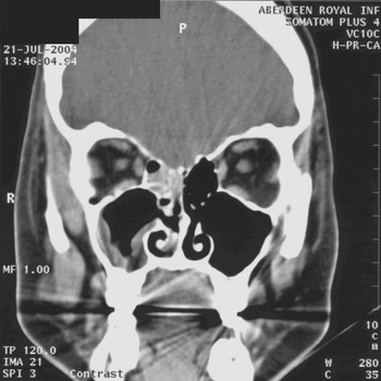
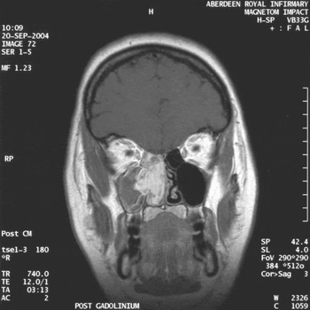
Computed tomography (CT) and subsequent magnetic resonance imaging (MRI) scans (Figures 1 and 2 respectively) revealed a soft tissue mass involving the right posterior ethmoid sinuses abutting the medial orbital wall. There was marked post-contrast enhancement on the MRI films and the appearances were thought to represent a vascular tumour. No extension into the orbit, skull base or sphenoid sinus was seen.
Coronal CT scan showing abnormal soft tissue in right posterior ethmoid.

T2 weighted MRI showing extensive abnormal soft tissue in right ethmoid complex and nasal cavity.

Examination under anaesthetic confirmed a bulky, friable haemorrhagic mass suspicious of sinonasal malignancy and biopsies were taken for histopathological examination.
Out of keeping with the clinical picture, histopathology suggested the lesion to be a non-tumorous lesion consisting of thrombosed blood vessels with recanalisation by smaller blood vessels.
In view of the high clinical suspicion of neoplasia a second biopsy was taken that was similarly reported as vascular, loose fibroconnective tissue with cleft-like spaces lined by endothelium associated with organising thrombus and blood, but also with prominent vascular channels out with areas associated with haemorrhage. After an interval of watchful waiting during which the patient had minor ongoing epistaxis repeat CT and MRI scans were performed demonstrating a marked increase in the size of the mass, raising the possibility of angiosarcoma in its pre-invasive state.
Elective resection was planned via a Denker's medial maxillectomy. Prior to surgery the patient was readmitted following several further episodes of brisk epistaxis and was observed on the ward. The haemoglobin remained stable, not requiring blood transfusion, and surgery was performed as planned.
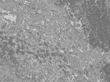
Histology revealed similar features within the biopsy and resection material. Strikingly haemorrhagic sinonasal mucosa presented numerous areas of vascular infiltration, with focal overlying ulceration. The vessels formed a sinusoidal network of interconnecting spaces lined by flattened or plump spindle cells with occasional normal mitotic figures and only mild atypia (Figure 3). Although the vascular areas did not obviously reside within a single large vessel, the appearances were entirely consistent with intravascular papillary endothelial hyperplasia although a pre-existing haemangioma could not be ruled out.
Histopathological appearance of soft tissue from right nasal cavity demonstrating intravascular papillary endothelial hyperplasia (Masson's pseudotumour) (H&E; ×200).

The patient made an uneventful recovery and has had no evidence of recurrent disease in 18 months of follow up.
Discussion
Intravascular papillary endothelial hyperplasia is a rare, benign reactive lesion, which can mimic angiosarcoma. There is a myriad of presentations dependent on the site and the extent of the disease. Intravascular papillary endothelial hyperplasia is described in the literature as arising at many different sites including intracranially,Reference Lee, Hui and Sitoh3 in the peri-orbita,Reference Werner, Hornblass, Reifler, Dresner and Harrison4 on the limbs and trunkReference Clifford, Temple, Jorda and Marecos5 and the head and neck region.Reference Tosios, Koutlas and Papanicolaou6 Although reports of intravascular papillary endothelial hyperplasia in the literature are rare there is a suggestion that these lesions have a tendency to bleed with subsequent haematoma formation.Reference Eichhorn and Rosenberg7, Reference Novak, Ferguson and Komorowski8, Reference Lee, Suh, Lim, Yang and Shin9
Intravascular papillary endothelial hyperplasia within the paranasal sinuses has only been described in two previous cases although we report the first arising from the ethmoid sinus. The presentation has been different in each case i.e. epistaxis in our case, nasal obstructionReference Moon, Chung and Hong10 or neurological and endocrine deficit due to sphenoid erosion.Reference Lancaster, Anderson, Sherman and Clark11
• This paper describes a case of Masson's pseudotumour or intravascular papillary endothelial hyperplasia presenting in the ethmoid sinus
• Intravascular papillary endothelial hyperplasia is a benign, reactive lesion first described by the French pathologist Pierre Masson in 1923
• The neoplasm can lead to diagnostic uncertainty due to its close resemblance clinically, radiologically and histopathologically to angiosarcoma
• Although surgery is usually necessary for cure a high index of clinical suspicion is needed to avoid surgical over treatment
In one pathological series, 10 of 44 cases of intravascular papillary endothelial hyperplasia arose in the head and neck region, the remainder arising on the fingers and trunk.Reference Clearkin and Enzinger12 There are two distinct forms of intravascular papillary endothelial hyperplasia: one form arising as a de novo lesion and the other as a focal condition in a pre-existing vascular process, such as pyogenic granuloma or haemangioma.Reference Kuo, Sayers and Rosai13 The microscopic appearances of our specimen were most consistent with the classically described de novo form demonstrating papillary proliferation of endothelial cells that was nearly always intimately associated with a thrombus, although it was difficult to entirely exclude a pre-existing haemangioma. Intravascular papillary endothelial hyperplasia seems to represent an exaggeration of the normal endothelial response to thrombosis. Whilst similar pathological features are often observed in angiosarcoma, the distinguishing features of intravascular papillary endothelial hyperplasia are that the proliferation occurs strictly within the vascular lumen, there is a lack of significant atypia and a benign clinical course.Reference Stewart and Smoller14
Although intravascular papillary endothelial hyperplasia is rare, it should be considered in the differential diagnosis of a vascular neoplasm of the nose and paranasal sinuses in order to prevent radical over treatment for a benign lesion.
Conclusion
Masson's pseudotumour or intravascular papillary endothelial hyperplasia is rare. The clinical, radiological and histopathological features may mimic angiosarcoma and although surgery is usually necessary for cure a high index of clinical suspicion is needed to avoid surgical over treatment.