Introduction
Anhydrous fluorosulfate minerals are few in number and very rare. Only seven natural hydrogen-free fluorosulfates without other species-defining anions have been described to date: kogarkoite Na3(SO4)F (Kogarko, Reference Kogarko1961; Pabst and Sharp, Reference Pabst and Sharp1973), grandreefite Pb2(SO4)F2, pseudograndreefite Pb6(SO4)F10 (Kampf et al., Reference Kampf, Dunn and Foord1989), thermessaite K2Al(SO4)F3 (Demartin et al., Reference Demartin, Gramaccioli, Campostrini and Orlandi2008), krasheninnikovite KNa2CaMg(SO4)3F (Pekov et al., Reference Pekov, Zelenski, Zubkova, Ksenofontov, Kabalov, Chukanov, Yapaskurt, Zadov and Pushcharovsky2012), kononovite NaMg(SO4)F (Pekov et al., Reference Pekov, Krzhizhanovskaya, Yapaskurt, Belakovskiy, Chukanov, Lykova and Sidorov2015), and shuvalovite K2(Ca2Na)(SO4)3F (Pekov et al., Reference Pekov, Zubkova, Britvin, Chukanov, Yapaskurt, Sidorov and Pushcharovsky2016). The last three mineral species, anhydrous fluorosulfates with species-defining both alkali and alkali-earth cations, are known only in high-temperature sublimates of active fumaroles at the Tolbachik volcano (Kamchatka, Russia). In this article we describe the fourth of such minerals, kantorite K2NaMg(SO4)2F.
Kantorite (Cyrillic: канторит) is named in honour of the Russian mineralogist Dr. Boris Zinovievich Kantor (1930–2022), a specialist in morphogenetic analysis of mineral crystals and aggregates (ontogeny of minerals). In particular, he made a significant contribution to the development of knowledge on the genesis of divergent crystals, spherulites of different types, and pseudo-stalactites (Kantor, Reference Kantor2003, Reference Kantor2021). Dr. Kantor was also a well-known mineral collector and made a great contribution to the popularisation of mineralogical knowledge and mineral collecting among amateurs in the Soviet Union and Russia in 1980s–2010s (Kantor, Reference Kantor1982, Reference Kantor1985, Reference Kantor1991, Reference Kantor2001, Reference Kantor2020).
Both the new mineral and its name have been approved by the Commission on New Minerals, Nomenclature and Classification of the International Mineralogical Association (IMA-CNMNC), as IMA2024–042 (Pekov et al., Reference Pekov, Britvin, Bulakh, Belakovskiy, Yapaskurt, Turchkova and Anikin2024). The accepted symbol is Kao. The holotype material is deposited in the systematic collection of the Fersman Mineralogical Museum, Moscow with the catalogue number 98804.
Occurrence and general appearance
Kantorite occurs at the Arsenatnaya fumarole located at the summit of the Second scoria cone of the Northern Breakthrough of the Great Tolbachik Fissure Eruption, Tolbachik volcano, Kamchatka peninsula, Far-Eastern Region, Russia. The Second scoria cone of the Northern Breakthrough of the Great Tolbachik Fissure Eruption is a monogenetic volcano formed in 1975 and still demonstrates strong fumarolic activity. The fumaroles here belong to the oxidising type and some of them are very richly mineralised (Fedotov and Markhinin, Reference Fedotov and Markhinin1983; Vergasova and Filatov, Reference Vergasova and Filatov2016; Pekov et al., Reference Pekov, Agakhanov, Zubkova, Koshlyakova, Shchipalkina, Sandalov, Yapaskurt, Turchkova and Sidorov2020). Arsenatnaya is a large, active oxidising-type fumarole. The temperature of gases, measured by us in the period 2012–2023, reached 500°C. It is a prolific mineral locality: more than 200 mineral species have been reliably identified here, including 72 new minerals. The general data on the Arsenatnaya fumarole and its mineral associations were reported by Pekov et al. (Reference Pekov, Koshlyakova, Zubkova, Lykova, Britvin, Yapaskurt, Agakhanov, Shchipalkina, Turchkova and Sidorov2018) and Shchipalkina et al. (Reference Shchipalkina, Pekov, Koshlyakova, Britvin, Zubkova, Varlamov and Sidorov2020).
Kantorite is one of the rarest minerals at the Arsenatnaya fumarole. It was found in only two specimens collected by us in July 2014 from a hot pocket located ∼1 m below the day surface. The temperature measured when collected in this area using a chromel–alumel thermocouple was ∼300°C. The minerals associated with kantorite are aphthitalite, langbeinite (including its Zn-bearing variety), arcanite, krasheninnikovite, vanthoffite, kononovite, wulffite, halite, sylvite, flinteite, fluoborite, chubarovite, johillerite, urusovite, zincite, tenorite, pseudobrookite, hematite, sanidine and fluorophlogopite.
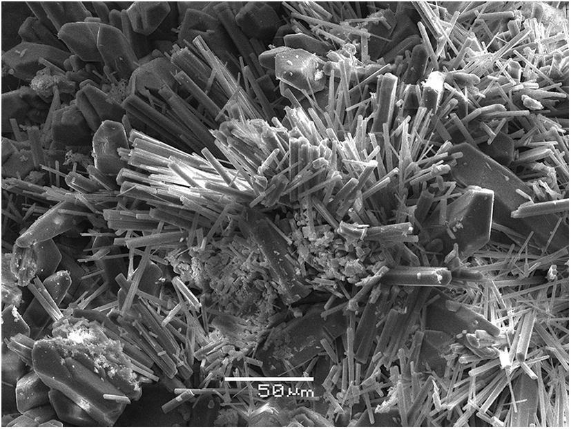
Kantorite occurs as long-prismatic to acicular crystals up to 0.1 mm long and up to 10 μm (typically up to 5 μm) thick, elongated along [100]. The main crystal faces, forming the prismatic zone, belong to the {0kl} zone; crystals are terminated by the pinacoid {100}. The crystals form near-parallel and bush-like open-work clusters up to 0.2 mm across, typically together with aphthitalite (Fig. 1), on the surface of basalt scoria altered by fumarolic gas.
Clusters of long-prismatic to acicular crystals of kantorite associated with pyramidal-prismatic (resembling typical quartz crystals in shape) aphthitalite crystals. SEM image, secondary-electron mode.

We suggest that kantorite was formed as a result of the interaction between hot volcanic gas and basalt scoria at the temperatures not lower than 300°C. Fumarolic gas is an obvious carrier of S, F, Na and K, whereas basalt can be a source of Mg which has low volatility in volcanic gases (Symonds and Reed, Reference Symonds and Reed1993).
Physical properties and optical data
Kantorite is transparent, colourless, with white streak and vitreous lustre. It is brittle. One direction of perfect cleavage was observed under both the optical microscope and the scanning electron microscope; based on the crystal structure data (see below), we suggest the (010) cleavage. The fracture is stepped. Density calculated using the empirical formula and unit-cell volume found from single-crystal X-ray diffraction (XRD) data is 2.498 g cm–3.
Kantorite is optically biaxial (+), α = 1.447(2), β = 1.449(2) and γ = 1.452(2) (589 nm). The angle 2V could not be measured due to small size of the crystals, their acicular habit and preferred orientation in immersion liquid prevented observation at the cross sections perpendicular to optical axes; 2V (calc.) = 79°. In transmitted plane-polarised light, kantorite is colourless and non-pleochroic.
Raman spectroscopy
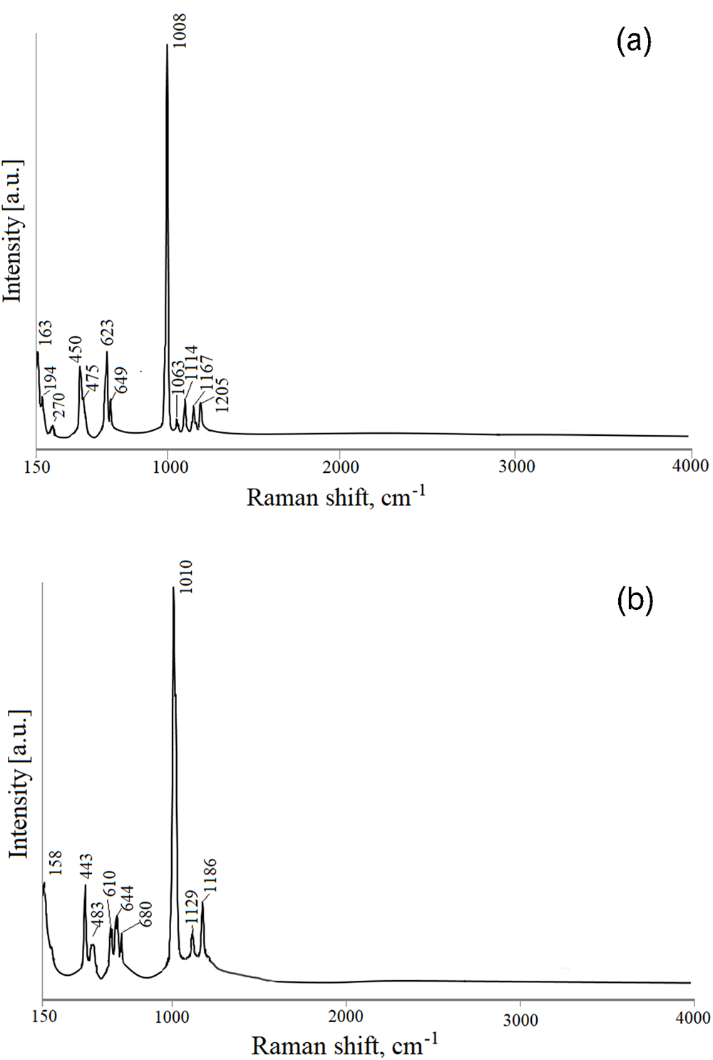
The Raman spectrum of kantorite (Fig. 2a) was obtained using an EnSpectr R532 spectrometer with a green laser (532 nm) at room temperature. The output power of the laser beam was ∼4 mW. The spectrum was processed using the EnSpectr expert mode program in the range 150 to 4000 cm–1 with a resolution of 6 cm–1. The back-scattered Raman signal was collected with a 40× PlanCN objective (numerical aperture 0.65). Signal acquisition time for a single scan was 1 s and the signal was averaged over 200 scans. The diameter of the focal spot on the sample was ∼5 μm. The spectrum was obtained for randomly orientated crystal.
The Raman spectra of kantorite (a) and krasheninnikovite (b).

The main bands in the Raman spectrum of kantorite correspond to S–O vibrations in (SO4)2– groups (assignments are according to Nakamoto, Reference Nakamoto1986, and Prieto-Taboada et al., Reference Prieto-Taboada, Fdez-Ortiz de Vallejuelo, Veneranda, Lama, Castro, Arana, Larrañag and Madariaga2019): (1) weak bands in the range 1050–1220 cm–1 correspond to ν3 antisymmetric stretching vibrations; (2) strong band of ν1 symmetric stretching vibrations near 1000 cm–1; (3) bands in the range 600–660 cm–1 are assigned to ν4 symmetric stretching vibrations; and (4) bands in the range 430–500 cm–1 belong to ν2 antisymmetric bending vibrations. Bands lower 300 cm–1 are mainly classified as bands corresponding to Mg–O, Mg–F, Na–O and K–O vibrations, as well as lattice modes. The lack of bands higher than 1220 cm–1 indicates the absence of groups with O–H, C–H, C–O, N–H, N–O and B–O bonds in kantorite.
The Raman spectrum of krasheninnikovite KNa2CaMg(SO4)3F (Fig. 2b), the mineral related to kantorite (see below), was obtained under the same conditions. Both spectra are similar in general pattern, however, demonstrate significant individual features in the number and position of bands that helps distinguish these minerals from one another.
Chemical composition
The chemical composition of kantorite was studied by electron microprobe using a Jeol JSM-6480LV scanning electron microscope (SEM) equipped with an INCA-Wave 500 wavelength-dispersive spectrometer (Laboratory of Analytical Techniques of High Spatial Resolution, Dept. of Petrology, Moscow State University), with an acceleration voltage of 15 kV and a beam current of 10 nA; the electron beam spot area was 3 μm2.
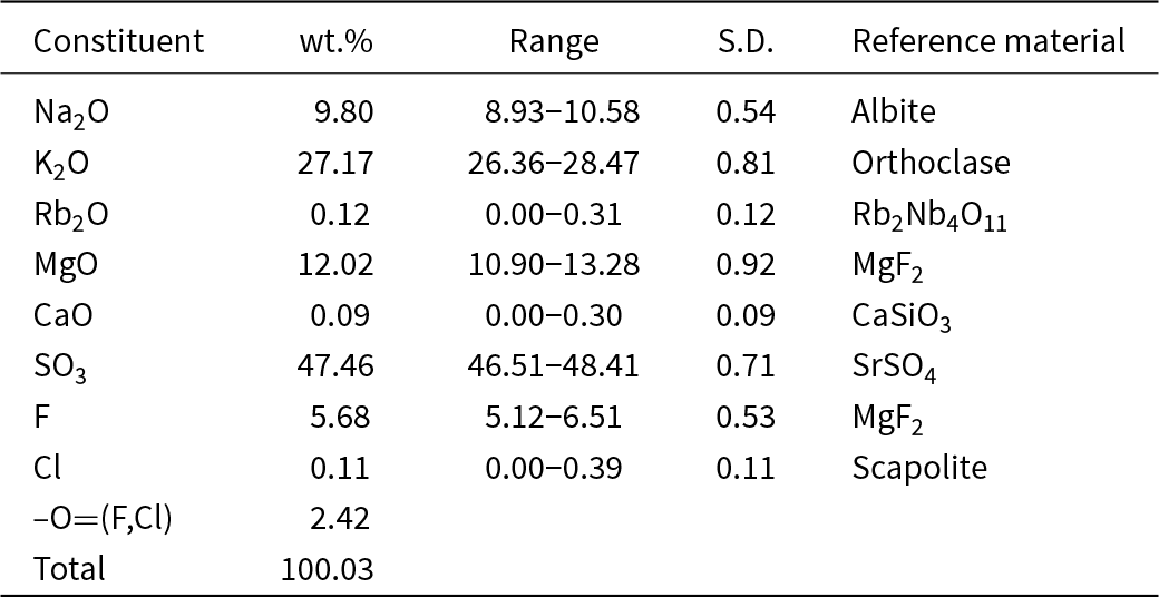
The chemical data in wt.% are given in Table 1. Contents of other elements with atomic numbers >6 are below detection limits.
Chemical composition of kantorite

Note: S.D. = standard deviation
The empirical formula calculated on the basis of (O+F+Cl) = 9 atoms per formula unit is (K1.94Na0.06)Σ2.00(Na1.00Ca0.01)Σ1.01Mg1.00(SO4.01)1.99(F1.01Cl0.01)Σ1.02. The idealised formula is K2NaMg(SO4)2F which requires Na2O 9.21, K2O 27.98, MgO 11.97, SO3 47.58, F 5.64, –O=F 2.38, total 100 wt.%.
The Gladstone–Dale compatibility index (Mandarino, Reference Mandarino1981), 1 – (K p/K c) = 0.008, is superior.
X-ray crystallography and crystal structure determination details
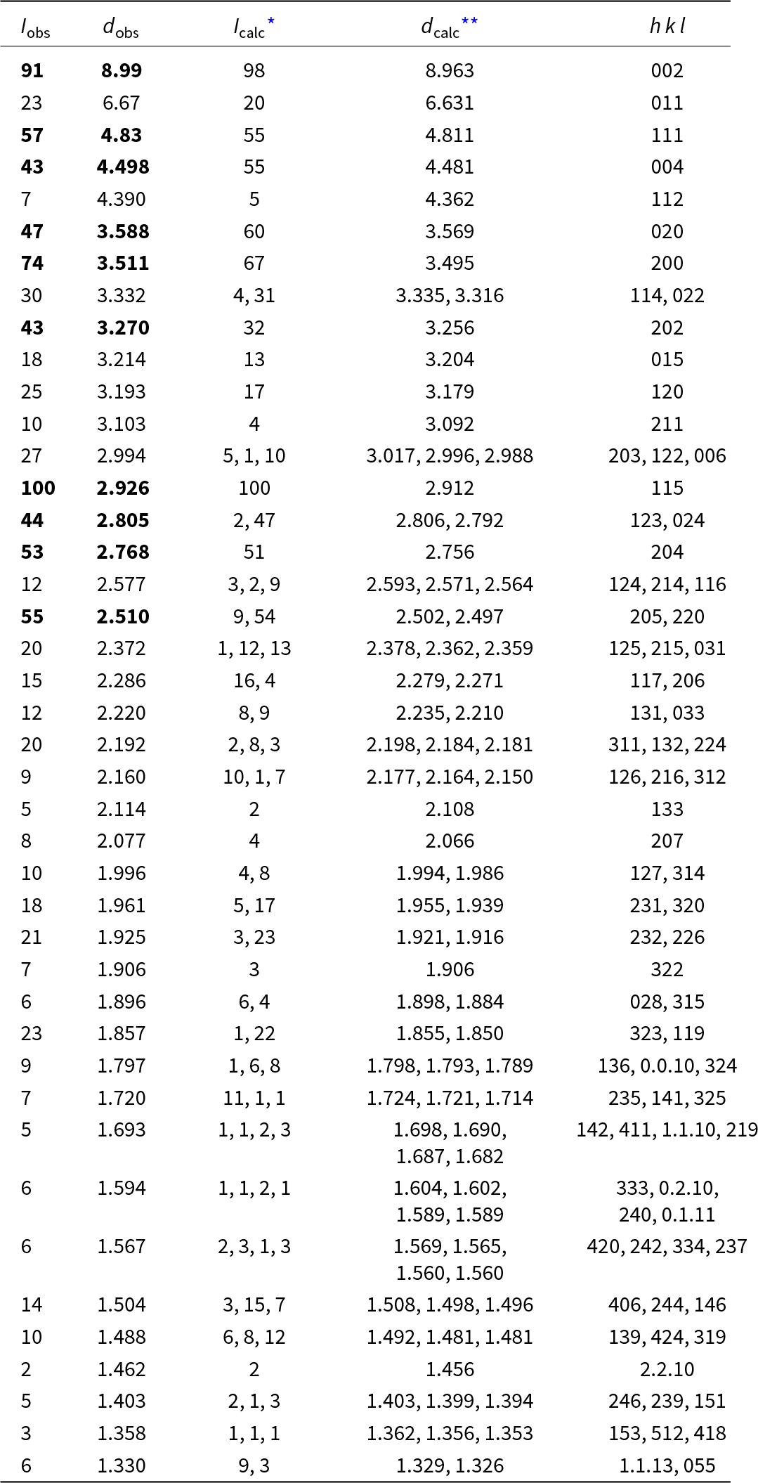
Powder XRD data for kantorite (Table 2) were collected in Debye-Scherrer geometry by means of a Rigaku RAXIS Rapid II diffractometer equipped with curved (semi-cylindrical) imaging plate detector (r = 127.4 mm), using CoKα radiation (λ = 1.79021 Å) generated by a rotating anode (40 kV, 15 μA) with microfocus optics; exposure time was set to 15 min. The image plate was calibrated against the NIST Si standard. The image-to-profile data processing was performed using osc2xrd software (Britvin et al., Reference Britvin, Dolivo-Dobrovolsky and Krzhizhanovskaya2017). Parameters of the orthorhombic unit cell refined from the powder data are: a = 7.008(2), b = 7.146(2), c = 17.961(4) Å and V = 899.5(7) Å3.
Powder X-ray diffraction data (d in Å) of kantorite

* Notes:For the calculated pattern, only reflections with intensities ≥1 are given; **for the unit-cell parameters obtained from single-crystal data. The strongest reflections are marked in boldtype.
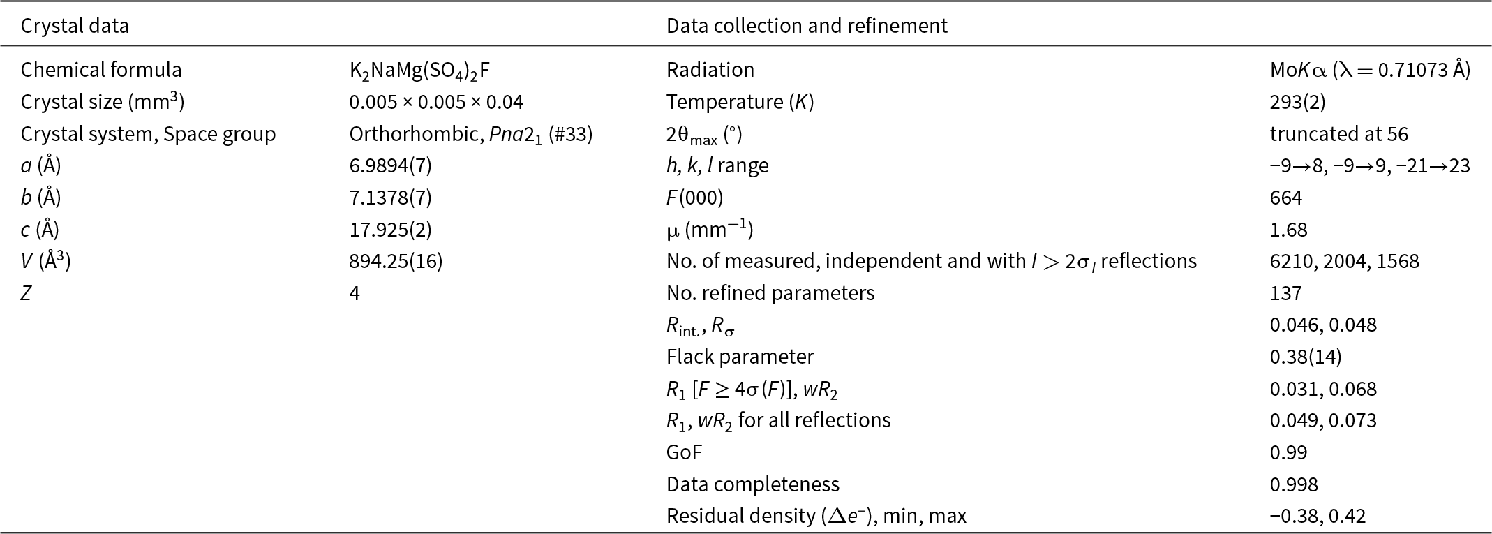
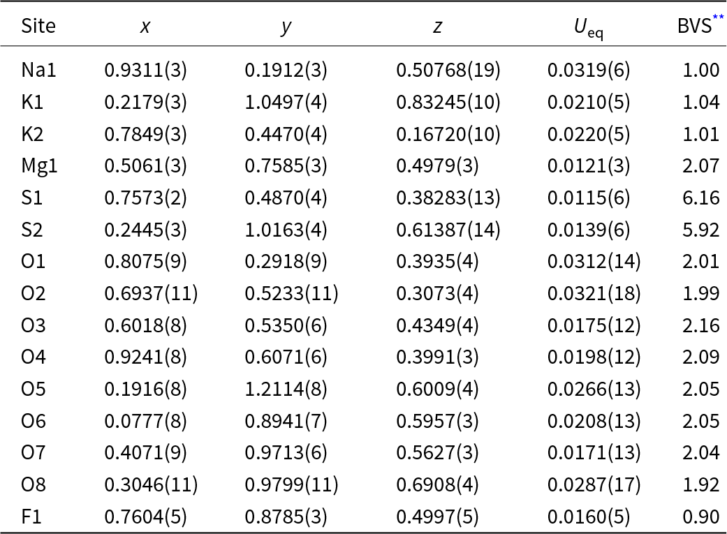
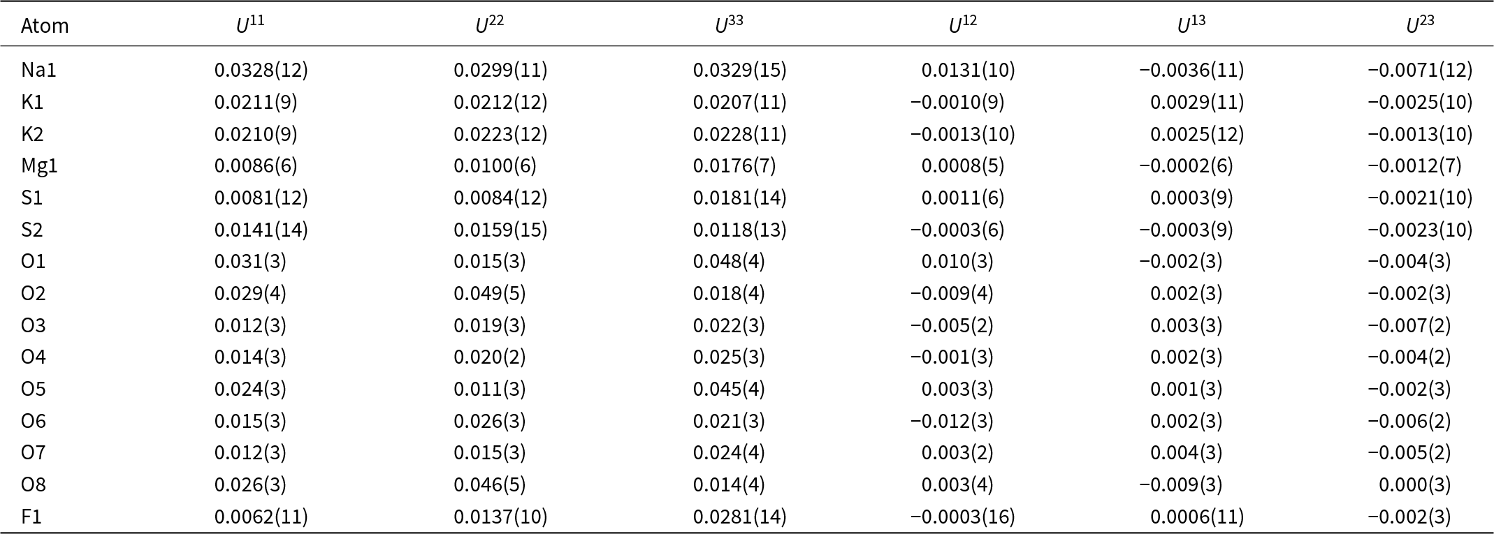
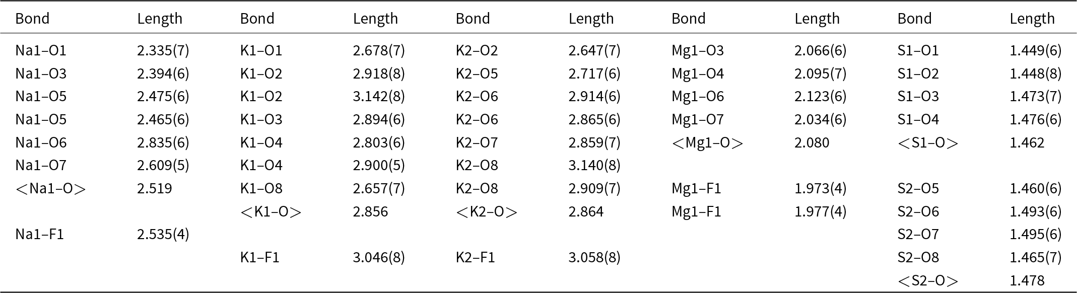
Single-crystal XRD studies of kantorite were carried out at room temperature using a Rigaku-Oxford diffraction XtaLAB Synergy-S diffractometer equipped with a microfocus X-ray tube (MoKα radiation) and HyPix-6000 hybrid photon counting detector. Data collection and reduction procedures were performed using CrysAlisPro, version 1.171.37.34 (Rigaku Oxford Diffraction). The crystal structure was solved and refined using the SHELX-2018 software package (Sheldrick, Reference Sheldrick2015) incorporated into the Olex2 v.1.5 graphical user environment (Dolomanov et al., Reference Dolomanov, Bourhis, Gildea, Howard and Puschmann2009). Data collection information and basic structure refinement details are presented in Table 3, coordinates and equivalent displacement parameters of atoms and bond-valence sums are given in Table 4, anisotropic displacement parameters in Table 5, and selected interatomic distances in Table 6. The detailed information on the data collection, reduction procedures, structure solution and refinement can be retrieved from the Crystallographic Information File (CIF) included in Supplementary materials.
Crystal parameters, data collection and structure refinement details for kantorite

Fractional coordinates and equivalent displacement parameters (U eq, Å2) of atoms and bond-valence sums (BVS, valence units) for kantorite*

* Crystallographic tables were created using PublCIF software (Westrip, Reference Westrip2010).
** Bond-valence sums were calculated using ECoN21 software (Ilinca, Reference Ilinca2022).
Anisotropic displacement parameters (Å2) of atoms for kantorite

Selected bond lengths (Å) in the structure of kantorite*

* Average atom–oxygen bond lengths were calculated separately from atom–fluorine bond lengths, as oxygen and fluorine represent the different chemical elements.
We attempted to refine the structure of kantorite in the centrosymmetric space group Pbcn which also gives reasonable results but with a disordered Na site. However, the free refinement in the space group Pna21 shows that Na is perfectly ordered with site occupancy factor = 1. This is concordant with the bond-valence sum for Na = 1.00 (Table 4). Therefore, the data available imply that the centrosymmetric space group describes only a simplified, ‘averaged’ structural model rather than a real one.
Discussion
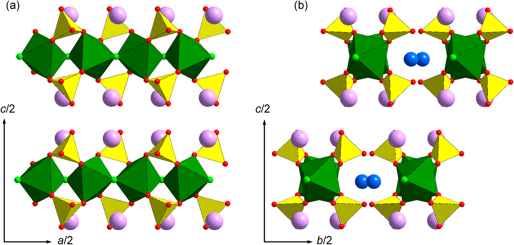
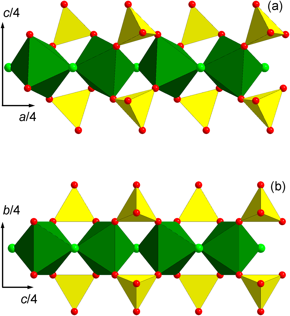
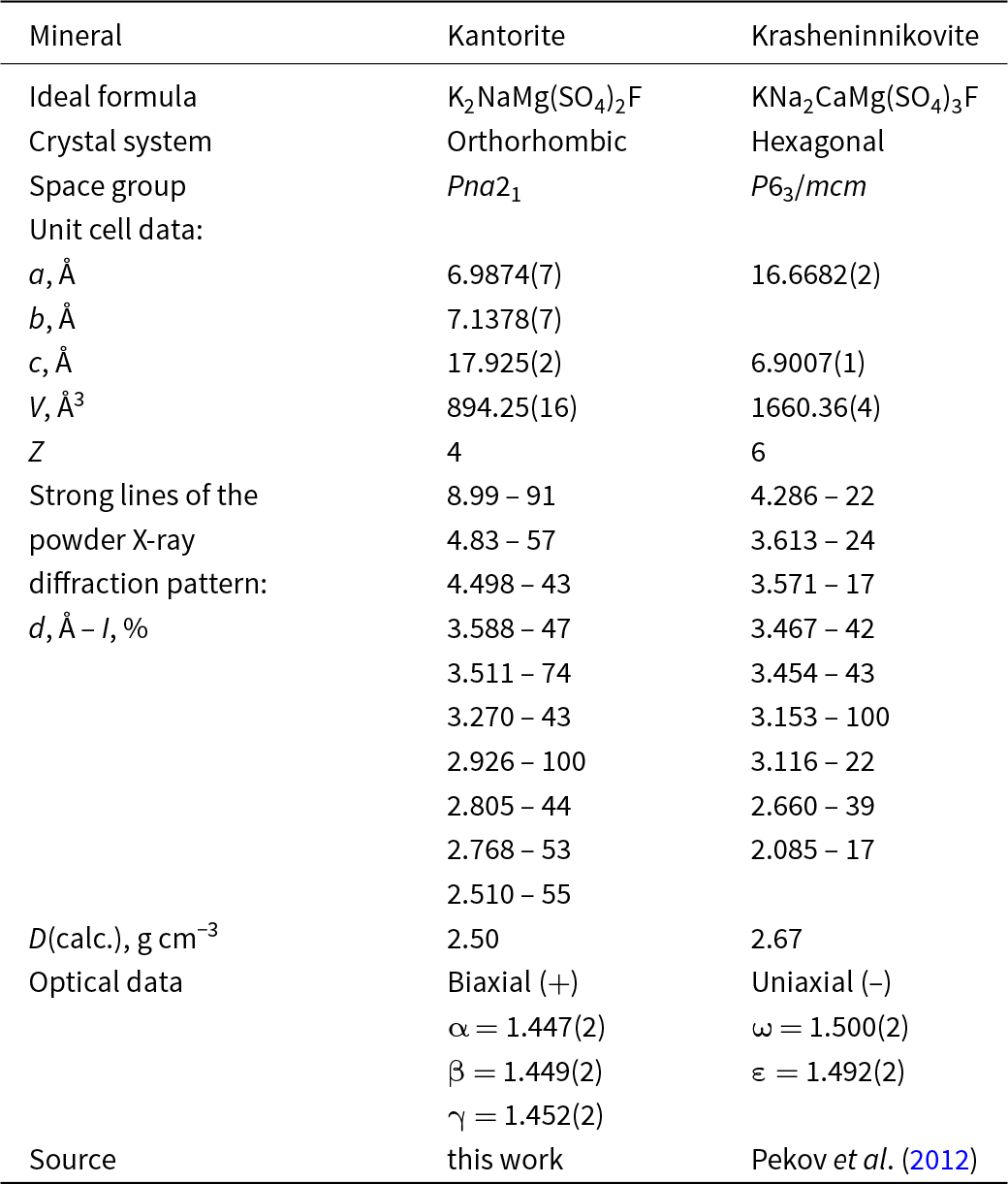
The crystal structure of kantorite is unique. It represents a quasi-framework in which the basic units are octahedra [MgO4F2] (Table 6). These octahedra are linked via bridging F atoms to form infinite chains running along the a-axis (Fig. 3). The chains of octahedra are encrusted by [SO4] tetrahedra via common corner-sharing oxygen atoms and assembled into the quasi-framework by alkali cations. The sulfate-encrusted chains of octahedra [MgO4F2] are topologically identical in the structures of kantorite and another fluorosulfate mineral, krasheninnikovite KNa2CaMg(SO4)3F (Pekov et al., Reference Pekov, Zelenski, Zubkova, Ksenofontov, Kabalov, Chukanov, Yapaskurt, Zadov and Pushcharovsky2012), Fig. 4. This determines the similarity of unit-cell parameters of these minerals: the a and c orthorhombic unit cell parameters of kantorite are comparable to the c and a hexagonal unit cell parameters of krasheninnikovite, respectively (Table 7). However, both chemical and structural differences between kantorite and krasheninnikovite are significant. The latter contains ‘additional’ sulfate groups which are not involved into the coordination of octahedral chains. These sulfate groups (SO4)2– are charge-balanced by the introduction of Ca2+ cations into the structure of krasheninnikovite. The relationship between the formulae of both minerals can be schematically represented as follows:
 \begin{equation*}\begin{array}{*{20}{c}}
\begin{gathered}
{{\text{K}}_{\text{2}}}{\text{NaMg(S}}{{\text{O}}_{\text{4}}}{{\text{)}}_{\text{2}}}{\text{F}} \hfill \\
{\text{kantorite}} \hfill \\
\end{gathered} &\begin{gathered}
\ \ {\text{K}} \leftrightarrow {\text{Na + CaS}}{{\text{O}}_{\text{4}}} \hfill \\
\ \ \xrightarrow{{\,\,\,\,\,\,\,\,\,\,\,\,\,\,\,\,\,\,\,\,\,\,\,\,\,\,\,\,\,\,\,\,\,\,\,\,\,\,\,\,\,\,}} \hfill \\
\end{gathered} &\begin{gathered}
\ \ {\text{KN}}{{\text{a}}_{\text{2}}}{\text{CaMg(S}}{{\text{O}}_{\text{4}}}{{\text{)}}_{\text{3}}}{\text{F}} \hfill \\
\ \ {\text{krasheninnikovite}} \hfill \\
\end{gathered}
\end{array}\end{equation*}
Crystal structure of kantorite. Octahedra [MgO4F2] are linked by bridging F atoms to form infinite chains running along the a axis, in two projections. The octahedral chains are encrusted by [SO4] tetrahedra via common corner-sharing oxygen atoms, and connected by alkali cations to form a quasi-framework. Legend: green octahedra = [MgO4F2]; yellow tetrahedra = [SO4]; red balls = oxygen; green balls = fluorine; blue balls = sodium; purple balls = potassium. Drawn using Diamond v.3 (Crystal Impact software).


Comparative data for kantorite and krasheninnikovite

Kantorite and krasheninnikovite belong to different crystal systems and easily can be distinguished by powder XRD patterns and optical data (Table 7).
Other chemically related hydrogen-free fluorosulfate minerals, kononovite NaMg(SO4)F (Pekov et al., Reference Pekov, Krzhizhanovskaya, Yapaskurt, Belakovskiy, Chukanov, Lykova and Sidorov2015) and shuvalovite K2(Ca2Na)(SO4)3F (Pekov et al., Reference Pekov, Zubkova, Britvin, Chukanov, Yapaskurt, Sidorov and Pushcharovsky2016), are quite different in terms of crystal structure from one another and from kantorite and krasheninnikovite. All four of these fluorosulfates occur in the Arsenatnaya fumarole, of which kononovite, shuvalovite and kantorite are endemic minerals of this remarkable locality.
Supplementary material
The supplementary material for this article can be found at https://doi.org/10.1180/mgm.2025.23.
Acknowledgements
We are grateful to three anonymous referees for valuable comments. The study was conducted under the state assignment of Lomonosov Moscow State University (state task No. 121061600049-4). XRD investigation was carried out at the Research Centre for X-ray Diffraction Studies of St Petersburg University in accordance with the state task of the Russian Federation No. AAAA-A19-119091190094.
Competing interests
The authors declare none.