Introduction
Recent neuroimaging studies of elderly people have revealed that not only dementia or mild cognitive impairment (MCI), but also normal aging is associated with neuronal loss and functional degeneration in the human brain (Pakkenberg and Gundersen, Reference Pakkenberg and Gundersen1997). Some previous morphological studies by magnetic resonance imaging (MRI) have shown that a decreased gray matter (GM) volume correlates with advanced aging (Sowell et al., Reference Sowell, Peterson, Thompson, Welcome, Henkenius and Toga2003). Furthermore, the decreased GM volumes of regions, including the hippocampus and prefrontal and temporal cortices, have been reported to be associated with several types of cognitive decline, such as the decline in attention, executive function, or memory in healthy elderly people (Salat et al., Reference Salat, Kaye and Janowsky2002; Convit et al., Reference Convit, Wolf, Tarshish and De Leon2003; He et al., Reference He2012). Consistently, several cognitive domains, such as attentional function, episodic memory, processing speed, and executive function, have been reported to decline with advanced aging (Baudouin et al., Reference Baudouin, Clarys, Vanneste and Isingrini2009; Park and Reuter-Lorenz, Reference Park and Reuter-Lorenz2009). On the other hand, a recent review by Kaup et al. (Reference Kaup, Mirzakhanian, Jeste and Eyler2011) revealed that in healthy elderly people, the results regarding the relationship between decreased regional GM volumes and cognitive decline are inconsistent among the above-mentioned previous studies. Thus, further neuroimaging research is required to investigate the relationship between neuronal loss and cognitive decline, which worsen with aging.
In most of the morphological in vivo studies on human brain aging, the neuronal loss in GM is evaluated on the basis of the degree of severity or subcortical atrophy, which is calculated from the regional GM volume detected by structural MRI (Killiany et al., Reference Killiany2000). However, other research studies suggest that receptors ubiquitously expressed in neurons, e.g. the γ-aminobutyric acid type A (GABAA) receptor, can be more sensitive markers of neuronal loss than regional GM volume (La Fougère et al., Reference La Fougère2011; Pascual et al., Reference Pascual2012). The GABAA receptor complex, which is the main part of the central benzodiazepine binding site, is a target of radioligands in in vitro and in vivo positron emission tomography (PET)/single-photon emission computed tomography (SPECT) (Pappatà et al., Reference Pappatà2010). In vivo PET/SPECT studies using the central GABAA/benzodiazepine antagonist 11C-flumazenil and the partial agonist 123I-iomazenil (IMZ) have demonstrated that patients with early stage Alzheimer's disease (AD) showed a decreased binding affinity of the above-mentioned radioligands in several cortical regions such as the frontal and parietotemporal areas (Pascual et al., Reference Pascual2012). In addition, the 11C-flumazenil binding affinity in the hippocampus and posterior cingulate cortex correlated with the memory performance of patients with AD (Pascual et al., Reference Pascual2012). Although this finding indicates that GABAA receptor imaging is highly useful for assessing neuronal loss associated with cognitive decline, knowledge of the relationship between decreased GABAA receptor binding affinity and several types of cognitive decline, e.g. the decline in attention or working memory function, with advancing age is still limited particularly at the human senium.
In this study with a cross-sectional design, we investigated the association of the performance of healthy elderly people in the attentional function test with regional GM volume, regional cerebral bold flow (rCBF), and GABAA receptor binding affinity by T1-weighted MRI, arterial spin labeling (ASL), and 123I-IMZ SPECT in the resting state. The rapid visual information processing (RVP) test of the Cambridge neuropsychological test automated battery (CANTAB) was performed to assess visual sustained attention and working memory processing (Bakan, Reference Bakan1959). For the analysis of correlation between the score of the attentional function test and MRI/SPECT data, we carried out small-volume corrections, focusing on the bilateral inferior frontal gyri, of which the right gyrus is critical to the inhibition of responses and attentional control (Aron et al., Reference Aron, Robbins and Poldrack2014). Patients with damage to the right inferior frontal gyrus have been reported to show disruption of stop-signal inhibition (Aron et al., Reference Aron, Fletcher, Bullmore, Sahakian and Robbins2003). A previous PET study using the RVP and simple sustained attention control tests showed that rCBF in the right inferior frontal gyrus increased during both tests compared with the rest condition, suggesting that this GM area is associated with sustained attention (Coull et al., Reference Coull, Frith, Frackowiak and Grasby1996).
Methods
Subjects
Thirty-seven individuals (aged 50–77 years (mean, 64.5 ± 7.3 years); 15 males and 22 females) were enrolled in this study in accordance with the following exclusion criteria: history of suffering from a type of dementia, neurological or psychiatric illnesses, diabetes mellitus, chronic alcoholism, chronic smoking, and obesity with a body mass index above 25. All the subjects enrolled in this study were right-handed, as assessed using the Edinburgh handedness inventory (Oldfield, Reference Oldfield1971). All the subjects provided their written informed consent. The study protocol was approved by the Ethics Committee of Gunma University.
Psychological measurements
The Montreal Cognitive Assessment (MoCA) was administered to all the subjects to assess their cognitive function (Nasreddine et al., Reference Nasreddine2005); we used scores ≥ 20 to exclude subjects with dementia and cognitive impairments, in accordance with a previous report (Waldron-Perrine and Axelrod, Reference Waldron-Perrine and Axelrod2012). The National Adult Reading Test was then conducted to estimate premorbid IQs (Nelson, Reference Nelson1982). The RVP test of CANTAB (Cambridge Cognition Ltd., Cambridge, UK) was administered to all the subjects of this study to assess their attentional function and working memory function, which is a visual continuous performance test using digits instead of letters.
MRI and 123I-IMZ SPECT
T1-weighted anatomical imaging and ASL acquisitions
MRI scans were acquired on a 3T Siemens Prisma scanner with a 24-channel phased-array head coil. We scanned high-resolution T1-weighted images for GM morphometry. MP-RAGE sequencing yielding 176 continuous slices of 1.0 mm thickness was carried out in the sagittal plane. Parameters for the scanning were as follows: echo time = 2 ms; repetition time = 2,000 ms; inversion time = 990 ms; flip angle = 9°; field of view = 256 mm; matrix size = 256 × 256; voxel size = 1 × 1 × 1 mm3. For the ASL acquisition, we used a pulsed PICORE (proximal inversion with a control for the off-resonance effect) sequence with the Q2TIPS (QUIPSS II with thin-slice TI1 periodic saturation) scheme using the following parameters: repetition time/echo time /inversion time 1/ inversion time 2/ = 4,000 ms/ 22 ms/1,800 ms/1,675 ms; matrix = 64 × 64; voxel size = 3.5 × 3.5 × 4.0 mm3; and 38 slices with an interslice gap of 1.0 mm.
123I-IMZ SPECT acquisition
All SPECT studies were performed using a dual-head rotating gamma camera (E.CAM Signature; Toshiba Medical Systems, Japan) with a low-and middle-energy general purpose (LMEGP) collimator providing a spatial resolution of 3.8 mm full width at half-maximum (FWHM). Images of the cerebral distribution of 123I-IMZ were acquired over 22 minutes, starting 180 minutes after a slow bolus intravenous injection of 167 MBq of the radiotracer (Lassen, Reference Lassen1992). Conditions for the acquisition were as follows: 30 s/step, 4°/step, continuous.
A collimator specifically designed for 123I studies was used for acquisition of 123I-IMZ data. The images were reconstructed by the filtered backprojection method using Butterworth (cutoff, 0.45 cycles/cm; order, 8.0) and Ramp filters with attenuation correction (Chang, 0.140/cm). The matrix size and slice thickness of SPECT images were 128 × 128 (2.20 mm/pixel) and 6.6 mm, respectively.
Imaging data processing
For regional GM volumetric analysis, T1-weighted images were analyzed using SPM8 (Wellcome Department of Cognitive Neurology, London, UK) implemented in Matlab 2014a (MathWorks, Natick, MA, USA) using VBM8 (http://dbm.neuro.uni-jena.de/vbm/download) with default parameters. The DARTEL (diffeomorphic anatomical registration through exponentiated lie algebra) toolbox of SPM8, an algorithm for accurate diffeomorphic image registration, was used to generate a set of group-specific templates. After the T1-weighted brain images were segmented (GM, white matter, and cerebrospinal fluid), normalized, and modulated using these templates, they were additionally warped using the Montreal Neurologic Institute (MNI) space. Resulting images were smoothed using an isotropic Gaussian kernel of 8 mm FWHM.
The obtained relative ASL CBF maps were analyzed using SPM8. The maps were realigned and coregistered to the corresponding GM images obtained by T1-weighted images processing. To remove the non-brain voxel, maps were masked with a binarized image, which is the sum of GM, white matter, and cerebrospinal fluid (thresholded by voxel intensity > 0.1). Then, the maps were spatially normalized to the MNI space using estimated deformation fields obtained by T1-weighted images processing.
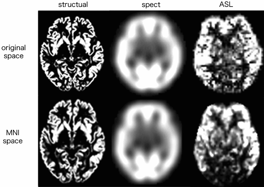
123I-IMZ SPECT images were spatially normalized to the MNI template by linear coregistration and non-linear warping based on the SPECT template bundled with the SPM8. All the normalized SPECT images were smoothed by convolution with an isotropic Gaussian kernel of 12 mm FWHM. This filtering technique for smoothing has been shown to be optimal (Merhof et al., Reference Merhof2011). The GM threshold was set at 0.8 and normalization of global CBF to 50 was performed by the ANCOVA model. The MPRAGE, SPECT, and ASL images of a sample subject in the original and MNI space are shown in Figure 1.
MPRAGE, SPECT, and ASL images of a sample subject in the original and MNI space at the same slice.

Statistical analyses
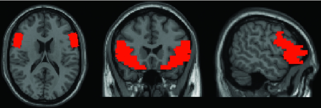
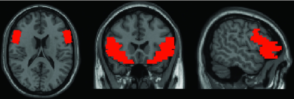
To determine the correlation of RVP score with regional GM volume, rCBF, and GABAA receptor binding affinity in the GM area, we performed whole-brain and volume-of-interest (VOI) analyses by small volume correction focusing on the bilateral inferior frontal gyri, of which the right gyrus is critical to inhibiting responses and attentional control (Aron et al., Reference Aron, Robbins and Poldrack2014), by automated anatomical labeling (AAL) of the Wake Forest University (WFU) Pickatlas (Maldjian et al., Reference Maldjian, Laurienti, Kraft and Burdette2003) (Figure 2). The statistical threshold was set at a familywise error rate of p < 0.05 (FWER-corrected p < 0.05) at the voxel level with adjustments for age, gender, and total GM volume. In both whole-brain and small volume correction analyses, the cluster extent correction procedure implemented in SPM8 was used, which computes the number of expected voxels per cluster according to the random field theory (Hayasaka and Nichols, Reference Hayasaka and Nichols2004).
To assess the relationship among demographic features, scores of MoCA and RVP tests, and predicted IQs, Pearson's correlation test and partial correlation with adjustment for confounding factors (age and gender) were performed. A p-value of < 0.05 was set as statistically significant. The Student t-test was conducted to estimate gender differences in demographic characteristics. All the statistical analyses were carried out using SPSS version-24 (IBM Corporation).
Results
The demographic features (mean ± SD) were as follows: years of education, 13.7 ± 2.0 years; predicted IQs, 110.9 ± 8.6 points; MoCA test score, 25.7 ± 2.9; and RVP test score (expressed in accuracy rate), 0.90 ± 0.04. All the subjects showed MoCA test scores > 20 and predicted IQs higher than 90. No significant gender difference was observed between age and MoCA and RVP test scores, and predicted IQ. The RVP test score and MoCA test scores negatively correlated with age (p < 0.01).
Correlation analysis between score of attentional function test and brain parameters determined from whole-brain T1-weighted, ASL, and 123I-IMZ SPECT images
In the analysis of regional GM volume, rCBF, and GABAA receptor binding affinity in the entire brain determined from T1-weighted, ASL, and 123I-IMZ SPECT images, respectively, the RVP test score showed no significant positive or negative correlation in any brain area with adjustment for age, gender, and total GM volume (FWER-corrected p > 0.05).
Correlation analysis between score of attentional function test and brain parameters determined from T1-weighted, ASL, and 123I-IMZ SPECT images focusing on bilateral inferior frontal gyri and temporal-parietal junction by small volume correction
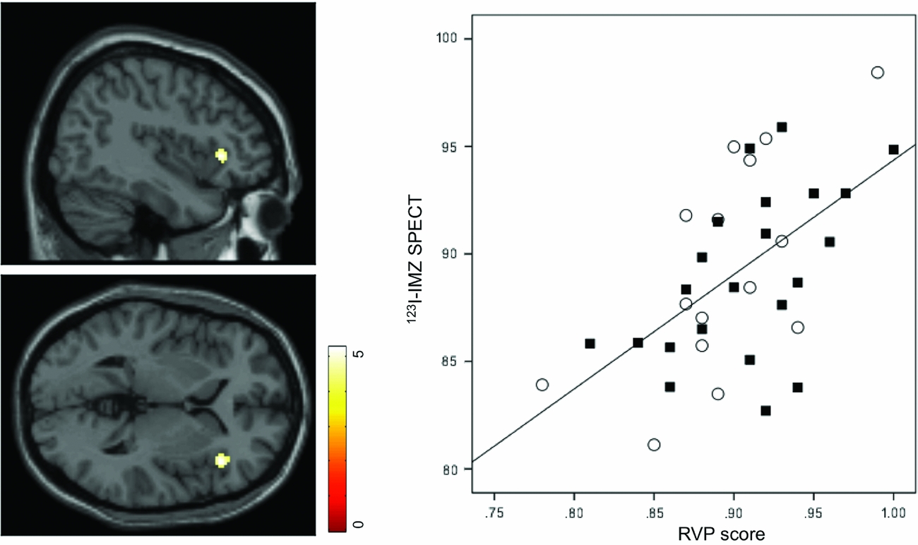
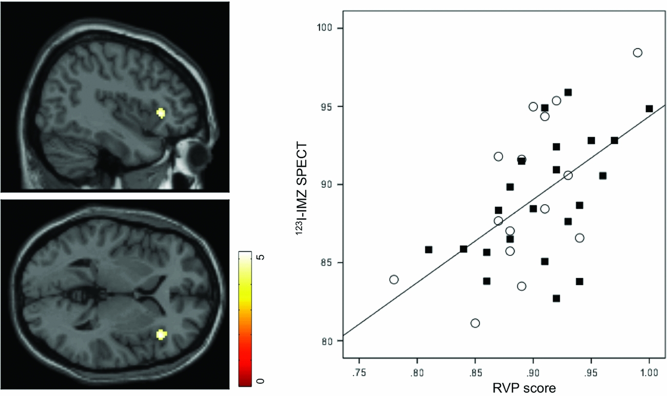
In the analysis of GABAA receptor binding affinity in the bilateral inferior frontal gyri and the temporal-parietal junction, the RVP test score showed a significant positive correlation in the right inferior frontal gyrus with adjustment for age, gender, and total GM volume (k = 61; peak MNI coordinates, x = 42, y = 26, and z = 2; t-value = 5.22; uncorrected p < 0.000 at voxel level; FWER-corrected p = 0.009 at cluster level) (Figure 3). No significant negative correlation was found between RVP test score and GABAA receptor binding affinity in any brain area (FWER-corrected p > 0.05). Moreover, no significant positive or negative correlation was found between RVP test score and regional GM volume or rCBF in any brain area (FWER-corrected p > 0.05).
Red area showing binary mask of bilateral inferior frontal gyri determined by AAL.
The left panel shows the brain region where regional GABAA receptor binding affinity (i.e. 123I-IMZ SPECT) positively correlated with attentional function test score (i.e. RVP test score) estimated by SPM with small volume correction focusing on the bilateral inferior frontal gyri. The right panel shows the association between RVP test score and the mean intensity of 123I-IMZ SPECT within this brain region (r 2 = 0.316, β = 0.562, p = 0.0003). Squares indicate male subjects and circles indicate female subjects.
Discussion
Analysis of 123I-IMZ SPECT images in the resting state showed that RVP test score significantly positively correlated with GABAA receptor binding affinity in the right inferior frontal gyrus in the healthy elderly people without dementia. On the other hand, analysis of T1-weighted images or ASL in the resting state showed no significant correlation between RVP test score and regional GM volume or rCBF in any brain area.
The regional GABAA receptor affinity on 123I-IMZ SPECT images correlates with brain neuronal density and a reduction in cortical GABAA receptor affinity indicates cortical neuronal damage or loss (Nakagawara et al., Reference Nakagawara, Sperling and Lassen1997). Consistently, several previous studies have revealed decreased 123I-IMZ binding in several cortical areas in patients with neurodegenerative diseases, such as AD (Pascual et al., Reference Pascual2012), vascular dementia (Hanyu et al., Reference Hanyu2012), and MCI (Pappatà et al., Reference Pappatà2010), compared with controls. In addition, some previous PET and functional MRI studies using the RVP test have revealed the activation of the GM area of the prefrontal cortex including the right inferior frontal gyrus during this test (Coull et al., Reference Coull, Frith, Frackowiak and Grasby1996; Lawrence et al., Reference Lawrence, Ross and Stein2002). Over the last two decades, the right inferior frontal gyrus has been widely recognized to be critical to the inhibition of responses and attention control (Aron et al., Reference Aron, Robbins and Poldrack2014). More recently, it has been suggested that the right inferior frontal gyrus plays a key role in the integration of bottom-up, sensory information with top-down, response-related information to facilitate flexible, and goal-directed behavior (Dodds et al., Reference Dodds, Morein-Zamir and Robbins2010). Considering the above-mentioned previous studies, the findings of our study, particularly the significant positive association between RVP test score and regional GABAA receptor binding affinity in the right inferior frontal gyrus, suggest that age-related neuronal loss in that GM area, as measured from 123I-IMZ SPECT images, contributes to the decline in attentional function in healthy elderly people.
We found a significant correlation of RVP test score with GABAA receptor binding affinity, but not with regional GM volume or rCBF in any brain areas. SPECT imaging of GABAA receptor binding distribution has been suggested to reflect neuronal integrity in the cerebral cortex (Ruan et al., Reference Ruan, D'Onofrio, Sancarlo, Bao, Greco and Yu2016). Thus, this SPECT imaging method might enable the detection of cortical abnormalities associated with attentional function, prior to abnormal structural and rCBF changes. Interestingly, recent images acquired using 123I-IMZ and hexamethyl propylene amine oxime (HMPAO) indicate that in patients with probable AD, the cortical area with decreased GABAA receptor binding affinity overlapped with that with decreased rCBF, with the former being larger than the latter (Varrone et al., Reference Varrone, Soricelli, Postiglione and Salvatore1999).
There are some limitations of this study. The major limitations are the small number of subjects, and the lack of data of subjects with age-related neurodegenerative disorders, i.e. MCI or AD. Also, younger subjects should also be included in future studies. Regarding methodological limitation, we must also mention the reliability of ASL. Previous investigations have revealed that the rCBF measured by ASL is similar to that measured by SPECT or PET (Johnson et al., Reference Johnson2006). The clinical use of ASL has increased in popularity compared with SPECT or PET, which requires the use of several radioligands such as HMPAO or H2(15)O. However, further in vivo research is needed to assess the usefulness and potential of ASL for rCBF measurement. Further studies including subjects in other age ranges with age-related neurodegenerative disorders will reinforce our findings in the future.
In conclusion, RVP test score was positively associated with GABAA receptor binding affinity in the right inferior frontal gyrus in healthy elderly people without dementia. Because the GABAA receptor binding distribution reflects neuronal integrity in the cerebral cortex (Ruan et al., Reference Ruan, D'Onofrio, Sancarlo, Bao, Greco and Yu2016), age-related cortical neuronal damage or loss in the right inferior frontal gyrus might lead to the decline in attentional function. These findings may lead to a better understanding of the role of the right inferior frontal gyrus in attentional function during normal aging.
Conflict of interest
None.
Description of authors’ roles
M. Kasagi collected and analyzed the data and wrote the paper. K. Narita was responsible for the statistical design of the study and for carrying out the statistical analysis. T. Motegi, K. Fujihara, Y. Suzuki, and M. Tagawa collected the data and assisted with the analysis of the data. K. Ujita and H. Shimada helped in carrying out the study. M. Fukuda supervised the data collection and assisted with the writing of the paper.