Introduction
Traditionally, in children, lung ultrasound was mainly used as an aid in the diagnosis of pleural effusion, thoracic masses, or assessment of diaphragmatic motion. However, in the past decade, its use has expanded, and it is now used in a variety of settings to answer a wide range of clinical questions. These include evaluating the cause of respiratory distress in the neonatal intensive care unit, to help in the diagnosis of pneumonia and bronchiolitis, detection of pneumothorax, assessment of the position of the endotracheal tube in the neonatal intensive care unit, assessment of extravascular lung water in children on dialysis or after cardiac surgery. Daniel Lichenstein published a series of articles on lung ultrasound in the 1990s which culminated in the development of the Blue Protocol for the use of lung ultrasound in acute respiratory failure in 2009. Reference Lichtenstein1 In the same year, he also described the use of lung ultrasound in neonates in the critical care units. Reference Lichtenstein2 Since then, the field of paediatric lung ultrasound has expanded rapidly and in the past 10 years, a huge body of literature has been published on this subject. Reference Pereda, Chavez and Hooper-Miele3–Reference Musolino, Tomà and De Rose6
B-lines are discrete vertical hyperechoic reverberation artifacts seen in lung ultrasound that arise from the pleura and extend to the bottom of the screen without fading, and move synchronously with lung sliding and have been used to diagnose cardiogenic pulmonary oedema. Reference Volpicelli, Elbarbary and Blaivas7 The role of B lines detected in lung ultrasound in the assessment of pulmonary congestion and heart failure has already been established in adults with heart failure. Reference Mézière, Biderman, Gepner and Barré8,Reference Lichtenstein9 B lines in lung ultrasound has also been compared to NT pro-BNP or New York Heart Association (NYHA) status in this population. Reference Li, Li and Zhu10
The B-line artifacts, their number, and distribution can be semi-quantitatively analysed in the form of a lung ultrasound score. Lung ultrasound score calculated by a variety of methods has been found to correlate with extravascular lung water in animal studies, Reference Zong, Guo, Liu, Bao and Yang11 in patients on haemodialysis, Reference Noble, Murray, Capp, Sylvia-Reardon, Steele and Liteplo12 with inotropic score and duration of ventilation in the postoperative cardiac intensive care unit Reference Girona-Alarcón and Cuaresma-González13,Reference Kaskinen, Martelius, Kirjavainen, Rautiainen, Andersson and Pitkänen14 and with severity of pulmonary congestion in adults with heart failure. Reference Li, Li and Zhu10,Reference Picano and Pellikka15 However, paediatric heart failure has varied pathophysiology, and the use of lung ultrasound in paediatric heart failure is still evolving.
CHD with left to right shunts is an important cause of heart failure in children. The pathophysiology of symptoms related to left to right shunt is partly related to elevated pulmonary capillary pressure. B lines generated due to the elevated pulmonary capillary pressure and pulmonary congestion may be an accurate marker of the severity of heart failure in this population.
The symptoms related to pulmonary overflow and its effect on the heart are collectively quantified by the Ross score. The Laer-modified Ross score is a commonly used clinical score to classify paediatric heart failure. It has two components related to history and four components from physical examination and is easy to use in a variety of clinical settings. The latest version of the Ross score incorporates NT pro-BNP as one of its components, but its use may be limited as NT pro-BNP is an invasive (blood) test. Reference Ross16 Lung ultrasound will have added value if there is good correlation between the severity of pulmonary overflow ( as seen by B line score) and heart failure severity (as judged by Ross score and/or NT pro- BNP). Compared to NT pro-BNP, lung ultrasound has the advantage of being a non-invasive bedside tool, and lung ultrasound score may be a valuable addition as a marker of heart failure in children with CHD.
Two studies have recently evaluated the role of lung ultrasound in diagnosing pulmonary overflow in unoperated CHD recently. Reference Rodríguez-Fanjul, Llop, Balaguer, Bautista-Rodriguez, Hernando and Jordan17,Reference Wu, Hou and Lu18 The present study was conducted to determine whether a semiquantitative lung ultrasound score correlates with severity heart failure (as assessed by modified Ross score and NT pro-BNP) in infants with left to right shunts.
Materials and methods
This was a cross sectional, observational, diagnostic study conducted in a tertiary care teaching hospital. The study was approved by the Institutional Ethics committee. Patients attending the outpatient clinic of the Division Paediatric Cardiology were assessed for enrolment. The parents of infants with known acyanotic CHD fulfilling the inclusion criteria and not having any exclusion criteria were enrolled after taking informed consent from the parents.
Sample size calculation
The sample size was calculated based on a previous study by Li et al, Reference Li, Li and Zhu10 who found a correlation coefficient of 0.44 between the Lung Ultrasound Score (LUS) and the NYHA functional class in adults with heart failure. Using alpha error = 0.05, correlation coefficient under null hypothesis = 0, correlation coefficient under alternate hypothesis = 0.44, a minimum sample size of 44 was needed to achieve a power of 85% and a sample size of 51 was needed for a power of 90 %. Therefore, we decided to enrol 50 infants in this study.
Inclusion criteria
-
Infants with CHD and post tricuspid left to right shunt, attending presenting in paediatric cardiology clinic.
Exclusion criteria
-
Patients having respiratory tract infection recently (within 10 days)
-
Liver or renal disorders causing fluid retention
-
Acute respiratory distress syndrome, asthma, bronchiolitis, or patients on mechanical ventilation.
-
Postoperative CHD, associated mitral valve stenosis, cor tiatriatum
-
Severe anaemia, any ongoing febrile illness
-
Congenital malformations of the lung such as cystic malformations and congenital diaphragmatic hernia
Assessment of modified ross score
The Ross score is a clinical score commonly used to assess the severity of heart failure in children. It has a scoring system that can be used either as a continuous score or an ordinal score (mild, moderate, or severe heart failure), similar to NYHA classification used in adults with heart failure. Reference Kaskinen, Martelius, Kirjavainen, Rautiainen, Andersson and Pitkänen14 The Ross score was assessed preferably, while the infant was asleep at least one hour after the last feed and before the blood sampling or lung ultrasound was performed. Although the latest version of Ross score also includes the NT pro BNP as one of its components, we used the older Laer-modified version in our study as it is easy to use in bedside.
Measurement of NT pro-BNP
NT pro BNP has emerged as an important biochemical marker of heart failure, and its level increases with the severity of heart failure as seen in heart failure in children with left to right shunt. Reference Rodríguez-Fanjul, Llop, Balaguer, Bautista-Rodriguez, Hernando and Jordan17
After clinical assessment, 3 ml peripheral venous sample was drawn for the measurement of NT pro-BNP. For the quantitative measurement of N-terminal pro-brain natriuretic peptide (NT pro-BNP) in human serum and plasma (EDTA or heparin), we used the VITROS ECi/ECiQ/3600 Immunodiagnostic Systems. (ortho clinical diagnostics, Rochester, NY, USA).
Lung ultrasound method
Lung ultrasound was done by a single operator using a linear transducer (frequency 6-14 MHz), using Mindray M7 ultrasound machine (Shenzhen Mindray Bio-Medical Electronics Co., Ltd Shenzhen, China) with lung ultrasound pre-set. The probe was kept perpendicular to the ribs, and imaging was done in a vertical direction to include the maximum number of intercostal spaces in each image. The scanning depth was kept at 2-4 cm and gain up to 50%, depending on patients’ features, harmonic imaging was kept off, focal point was single and kept near the pleural surface. MI was kept at 0.1. The operator was blinded to the clinical diagnosis when LUS was performed.
There are several protocols for lung ultrasound that have been used in previous studies using anywhere between 4 and 28 zones. The choice of a particular protocol usually depends on the clinical setting. In the emergency and ICU settings, 3- 6 zones of the anterior and lateral chest have been used. Reference Lichtenstein9,Reference Li, Li and Zhu10,Reference Kaskinen, Martelius, Kirjavainen, Rautiainen, Andersson and Pitkänen14 On the other hand, studies in patients on haemodialysis have used a more extensive 28-zone protocol. Reference Noble, Murray, Capp, Sylvia-Reardon, Steele and Liteplo12 Previous studies of patient with unoperated CHD have excluded the posterior chest wall. Reference Rodríguez-Fanjul, Llop, Balaguer, Bautista-Rodriguez, Hernando and Jordan17,Reference Wu, Hou and Lu18 14-zone protocol was selected for in this study because we felt it was important to include the posterior lung zones. It was thought that pathology related to pulmonary overflow is also more likely to manifest in these dependent regions in the posterior part of the lung.
A 14-zone protocol (7 in each lung), as initially described by Laursen et al, was used. Reference Laursen, Sloth and Lambrechtsen19 This protocol has been used in other studies subsequently and has also been proposed for use to achieve international standardisation of lung ultrasound in COVID-19 patients. Reference Davidsen, Laursen and Højlund20,Reference Haurylenka, Damantsevich, Filustsin and Damantsevich21
According to this protocol, the anterior chest wall is delineated from the sternum to the anterior axillary line and from the clavicle to the diaphragm. The lateral chest wall is delineated from the anterior to the posterior axillary line. Both anterior and lateral chest walls are subdivided into upper and lower zones (the area above the fourth intercostal space was defined as the upper zone). The posterior chest wall is divided into 3 equal zones.
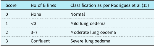
Posterior zones were scanned with the infant lying in the left and right lateral position while the anterior and lateral zones were scanned with the infant lying in the supine position. (Supplementary figure S1) The scoring of each zone was done according to the maximum number of B lines visible in one intercostal space as described below. The scoring system is based on the classification of pulmonary congestion as described in a previous study by Rodriguez et al. Reference Rodríguez-Fanjul, Llop, Balaguer, Bautista-Rodriguez, Hernando and Jordan17 We gave a numerical score to the classification (see Table 1) which they used in their study (Supplementary figure S2).
Lung Ultrasound B Line classification

Each intercostal space was examined thoroughly, and the image with the highest score in each zone was used for scoring. The score in all 14 zones was added to find a total lung ultrasound score. Another score, a modified lung ultrasound score, was calculated by excluding scores of 1. This was done because up to 3 B lines are thought to be present even in normal lungs. Ross scoring, and blood sampling (for measurement of NT pro-BNP) and Lung ultrasound were done within 1–2 hours of each other.
Primary outcome was the correlation between LUS score and the modified Ross score. Secondary outcome was correlation between LUS score and NT pro-BNP. Intra and interobserver variation of LUS detected B lines has been shown to be low; therefore, we did not re-evaluate our results with a second blinded reviewer. Reference Haurylenka, Damantsevich, Filustsin and Damantsevich21
Result
The median (Inter Quartile Range (IQR) [range]) age of the patients was 4 months (IQR [1-11]), 20 (40.0%) of patients were below the age of 3 months and 19 (38%) of the patients were female. The median weight of the group was 4.6 kg (range 2.8-7.2 kg). 82% of the infants had ventricular septal defect and the rest had a patent ductus arteriosus. Among the patients with ventricular septal defect (VSD), 26% had small, 20% had moderate, and 32 % had large defects, while 22% had small and 22% had moderate PDA. Modified Ross score was calculated for all the patients. Median value of modified Ross score of study subjects was 8(IQR [7–11]).
Lung ultrasound was performed in the patients according to a 14-zone protocol and scoring of individual zones was done and the total score was calculated. All zones were scanned in all 50 patients.
The median (IQR) total lung ultrasound scores on the right and the left sides were 7.66 (5.6–8.7) and 7.26 (5.8–8.2) respectively. Most positive scores were observed in posterior, gravity dependent lung zones compared to anterior or lateral zones. The median (IQR) lung ultrasound scores in anterior, lateral, and posterior zones were 4.20 (3.8–5.21), 3.60 (3.51–3.75) and 8.04 (5–11.75). The lung ultrasound scores of the patients with mild heart failure were not statistically different from those with moderate to severe heart failure. The mean lung ultrasound scores in the anterior, lateral, and posterior regions were 3.14 versus 3.64 (p = 0.44), 2.71 versus 3.37 (p = 0.30), and 7.57 versus 9.00 (p = 0.25), respectively, in mild versus moderate/severe heart failure.
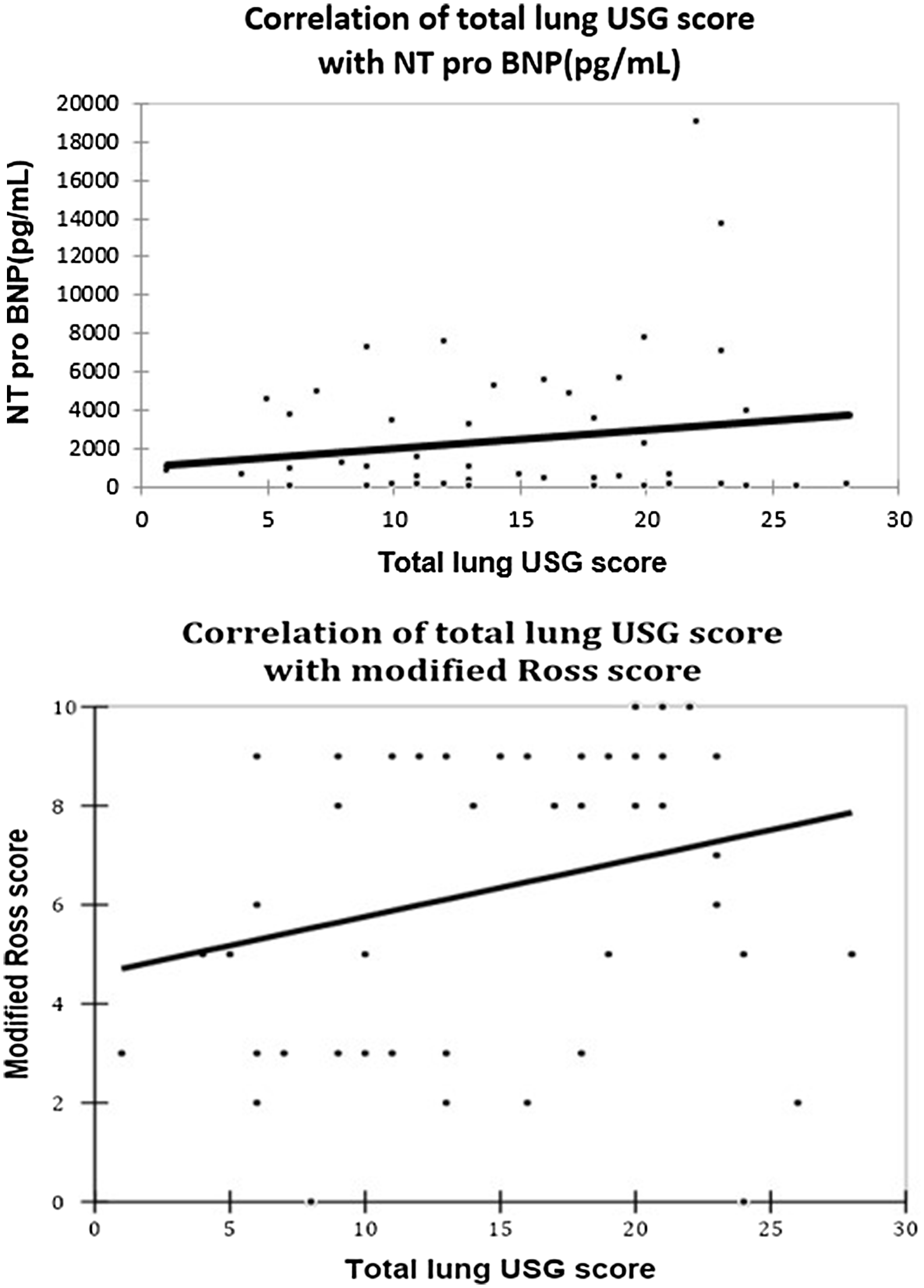
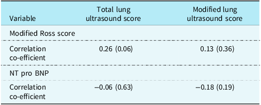
No significant correlation was seen between lung ultrasound scores and Ross score or NT pro BNP levels as shown in Table 2 and Figure 1.
Shows the correlation between total lung ultrasound score and modified Ross score and NT pro BNP in a graphical form.

Showing distribution of lung score and correlation of total and modified lung score with modified Ross score and NT pro-BNP

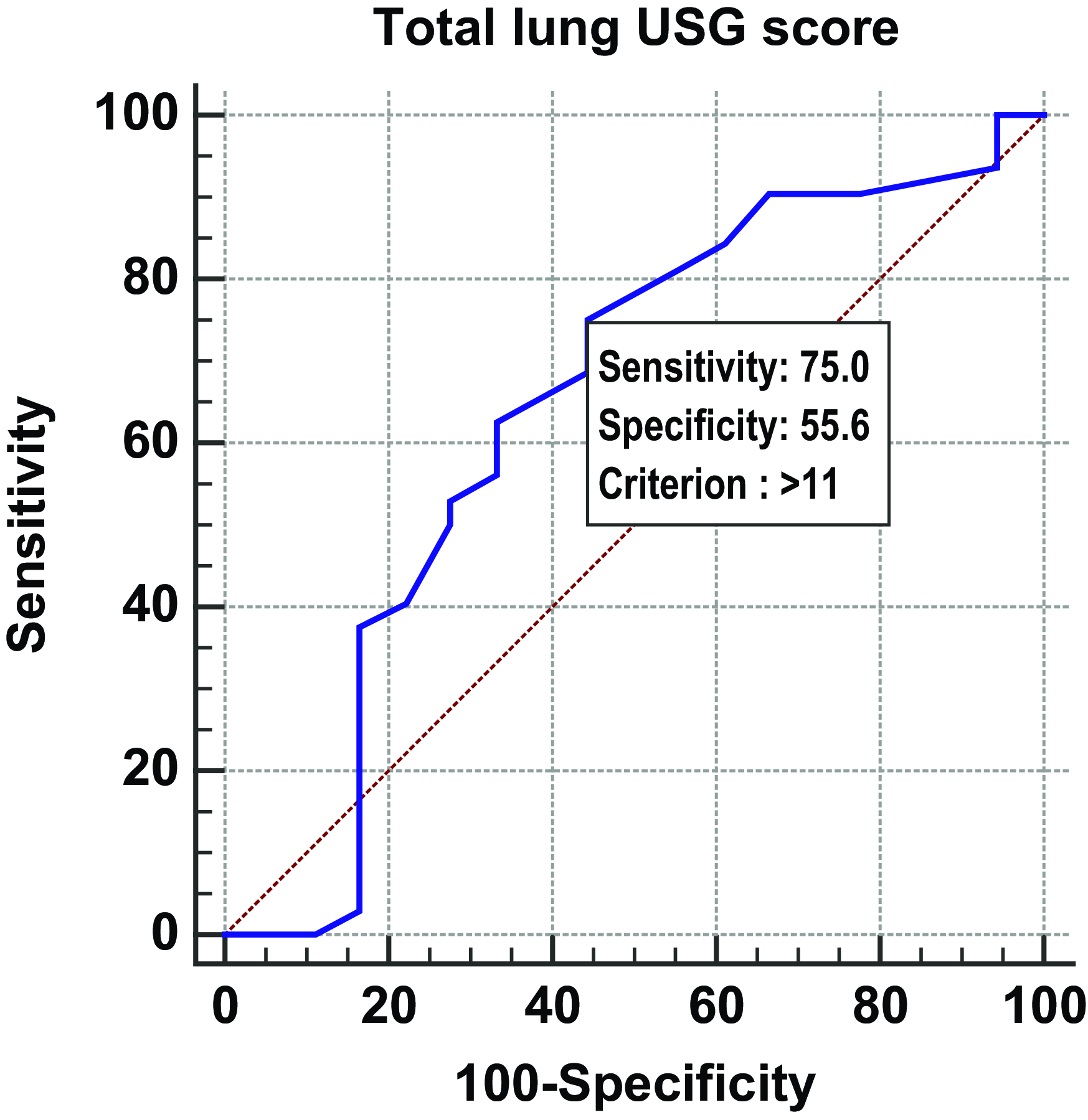
Receiver operator curve (ROC) was plotted to find the optimal lung ultrasound score to identify heart failure (Fig 2). Lung ultrasound score cut-off of 11 had a sensitivity of 75 % and specificity of 55% in detecting moderate to severe heart failure with area under the curve (AUC) of 0.64. The PPV, NPV and accuracy was 75.0%, 55.6% and 68.0 % respectively. (Moderate and severe congestive heart failure were defined as Ross scores of 6 and >10 respectively)
Showing the ROC curve for the optimal cut-off of lung ultrasound to detect moderate to severe CHF.

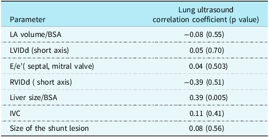
Various Echocardiographic parameters were also correlated with the Lung ultrasound score, as shown in Table 3. No correlation was seen with any parameters except the Body surface area indexed liver size.
Showing the correlation co-efficient of various echocardiographic parameters with the lung ultrasound score

LA: left atrium, BSA: body surface area, LVIDd: left ventricular internal dimension in diastole, RVIDd: right ventricular internal dimension in diastole, IVC: inferiorcaval vein.
Discussion
This is the first study to examine the relationship of a semi-quantitative lung ultrasound score with severity of heart failure in children with unoperated CHD. In a previous study in adults with heart failure, a significant linear correlation was found between the simplified ultrasound score, NT pro-BNP values Reference Sugimoto, Manabe and Nakau22 (r = 0.50; P < 0.0001), and NYHA functional class (r = 0.44, P < 0.0001). Reference Li, Li and Zhu10 It was expected that similar correlation would be present between LUS score, modified Ross score, and NT pro BNP in children with left to right shunt. In this study, though there was a positive correlation between LUS score and modified Ross score, the correlation coefficient was low (r = 0.262) and did not reach significance (p = 0.06). The AUC generated by the ROC for detecting moderate to severe heart failure was also low (0.64) and had a limited sensitivity, specificity, and diagnostic accuracy. Moreover, correlation between the modified lung ultrasound score and NT pro-BNP was not significant.
Lung ultrasound has been used as a test to diagnose pulmonary overflow in children with unoperated CHD and has been found to have higher diagnostic accuracy than chest radiographs in previous studies. Reference Rodríguez-Fanjul, Llop, Balaguer, Bautista-Rodriguez, Hernando and Jordan17,Reference Wu, Hou and Lu18 Pulmonary overflow or congestion was detected based on the presence of B lines in lung ultrasound in these studies. However, these studies did not attempt to correlate the degree of pulmonary congestion as seen in lung ultrasound with the clinical severity of symptoms or NT pro BNP.
The B lines can appear in lung ultrasound due to a variety of causes; however, the exact pathophysiology of B lines seen in CHD with left to right shunt is not clear. Presumably increased pulmonary blood flow due to left to right shunt causes an increase in pulmonary capillary pressure. This may lead to increased fluid in the interstitium that could have been picked up as B lines in previous studies. Based on this premise, the larger the left to right shunt, the greater the hydrostatic pressure and therefore a greater number of B lines would be expected compared to a smaller shunt. However, our study does not demonstrate this. One possible reason for this is as follows. Apart from an increase in pulmonary capillary pressure, other pathophysiologic mechanisms also occur concurrently in children with shunt lesions which can leads to the cardio-respiratory symptoms. Pulmonary atelectasis, bronchial mucosal congestion, bronchial compression, air trapping, and collapse of the lungs are other factors apart from increased pulmonary capillary pressure which contribute to the symptoms in these children. Reference Demi, Mento and Di Sabatino23 Only some of these factors may manifest as B lines on lung ultrasound. The relative proportion of each of these components in a particular patient could have confounded the measurement of the lung ultrasound score as well as the Ross score and the NT pro-BNP. This could have resulted in the lack of correlation between these parameters. The fact that the LUS score did not show a correlation with NT pro-BNP levels and echocardiographic parameters of Left sided volume overload, suggests that elevated pulmonary capillary pressure may not be the predominant mechanism for producing the B lines seen in these infants.
Most of the B lines seen in these children were in the posterior chest wall. The predominance of pathology in the dependent lung zones has been seen in previous studies in neonates/infants. Reference Jambrik, Monti and Coppola24,Reference Cochran, Gyepes and Smith25 However, lung ultrasound findings related to pulmonary overflow are also likely to be affected by gravity and more likely to be found in the dependent regions of the lung. Higher B-line score in the posterior lung fields of children with left to right shunt as seen in this study has an implication for physiotherapy and patient positioning and are important to take note of.
Limitaion
There were several limitations of this study. This study was done in a small population based in a single institution and its results may not be generalisable. The appearance of ultrasound artifacts and B line counting is an operator-dependent semi-quantitative process. Intra or inter-observer variation was not assessed for the lung ultrasound scoring, which is a limitation of this study. The effect of diuretics/ fluid restriction on lung ultrasound score and Ross score was not looked into in this study, ideally this should have been accounted for. However, any effect of these factors would be similar on the lung ultrasound score as well as the modified Ross score also. Therefore, the extent of confounding by this factor might be limited. Reference Liang, Liang and Chen26
There was lack of correlation between modified Ross score and NT pro BNP in our study. Though, our study was not powered to test this correlation, this makes our interpretation less convincing. 40 % of our population were less than 3 months. As reported in the literature, there is marked and rapid increase in NT pro BNP after birth, which gradually decreases in the first few months, and there is wider variability of NT pro BNP in this age group. Reference Lam, Higgins and Zhang27 This could be one of the reasons for the poor correlation.
Finally, the failure to exclude the presence of subclinical infection or atelectasis is a major limitation of this study. Reference Song, Kim, Lee, Ro, Kim and Kim28 These factors could have resulted in the lack of correlation between lung ultrasound scores and the clinical scores of heart failure. Though infants with clinically active infection or those with fever within 10 days prior to enrolment were excluded from the study, these criteria might not be sufficient to exclude subclinical respiratory infection. In future, studies on this subject should also comment on the pleural abnormalities along with the B lines to characterise the lung abnormalities fully.
Conclusion
In this study, lung ultrasound scores derived from B lines did not have a correlation with the modified Ross score or NT pro-BNP. B lines in children with left to right post tricuspid shunts are present more often in the dependent regions of the chest. More research is needed on this topic before lung ultrasound can be used to detect the severity of heart failure in infants with left to right shunts.
Supplementary material
The supplementary material for this article can be found at https://doi.org/10.1017/S1047951125001738.
Acknowledgements
None.
Competing interests
None.
Ethical standards
The authors assert that all procedures contributing to this work comply with the ethical standards of the relevant national guidelines on human experimentation (please name) and with the Helsinki Declaration of 1975, as revised in 2008, and has been approved by the institutional committee.