Introduction
Microphallid infections are the dominant trematode infections among intertidal snails in many marine systems (Jokela & Lively, Reference Jokela and Lively1995; Madhavi et al., Reference Madhavi, Umadevi and Swarnakumari1997; Al-Kandari et al., Reference Al-Kandari, Abdul-Salam, Moussa and Sreelatha2007). Maritrema eroliae and Probolocoryphe uca (Digenea: Microphallidae) are considered to be the most common microphallid infections parasitizing the snails Clypeomorus bifasciata and Cerithidea cingulata, respectively, at Kuwait Bay (Abdul-Salam et al., Reference Abdul-Salam, Sreelatha and Ashkanani1994; Abdul-Salam & Sreelatha, Reference Abdul-Salam and Sreelatha1998; Al-Kandari et al., Reference Al-Kandari, Abdul-Salam and Meakins2000, Reference Al-Kandari, Abdul-Salam, Moussa and Sreelatha2007). The flukes share sympatric geographic ranges in Kuwait Bay, using migratory birds as definitive hosts. Their larval stages utilize co-occurring snail intermediate hosts from Prosobranchia. The discrimination of adults of both genera is easy because of significant differences in size, shape and several other morphological characteristics (Abdul-Salam & Sreelatha, Reference Abdul-Salam and Sreelatha2000; Al-Kandari et al., Reference Al-Kandari, Abdul-Salam, Moussa and Sreelatha2007). Unlike adult worms, intramolluscan larval stages of both microphallids are morphologically similar and almost indistinguishable (Abdul-Salam & Sreelatha, Reference Abdul-Salam and Sreelatha1998; Al-Kandari et al., Reference Al-Kandari, Al-Bustan and Alnaqeeb2011). The morphological differences between their larval stages are often based on the size and shape of the cercariae and their stylets. Their association with the adult flukes by morphological characteristics alone is very difficult. The overlapping spectrum of intermediate hosts and geographical areas they inhabit makes discrimination between them even more difficult. The larval stages have ecological importance in their environment due to their ability to castrate their snail intermediate hosts and cause mortality in the second intermediate host, hence influencing the population dynamics of their hosts (Fredensborg et al., Reference Fredensborg, Mouritsen and Poulin2005; Fredensborg & Poulin, Reference Fredensborg and Poulin2006). Therefore, proper identification of the larval stages of M. eroliae and P. uca is desirable for ecological studies (Hust et al., Reference Hust, Frydenberg, Sauriau, Le Gall, Mouritsen and Jensen2004) as well as for understanding the transmission dynamics between primary, secondary and final hosts. Since morphological examination could not provide reliable differentiation of M. eroliae and P. uca in infected molluscs, molecular techniques have been developed for that purpose (Al-Kandari & Al-Bustan, Reference Al-Kandari and Al-Bustan2010; Al-Kandari et al., Reference Al-Kandari, Al-Bustan and Alnaqeeb2011).
Polymerase chain reaction (PCR)-based techniques and sequence analysis of selected genes is often used for reliable identification and differentiation of different trematode species (Barber et al., Reference Barber, Mkoji and Loker2000; Sugiyama et al., Reference Sugiyama, Morishima, Kameoka and Kawanaka2002; Kralova-Hromadova et al., Reference Kralova-Hromadova, Spakulova, Horackova, Turcekova, Novobilsky, Beck, Koudela, Marinculic, Rajsky and Pybus2008; Francisco et al., Reference Francisco, Almeida, Castro and Santos2010; Ichikawa & Itagaki, Reference Ichikawa and Itagaki2010). Recently, the authors deposited the complete sequences of internal transcribed regions ITS1 and ITS2 rDNA of M. eroliae and P. uca in GenBank (HQ650133 and GQ377842). The ITS2 sequences of M. eroliae and P. uca were found to be conserved at the species level, with fixed differences between the two microphallids, while their ITS1 sequences were far more variable, with intraspecific variations (Al-Kandari & Al-Bustan, Reference Al-Kandari and Al-Bustan2010; Al-Kandari et al., Reference Al-Kandari, Al-Bustan and Alnaqeeb2011). These sequences have been used for the accurate identification of both microphallids as well as in phylogenetic studies. The sequences were also helpful in resolving parts of the life-cycles of both microphallids (Al-Kandari & Al-Bustan, Reference Al-Kandari and Al-Bustan2010; Al-Kandari et al., Reference Al-Kandari, Al-Bustan and Alnaqeeb2011). Although DNA types based on sequences of ITS2 regions are of value to discriminate accurately between M. eroliae and P. uca, DNA sequencing is costly, time consuming and needs a sophisticated set-up. Therefore, the development of quick and cost-effective techniques is required for the accurate discrimination between the larval stages of the two microphallid species from Kuwait Bay. To the best of our knowledge, molecular discrimination between M. eroliae and P. uca microphallids has not been described to date. In the present study we describe PCR-restriction fragment length polymorphism (PCR-RFLP) and PCR with species-specific primers that allow quick and accurate differentiation between larval stages of M. eroliae and P. uca.
Materials and methods
Collection and examination of snails for sporocysts
The gastropods C. bifasciata and C. cingulata (Gastropoda: Prosobranchia) were collected at low tide from Shuwaikh Bay, located along the northern coast of Kuwait (29°21′N, 47°57′E). In total, 1000 snails were handpicked from mud and rocks covering the shore during the period from April 2009 to September 2009. In the laboratory, snails were maintained in aerated seawater at about 20°C. Snails were gently crushed with a hammer and their soft tissues were examined for the presence of microphallid sporocysts using a dissecting microscope. The identification of M. eroliae and P. uca was made with vitally stained (0.5% neutral red) specimens based on morphological characteristics, as described by Abdul-Salam & Sreelatha (Reference Abdul-Salam and Sreelatha1998) and Al-Kandari et al. (Reference Al-Kandari, Abdul-Salam, Moussa and Sreelatha2007). Morphological identification had been studied previously and confirmed by molecular identification using rDNA sequences (Al-Kandari & Al-Bustan, Reference Al-Kandari and Al-Bustan2010; Al-Kandari et al., Reference Al-Kandari, Al-Bustan and Alnaqeeb2011). For each M. eroliae or P. uca infection, the sporocysts were cleaned from snail tissues and stored in 95% ethanol for subsequent DNA extraction.
DNA extraction, PCR amplification and PCR-RFLP analysis
Individual parasite sporocysts were washed with milli-Q filtered water (mqH2O) to remove traces of ethanol. The genomic DNA (gDNA) was extracted using proteinase K digestion and organic/aqueous phase extraction (Barber et al., Reference Barber, Mkoji and Loker2000).
The ITS2 region of both microphallid species was amplified using the primer combination HC2/LC1 as described by Després et al. (Reference Després, Imbert-Establet, Combes and Bonhomme1992). The PCR reactions were performed as described by Al-Kandari & Al-Bustan (Reference Al-Kandari and Al-Bustan2010). DNA ladder markers of lengths 50 and 123 bp (Sigma, St. Louis, Missouri, USA) were used to estimate the size of the amplified products. PCR products were purified using Nucleospin Extract II kit (Macherey Nagel, Duren, Germany) according to the manufacturer's protocol.
The previously published ITS2 sequences of M. eroliae and P. uca (Al-Kandari & Al-Bustan, Reference Al-Kandari and Al-Bustan2010; Al-Kandari et al., Reference Al-Kandari, Al-Bustan and Alnaqeeb2011) were subjected to NEB cutter 2.0 (Vincze et al., Reference Vincze, Posfai and Roberts2003) to detect the candidate restriction site. A restriction site (CCACG) for AvaII was detected in the ITS2 of M. eroliae but not for P. uca. For restriction analysis, 1 μg of the PCR products were subjected to digestion overnight at 37°C with the enzyme in a total volume of 50 μl, containing 10 U of AvaII and NEBuffer 4 (New England Biolabs, Beverly, Massachusetts, USA). The restriction fragments were electrophoresed on a 4% Nusieve-agarose gel stained with 10 μg/ml ethidium bromide. Samples without DNA were included as no-DNA controls in each PCR amplification run to exclude potential carry-over contamination. Different numbers of sporocysts (10, 50, 100 sporocysts) were used for gDNA extraction in order to evaluate the sensitivity of the PCR-RFLP method.
PCR with species-specific primers
To establish a more direct PCR procedure for species discrimination and identification based on the previously published ITS2 sequences (Al-Kandari & Al-Bustan, Reference Al-Kandari and Al-Bustan2010; Al-Kandari et al., Reference Al-Kandari, Al-Bustan and Alnaqeeb2011), species-specific primers were designed using Primer3 (Rozen & Skaletsky, Reference Rozen, Skaletsky, Krawetz and Minsener2000). The following primers were designed to target unique regions of the ITS2 sequence of M. eroliae and P. uca, respectively:
MeR1: 5′GCCAACTGTGTGAATTAATGTG3′;
MeR2: 5′AATGAACCGAACAACCAGTGA3′;
PuR1: 5′CTGTGTATATGTGACTGC3′;
PuR2: 5′CAGGCCCTAAAGCGCACA3′.
The PCR reactions were performed in a total volume of 25 μl (2.5 μl of 10 × PCR buffer, 2.5 mm MgCl2, 10 μm of each deoxynucleoside triphosphate (dNTP), 1.5 μm of each primer and 1 U of Amplitaq Gold DNA polymerase). The PCR conditions used were: an initial denaturation step at 91°C for 12 min, followed by 35 cycles of 30 s at 91°C, 30 s at 58°C and 2 min at 72°C, followed by a final extension of 7 min at 72°C, and maintained thereafter at 4°C. The PCR amplicons were resolved on a 1.2% agarose gel stained with 10 μg/ml ethidium bromide. The primer set HC2/LC1 (Després et al., Reference Després, Imbert-Establet, Combes and Bonhomme1992) was used as a control for the presence of microphallid genomic DNA in each sample.
The PCR products were purified using Nucleospin Extract II kit (Macherey Nagel) according to the manufacturer's protocol. To confirm their identity to the ITS2 regions of M. eroliae and P. uca, the PCR products were sequenced using the corresponding specific primer for the forward and reverse sequencing in two separate reactions. The purified PCR products were then sequenced in a 3130XL Gene Analyzer using the Big Dye Terminator kit (Version 3.1) according to the manufacturer's instructions (Applied Biosystems, Foster City, California, USA). The sequences were first analysed using EMBOSS (Rice et al., Reference Rice, Longden and Bleasby2000) and aligned using ClustalW2 (Thompson et al., Reference Thompson, Higgins and Gibson1994). The obtained ITS2 sequences were compared with the corresponding sequences of M. eroliae and P. uca in GenBank (accession numbers HQ650133 and GQ377842) and to those published in earlier studies (Al-Kandari & Al-Bustan, Reference Al-Kandari and Al-Bustan2010; Al-Kandari et al., Reference Al-Kandari, Al-Bustan and Alnaqeeb2011).
Results
PCR-RFLP analysis
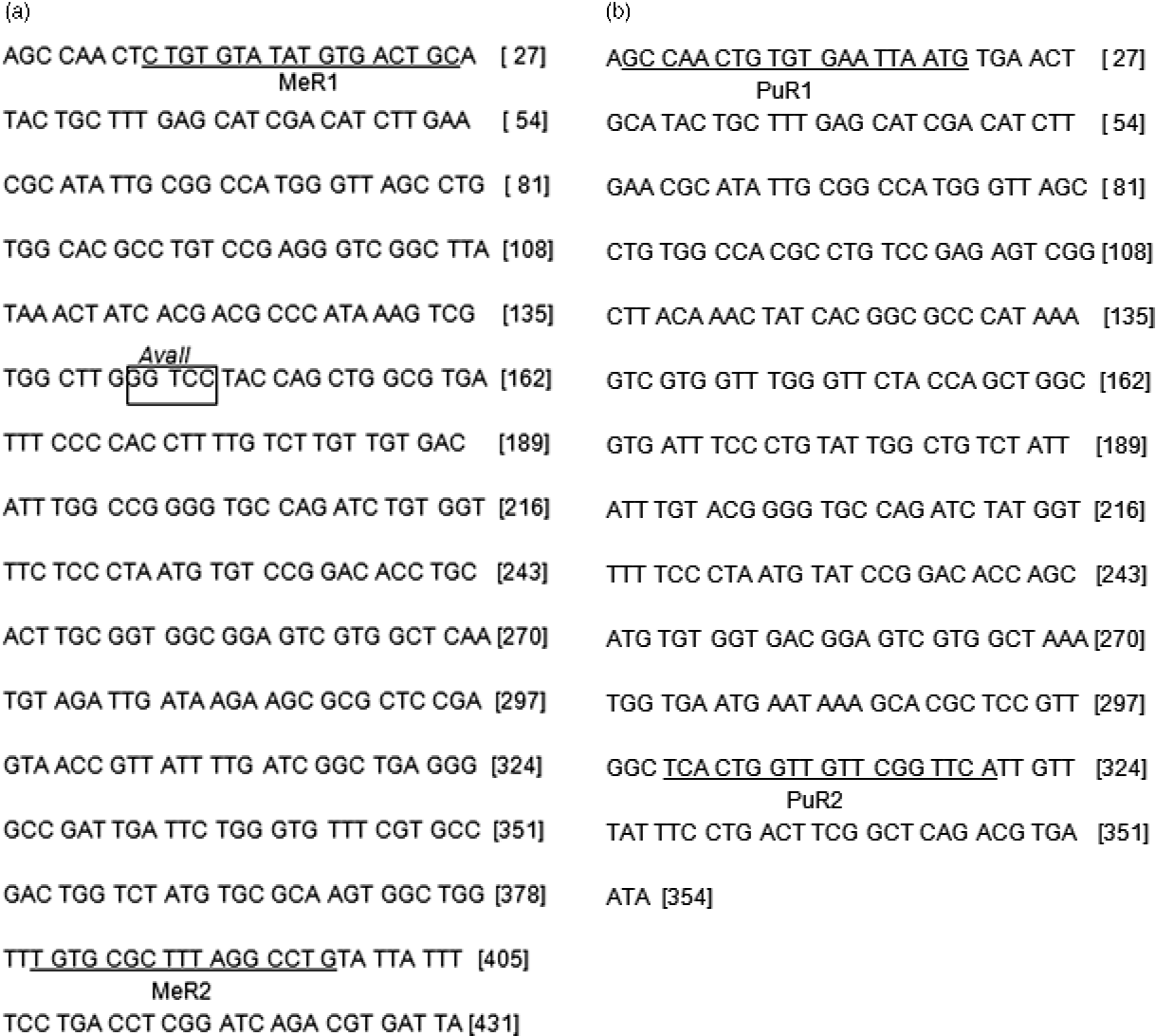
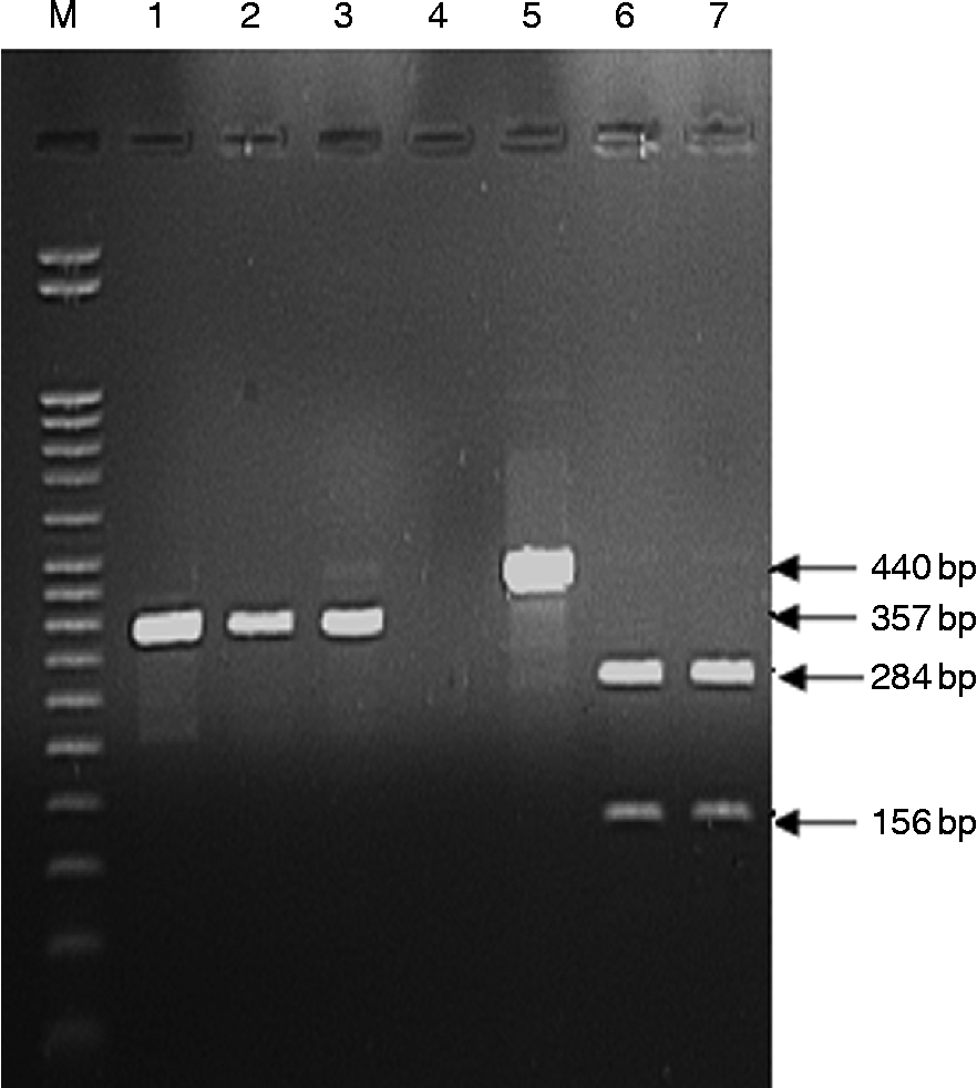
DNA fragments from the ITS2 regions of 47 samples of M. eroliae and 92 samples of P. uca were successfully amplified. The obtained sequences were compared for all samples of both microphallid species and were found to be identical in length and composition for each species. Search of the nucleotide databases revealed that the sequences of the ITS2 were identical to those deposited previously in GenBank with accession numbers HQ650133 for M. eroliae and GQ377842 for P. uca. The sequences thus obtained were analysed to select an appropriate restriction enzyme for the accurate discrimination between M. eroliae and P. uca sequences using the PCR-RFLP method. The endonuclease AvaII was selected on the basis of the theoretical restriction maps generated from the previously published sequences of ITS2 from M. eroliae and P. uca (Al-Kandari & Al-Bustan, Reference Al-Kandari and Al-Bustan2010; Al-Kandari et al., Reference Al-Kandari, Al-Bustan and Alnaqeeb2011) (fig. 1). The undigested PCR products from the PCR amplification were about 440 and 357 bp in length for all samples of M. eroliae and P. uca, respectively (fig. 2). When the PCR products were digested with AvaII, two fragments – 284 bp and 156 bp – were produced from the PCR product of M. eroliae samples. However, the PCR product from P. uca samples remained undigested with AvaII (fig. 2). No intraspecific variation in the RFLP patterns was detected for any of the DNA preparations from either microphallid when the PCR products were digested with AvaII enzyme. These results were reproduced successfully using the ITS2 sequences for all the 47 samples of M. eroliae and 92 samples of P. uca. The PCR-RFLP results were reproducible regardless of the numbers of sporocysts (10, 50, 100 sporocysts) from which gDNA was extracted for each species.
Sequences of the ITS2 rDNA region of the microphallid species. (a) Maritrema eroliae, where the box represents the recognition site of the AvaII restriction enzyme. The primer annealing sites of (MeR1/MeR2) are underlined. (b) Probolocoryphe uca, where the primer annealing sites of (PuR1/PuR2) are underlined.

RFLP patterns of ITS2 PCR products of Maritrema eroliae and Probolocoryphe uca from Kuwait Bay after digestion with AvaII enzyme and visualized on a 3:1 Nusieve-agarose gel stained with 10 μg/ml ethidium bromide. Lanes: M, 50 bp DNA marker; 1, PCR products of P. uca; 2 and 3, PCR products of P. uca after digestion with AvaII; 4, AvaII enzyme reaction with no DNA; 5, PCR products of M. eroliae; 6 and 7, PCR products of M. eroliae after digestion with AvaII.
PCR with species-specific primers
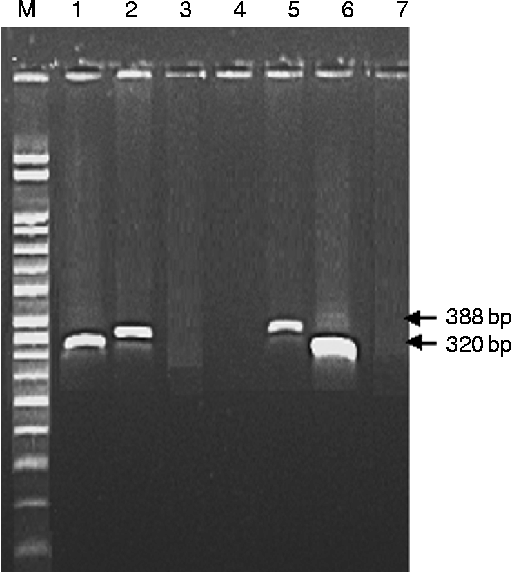
The M. eroliae-specific primers (MeR1/MeR2) and P. uca-specific primers (PuR1 /PuR2) were designed to target the ITS2 region in both microphallids (fig. 1). As expected, the primer set MeR1/MeR2 amplified a PCR product of about 388 bp from M. eroliae DNA but not from P. uca DNA (fig. 3). In contrast, PuR1/PuR2 amplified a product of about 320 bp from P. uca DNA but not from M. eroliae DNA (fig. 3). No intraspecific variation in the PCR patterns was detected. These PCR products were sequenced using the corresponding specific primers and were confirmed to be sequences from the ITS2 region of rDNA from the respective microphallid species. These results were reproduced successfully using the ITS2 sequences for all the 47 samples of M. eroliae and 92 samples of P. uca, regardless of the numbers of sporocysts used for genomic DNA extraction.
PCR amplification of genomic DNA of Maritrema eroliae and Probolocoryphe uca from Kuwait Bay using species-specific ITS2 primers and visualized on a 1.2% agarose gel stained with 10 μg/ml ethidium bromide. Lanes: M, 50 bp DNA marker; 1, PCR products of P. uca with trematode-specific primers; 2, PCR products of M. eroliae with MeR1–MeR2 primers; 3, PCR products of P. uca with MeR1–MeR2 primers; 4, reaction with no DNA; 5, PCR products of M. eroliae with trematode-specific primers; 6, PCR products of P. uca with PuR1–PuR2 primers; 7, PCR products of M. eroliae with PuR1–PuR2 primers.
Discussion
Information gained from DNA through the application of PCR and sequencing techniques is being increasingly applied as a supplement to traditional morphological approaches for trematode identification (Hust et al., Reference Hust, Frydenberg, Sauriau, Le Gall, Mouritsen and Jensen2004). Revealing the sequences of ITS1 and ITS2 enabled the distinction between closely related trematode species and the identification of trematode species at any stage of their life cycles (Morgan & Blair, Reference Morgan and Blair1995). Recently, there has been progress in developing molecular methods for the identification of two microphallid species, M. eroliae and P. uca from Kuwait Bay, using genetic markers identified in rDNA. The phylogenetic relationships of M. eroliae and P. uca were examined in earlier studies using sequences of the ITS region (Al-Kandari & Al-Bustan, Reference Al-Kandari and Al-Bustan2010; Al-Kandari et al., Reference Al-Kandari, Al-Bustan and Alnaqeeb2011). In these studies, complete sequences of the ITS1 and ITS2 regions were generated from the larval stages of both microphallids using direct sequencing methods. Although DNA typing based on ITS1 and ITS2 sequences enabled the discrimination between M. eroliae and P. uca, DNA sequencing remained time-consuming, laborious and required relatively sophisticated and expensive set-ups. On the other hand, PCR-RFLP and direct PCR amplification using species-specific primers promises rapid, inexpensive and simple species discrimination. Variations in the ITS1 sequences (0.2% for M. eroliae, 7% for P. uca) (Al-Kandari & Al-Bustan, Reference Al-Kandari and Al-Bustan2010; Al-Kandari et al., Reference Al-Kandari, Al-Bustan and Alnaqeeb2011) among members of the same species do not permit reliable species-specific primer design or the selection of restriction enzymes for RFLP. On the other hand, ITS2 sequences are well conserved among members of the same species and provide fixed differences between species (Hust et al., Reference Hust, Frydenberg, Sauriau, Le Gall, Mouritsen and Jensen2004). For that reason, ITS2 sequences were used in this study as genetic markers to discriminate between M. eroliae and P. uca using RFLP analysis and species-specific primers.
PCR-RFLP analysis of ITS sequences has received considerable attention as a reliable approach for discriminating closely related trematode species that lack pronounced morphological differences, such as Paragonimus westermani and Paragonimus miyazakii (Sugiyama et al., Reference Sugiyama, Morishima, Kameoka and Kawanaka2002), Fasciola hepatica and Fascioloides magna (Kralova-Hromadova et al., Reference Kralova-Hromadova, Spakulova, Horackova, Turcekova, Novobilsky, Beck, Koudela, Marinculic, Rajsky and Pybus2008), Fasciola hepatica and Fasciola gigantica (Ichikawa & Itagaki, Reference Ichikawa and Itagaki2010), Opisthorchis viverrini, Opisthorchis felinus and Clonorchis sinensis (Kang et al., Reference Kang, Sultana, Loktev, Wongratanacheewin, Sohn, Eom and Park2008) and Schistosoma haematobium and Schistosoma bovis (Barber et al., Reference Barber, Mkoji and Loker2000). In the current work, the PCR-RFLP-based approach has differentiated successfully between the larval stages of M. eroliae and P. uca. AvaII digested the ITS2 sequence of M. eroliae and produced two easily visualized bands sized 284 and 156 bp whereas the PCR products from P. uca remained uncut (fig. 2). However, this method does not seem to be suitable for surveying snails, such as C. cingulata and C. bifasciata, infected with other digenean parasites, because the primers used for the retrieval of the complete ITS2 region appear to anneal to highly conservative regions of 5.8S and 28S of most digenetic trematodes (Kralova-Hromadova et al., Reference Kralova-Hromadova, Spakulova, Horackova, Turcekova, Novobilsky, Beck, Koudela, Marinculic, Rajsky and Pybus2008) and, as such, they amplify the ITS2 region for all digenean parasites infecting the same snail host. It has been reported that the snail intermediate hosts of M. eroliae and P. uca may harbour between 12 (Abdul-Salam et al., Reference Abdul-Salam, Sreelatha and Ashkanani1994; Abdul-Salam et al., Reference Abdul-Salam, Sreelatha and Al-Kandari1997; Al-Kandari et al., Reference Al-Kandari, Abdul-Salam, Moussa and Sreelatha2007) and 15 (Abdul-Salam & Sreelatha, Reference Abdul-Salam and Sreelatha1998) different trematode infections, respectively. Thus it is possible that the digenean-specific primers used in this study to amplify the ITS2 region from M. eroliae and P. uca can also amplify the same region in other trematode species whose DNA sequences are still not available in GenBank and, thus, may contain the AvaII restriction site. In this case, the discrimination between those trematode species and M. eroliae and P. uca would be impossible using this technique.
This called for the more rigorous second approach proposed in this study, which used PCR with species-specific primers that allowed more distinctive differentiation between M. eroliae and P. uca (fig. 3). The primers were designed for the ITS2 region, which exhibits high interspecific variation sites and no variations within each species. The designed primers do not show any similarity to the ITS2 sequence of the unclassified microphallid species available in GenBank (accession number FJ966111). However, it is possible that there could be other undescribed microphallid species in related parasite–host systems that may show nucleotide sequences identical to one of the primers prepared in this study. Thus, the general applicability of our primers cannot be verified until more microphallid ITS2 sequences become available in GenBank.
Our approach to the molecular discrimination of M. eroliae and P. uca was twofold, either by PCR amplification of the complete ITS2 and its consequent RFLP analysis, or by PCR amplification of the selected ITS2 region with highly species-specific primers. In PCR-RFLP, two steps were required, PCR amplification and restriction of the PCR products with an endonuclease. In the direct PCR amplification using species-specific primers, only one step was required. The second approach is more advantageous because it enables more rapid discrimination of the microphallid species. Beside technical benefits, this method has the potential to be more specific in the case of hosts infected with a variety of parasite species. In future studies, we expect this approach to be more reliable in the elucidation of the definitive hosts for M. eroliae and P. uca from Kuwait Bay. We anticipate, with the continuing advances in next-generation sequencing methods, that microphallid species-specific primers would be a better and more reliable approach for species identification.
In conclusion, this is the first report of molecular discrimination between M. eroliae and P. uca using PCR-RFLP and PCR with species-specific primers. Amplifying the ITS2 region with species-specific primers allows quick and reliable differentiation between M. eroliae and P. uca from Kuwait Bay, where both microphallids coexist, eliminating the need for sequencing. Also, it facilitates the screening of a larger number of hosts from different geographical areas at a relatively low cost and within a shorter time-frame than sequencing, providing an ideal solution for wide-scale field and environmental studies.
Acknowledgements
This work was supported by Kuwait University Research Grant No. [SL08/08]. The authors gratefully acknowledge Dr Devon B. Keeney of the Department of Biological Sciences, Le Moyne College, New York, USA for reviewing the manuscript. Molecular analysis was performed with the support of the General Facility Project (GS 01/02) in the use of the ABI 3130Xl Gene Analyzer at the Biotechnology Center, Faculty of Science, Kuwait University.