INTRODUCTION
Recent advances in biomarkers have led to the definition of a preclinical stage of Alzheimer’s disease (AD), in which pathophysiologic changes (e.g., accumulation of amyloid in the neocortex and tau in the medial temporal lobe) occur many years before the onset of cognitive impairment (Jack et al., Reference Jack, Bennett, Blennow, Carrillo, Dunn and Haeberlein2018, Reference Jack, Knopman, Jagust, Shaw, Aisen, Weiner and Trojanowski2010). Given that individuals in the preclinical stage represent a group at high risk for cognitive decline, there is a need to characterize the cognitive performance of individuals in this stage of the disease and how cognitive performance relates to the earliest pathological changes in AD.
The study of families with autosomal dominant AD (ADAD) offers a unique opportunity to characterize the preclinical stage of AD, as carriers of ADAD are virtually guaranteed to develop the disease, allowing the investigation of the earliest cognitive changes of AD (Fuller et al., Reference Fuller, Cronin-Golomb, Gatchel, Norton, Guzmán-Vélez, Jacobs and Quiroz2019). Studies conducted in ADAD have suggested that changes in verbal episodic memory (VeM) can be observed approximately 10 years before the onset of clinical impairment (Aguirre-Acevedo et al., Reference Aguirre-Acevedo, Lopera, Henao, Tirado, Muñoz, Giraldo and Jaimes2016; Bateman et al., Reference Bateman, Xiong, Benzinger, Fagan, Goate, Fox and Network2012). In addition, it has been shown that greater cortical amyloid and higher levels of regional tau are associated with worse VeM performance in cognitively unimpaired ADAD carriers (Quiroz et al., Reference Quiroz, Sperling, Norton, Baena, Arboleda-Velasquez, Cosio and Johnson2018).
Evidence suggests that accumulation of pathology in the early stages of AD involves brain pathways and structures that support memory processing (Schultz et al., Reference Schultz, Gordon, Mishra, Su, Perrin, Cairns and Benzinger2018; Sperling et al., Reference Sperling, Mormino, Schultz, Betensky, Papp, Amariglio and Johnson2019). It has been observed, for example, that in cognitively unimpaired ADAD mutation carriers the earliest sites of tau accumulation involve regions of the medial temporal lobe (e.g., the entorhinal cortex) and inferior temporal lobe (Quiroz et al., Reference Quiroz, Sperling, Norton, Baena, Arboleda-Velasquez, Cosio and Johnson2018). In addition, results from human lesion studies and functional neuroimaging studies in cognitively normal adults suggest that these structures and their projections play a critical role not only in VeM but also in visual memory (ViM) processing (Barbeau, Pariente, Felician, & Puel, Reference Barbeau, Pariente, Felician and Puel2011; Braskie, Small, & Bookheimer, Reference Braskie, Small and Bookheimer2009; Fiebach, Rissman, & D’Esposito, Reference Fiebach, Rissman and D’Esposito2006; Ranganath, Cohen, Dam, & D’Esposito, Reference Ranganath, Cohen, Dam and D’Esposito2004).
In contrast to VeM, ViM and its relationship with markers of AD pathology have been less explored in preclinical ADAD. Studies conducted in subjects at risk of developing sporadic AD—in which ViM was included within a larger memory composite—indicated that relative to amyloid-negative older adults, amyloid-positive cognitively normal older adults exhibited greater memory impairment (Lim et al., Reference Lim, Kalinowski, Pietrzak, Laws, Burnham, Ames and Maruff2018; Pike et al., Reference Pike, Savage, Villemagne, Ng, Moss, Maruff and Rowe2007). However, it is unclear whether changes in ViM can be observed in individuals with ADAD who are still cognitively unimpaired, and whether these changes are associated with tau pathology, which has been closely associated with the onset of cognitive decline (Quiroz et al., Reference Quiroz, Sperling, Norton, Baena, Arboleda-Velasquez, Cosio and Johnson2018).
The Rey–Osterrieth Complex Figure test recall (ROCF-r) (Rey, Reference Rey2003) is among the most widely used tests of ViM. The alterations observed in this test have been well documented in patients with mild cognitive impairment (MCI) (Lekeu et al., Reference Lekeu, Magis, Marique, Delbeuck, Bechet, Guillaume and Salmon2010; Peter et al., Reference Peter, Sandkamp, Minkova, Schumacher, Kaller, Abdulkadir and Klöppel2018) and mild dementia (Salimi et al., Reference Salimi, Irish, Foxe, Hodges, Piguet and Burrell2018), but it is less known how ROCF-r performance relates to in vivo markers of AD pathology in the preclinical stage of AD.
The present study was conducted with young, cognitively unimpaired Presenilin1 (PSEN1) E280A mutation carriers from the world’s largest ADAD kindred who have previously shown to have accumulation of AD pathology and changes in VeM many years before the median age of onset of MCI in this cohort (Aguirre-Acevedo et al., Reference Aguirre-Acevedo, Lopera, Henao, Tirado, Muñoz, Giraldo and Jaimes2016; Fleisher et al., Reference Fleisher, Chen, Quiroz, Jakimovich, Gutierrez Gomez, Langois and Reiman2015; Quiroz et al., Reference Quiroz, Sperling, Norton, Baena, Arboleda-Velasquez, Cosio and Johnson2018). In this study, we extend previous results by exploring whether ViM impairment is evident in preclinical ADAD, and whether it is related to in vivo markers of AD pathology. We hypothesized that greater in vivo AD pathology and greater age would be related to worse ROCF-r performance in cognitively unimpaired mutation carriers.
METHODS
Participants
Forty-five cognitively unimpaired individuals from a Colombian kindred with the Presenilin 1 (PSEN1) E280A mutation (19 carriers and 26 age-matched noncarriers) were recruited from the Colombian Alzheimer’s Prevention Initiative (API) registry (Tariot et al., Reference Tariot, Lopera, Langbaum, Thomas, Hendrix, Schneider and Initiative2018). The clinical and preclinical courses of this cohort have been well characterized. Mutation carriers develop MCI around 44 years [95% CI 43–45] and dementia due to AD around the age of 49 [95% CI 49–50] (Acosta-Baena et al., Reference Acosta-Baena, Sepulveda-Falla, Lopera-Gómez, Jaramillo-Elorza, Moreno, Aguirre-Acevedo and Lopera2011). In mutation carriers, high levels of cortical amyloid burden are observed at an average age of 28 years (Fleisher et al., Reference Fleisher, Chen, Quiroz, Jakimovich, Gomez, Langois and Reiman2012), and elevated regional tau levels in the medial temporal lobe (e.g., entorhinal and inferior temporal cortices) at an average age of 38 years (Quiroz et al., Reference Quiroz, Sperling, Norton, Baena, Arboleda-Velasquez, Cosio and Johnson2018).
In this cross-sectional study, we included cognitively unimpaired individuals who had (1) a Mini-Mental State Examination (Folstein, Folstein, & McHugh, Reference Folstein, Folstein and McHugh1975) score of 26 or greater, (2) a Clinical Diagnostic Rating Scale (Morris, Reference Morris1993) score of 0, and (3) a Functional Assessment Staging Test (Reisberg, Reference Reisberg1988) score lower than 3 points. Patients with dementia or with other neurological or psychiatric disorders, such as clinical depression or anxiety, were excluded from the study.
This study was carried out in accordance with the principles of the Declaration of Helsinki and was approved by the Ethical Research Committee of University of Antioquia in Colombia and Massachusetts General Hospital (MGH) in Boston. All participants provided written informed consent. Participants and researchers were blind to genetic status.
Procedures
All the participants completed a comprehensive neuropsychological evaluation in Medellín, Colombia, at the University of Antioquia and traveled to Boston, USA, to undergo PET imaging at MGH. The data of this study are part of the Colombia–Boston (COLBOS) project, a longitudinal study aimed at studying biomarkers and cognitive changes in preclinical ADAD.
Visual memory: immediate recall of Rey–Osterrieth Complex Figure test (ROCF-r)
We measured ViM using the ROCF-r (Rey, Reference Rey2003). The ROCF test assesses both visuospatial and constructional abilities, as well as ViM. The test consists of a copy (ROCF-c) and immediate recall phase. Participants are first asked to copy a complex geometrical figure and immediately after the copy phase, they are asked to recall as much as possible. The maximum score for each phase is 36, and the scores were based on previously established criteria (Rey, Reference Rey2003). In this study, we analyzed the percent retention of the ROCF (ROCF-pr) in which the recall total score is divided by copy score and multiplied by 100. In contrast to the recall total score, ROCF-pr could be a more useful measure to detect alterations in ViM, given that it removes the effect of execution level from the copy phase (Boone, Lesser, Hill-gutierrez, Berman, & D’Elia, Reference Boone, Lesser, Hill-gutierrez, Berman and D’Elia1993).
To compare ViM with VeM, participants completed the word list memory test from the Colombian-normed Consortium to Establish a Registry for Alzheimer’s Disease (CERAD-m) (Aguirre-Acevedo et al., Reference Aguirre-Acevedo, Gómez, Moreno, Henao-Arboleda, Motta, Muñoz and Lopera2007). In this study, we included the word list learning and the word list free recall. The maximum number of correct responses of the learning phase and the recall phase is 30 and 10, respectively.
The ROCF-c and CERAD-m scores were transformed to Z scores based on the normative data of a larger sample of Colombians who were not ADAD mutation carriers (Torres et al., Reference Torres, Vila-Castelar, Bocanegra, Baena, Guzmán-Vélez, Aguirre-Acevedo and Lopera2019). We also calculated a CERAD-m total score from the average of the Z scores of each subtest. The percent retention’s Z score was calculated based on the data from the noncarriers in this present study, as Colombian norms were not available. Finally, a visual–verbal composite was calculated by averaging the Z scores of the ROCF-pr and of the two CERAD-m subtests. All neuropsychological tests were administered in Spanish, which was the primary language of all participants. The administration and scoring of these cognitive tests were carried out by clinical neuropsychologists of the Neurosciences Group at the University of Antioquia in Medellín, Colombia.
Brain imaging
The procedures for image acquisition and processing have been previously described in detail (Quiroz et al., Reference Quiroz, Sperling, Norton, Baena, Arboleda-Velasquez, Cosio and Johnson2018). In brief, cortical amyloid was measured using carbon 11C-Pittsburgh compound B (PiB) positron emission tomography (PET). PiB PET data were expressed as the distribution volume ratio (DVR) and the cerebellar gray matter was used as the reference tissue. PiB PET retention was measured using cortical regions of interest (ROIs) conformed by frontal, lateral temporal, and retrosplenial cortices. Regional Tau levels were assessed using 18F-flortaucipir (FTP) PET imaging. The FTP binding was expressed in Free-Surfer ROIs as standardized uptake value ratio (SUVR) and the cerebellar gray matter was used as the reference, as previously described (Quiroz et al., Reference Quiroz, Sperling, Norton, Baena, Arboleda-Velasquez, Cosio and Johnson2018). In this study, we examined tau levels in the bilateral entorhinal (EC) and inferior temporal (IT) cortices—regions which were characterized as the earliest sites of tau accumulation in this ADAD cohort (Quiroz et al., Reference Quiroz, Sperling, Norton, Baena, Arboleda-Velasquez, Cosio and Johnson2018). PET data were partial volume corrected.
Statistical Analysis
Demographic, clinical, memory, and biomarker data were analyzed using descriptive measures. Mann–Whitney U and Chi-square tests were used to test differences between cognitively unimpaired carriers and noncarriers. Nonparametric effect sizes (r) were calculated by dividing the Z score by the square root of the sample size. Spearman correlations were used to explore the relationship among memory scores (ROCF-pr, CERAD-m, and visual–verbal composite), in vivo PET, and age. Nonparametric partial correlations were performed between ViM and biomarkers adjusting for VeM and age. Confidence intervals were calculated for the correlation coefficients. Alpha values were set at p < .05. All statistical analyses were carried out on SPSS 24.0 statistical software (IBM, 2016).
RESULTS
Demographics and Clinical Data
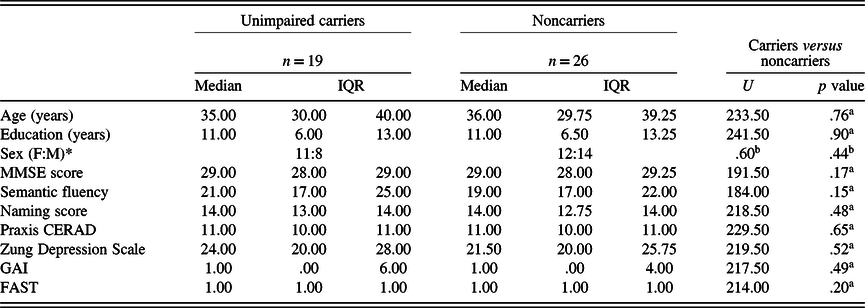
Demographics and clinical data are shown in Table 1. Cognitively unimpaired mutation carriers and noncarriers did not differ on age, level of education, sex distribution, and clinical or neuropsychological test scores. The median age of carriers was 35 years—approximately 9 years younger than the median age of MCI onset in this ADAD cohort (Acosta-Baena et al., Reference Acosta-Baena, Sepulveda-Falla, Lopera-Gómez, Jaramillo-Elorza, Moreno, Aguirre-Acevedo and Lopera2011).
Demographic data and clinical evaluation

Note. Values are expressed as median and interquartile range (IQR) with the exception of sex.
MMSE: Mini-Mental State Examination; GAI: Geriatric Anxiety Inventory; FAST: Functional Assessment Staging; CERAD: Consortium to Establish a Registry for Alzheimer’s Disease.
U: Mann–Whitney U test.
* Chi-square test (χ 2).
a p values were calculated through Mann–Whitney U test.
b p values were calculated through Chi-square test (χ 2).
In Vivo Markers of AD Pathology
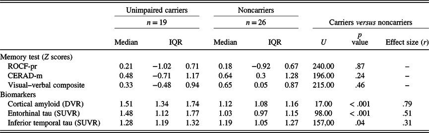
Compared with noncarriers, cognitively unimpaired carriers had greater cortical amyloid and regional tau burden, consistent with a previous report (Quiroz et al., Reference Quiroz, Sperling, Norton, Baena, Arboleda-Velasquez, Cosio and Johnson2018). Biomarker data are shown in Table 2.
Performance of each group on memory tests and biomarker data

Note. Values are expressed as median and interquartile range (IQR).
ROCF: Rey–Osterrieth Complex Figure test; ROCF-pr: ROCF percent retention; CERAD: Consortium to Establish a Registry for Alzheimer’s Disease; CERAD-m: CERAD word list memory test.
DVR: distribution volume ratio; SUVR: standardized uptake value ratio.
U: Mann–Whitney U test.
p values were calculated through Mann–Whitney U test.
ViM and VeM
There were no significant differences between cognitively unimpaired carriers and noncarriers on the ROCF-pr, CERAD-m, and the visual–verbal composite. In contrast with the noncarriers, mutation carriers showed greater variability in these measures. For further reference, see Table 2.
Association Among Cortical Amyloid, Regional tau, ViM, and VeM
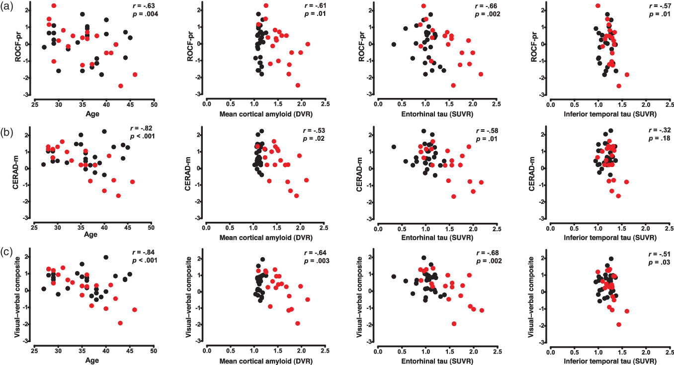
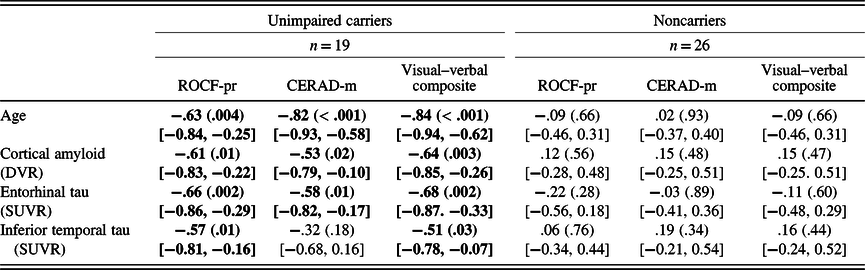
In cognitively unimpaired mutation carriers, greater mean cortical PiB retention was related to worse performance on ROCF-pr, CERAD-m, and the visual–verbal composite. Similarly, higher levels of FTP binding in EC and IT were associated with lower performance on ROCF-pr and the visual–verbal composite. The correlations for CERAD-m scores were only observed with tau in the EC, but not in the IT. As shown in Figure 1, the relation between VeM and ViM with the in vivo PET measurements shared a comparable distribution in the scatter plots. Table 3 lists the correlations between markers of AD pathology, ViM, and VeM. None of these associations were observed in noncarriers.
Correlations among in vivo biomarkers, visual and verbal memory, and age. Scatter plots show the negative correlations between in vivo biomarkers and (a) ROCF-pr and (b) CERAD-m, and (c) visual–verbal composite. Memory tests are expressed as Z score. Black circles represent noncarriers, red circles represent cognitively unimpaired PSEN1 mutation carriers. Statistical values included correspond to the correlation coefficients in the mutation carriers. Abbreviations: ROCF: Rey–Osterrieth Complex Figure test; ROCF-pr: ROCF percent retention; CERAD: Consortium to Establish a Registry for Alzheimer’s Disease; CERAD-m: CERAD word list memory test; DVR: distribution volume ratio; SUVR: standardized uptake value ratio.

Correlations among visual and verbal memory, age, and in vivo biomarkers

Note. The correlations were calculated through Spearman’s rho (r s). p values are reported in parentheses and 95% confidence intervals are shown in brackets.
ROCF: Rey–Osterrieth Complex Figure test; ROCF-pr: ROCF percent retention; CERAD: Consortium to Establish a Registry for Alzheimer’s Disease; CERAD-m: CERAD word list memory test; DVR: distribution volume ratio; SUVR: standardized uptake value ratio.
VeM and ViM were positively associated (r = .39, p = .01, 95% CI, [0.11, 0.61]) across all participants. In cognitively unimpaired mutation carriers, ViM was related to tau pathology, even after controlling for VeM (EC tau: r = −.49, p = .04, 95% CI, [−0.77, −0.04]; IT tau: r = −.50, p = .04, 95% CI, [−0.78, −0.05]). A moderate negative correlation between ROCF-pr and cortical PiB retention was observed when controlling for VeM, but this was not significant (r = −.44, p = .07, 95% CI, [−0.75, 0.02]).
Association Among Age, Cortical Amyloid, Regional tau, ViM, and VeM
We found that greater age in cognitively unimpaired carriers was associated with lower scores in ROCF-pr, CERAD-m, and the visual–verbal composite (Table 3). This association was not observed in noncarriers. When we examined the correlations between markers of AD pathology with both ViM and VeM adjusting for age, we did not find any significant associations (mutation carriers: ROCF-pr: PiB: r = −.25, p = .33, 95% CI, [−0.63, 0.23]; EC tau: r = −.40, p = .10, 95% CI, [−0.72, 0.07]; IT tau: r = −.30, p = .22, 95% CI, [−0.67, 0.18]; CERAD-m: PiB: r = .31, p = .22, 95% CI, [−0.17, 0.67]; EC tau: r = −.02, p = .93, 95% CI, [−0.47, 0.44]; IT tau: r = .37, p = .13, 95% CI, [−0.10, 0.71]; visual–verbal composite: PiB: r = .06, p = .81, 95% CI, [−0.40, 0.50]; EC tau: r = −.23, p = .35, 95% CI, [−0.62, 0.25]; IT tau: r = −.01, p = .98, 95% [CI, −0.46, 0.45]).
ViM Versus Visuo-Spatial Abilities
We used the ROCF-c to examine whether these associations with age and brain pathology were specific to VIM versus visuo-spatial abilities. No significant correlations were observed between ROCF-c and age (mutation carriers: r = −.06, p = .80, 95% CI, [−0.50, 0.41]). There was no relation between ROCF-c and brain pathology in either group (mutation carriers: PiB: r = .16, p = .52, 95% CI, [−0.32, 0.57]; EC tau: r = −.21, p = .39, 95% CI, [−0.61, 0.27]; IT tau: r = −.11, p = .65, 95% CI, [−0.54, 0.36]).
DISCUSSION
The present study examined the relation between ViM and in vivo AD pathology in preclinical ADAD. To this end, we administered the ROCF-r test and measured cortical amyloid and regional tau using PET in cognitively unimpaired PSEN1 E280A mutation carriers and noncarrier family members who belong to a large Colombian cohort with ADAD due to the PSEN1 E280A mutation. Our results showed that greater cortical amyloid burden, higher levels of regional tau, and older age were associated with lower ViM performance in cognitively unimpaired mutation carriers.
Our findings showed that the cognitively unimpaired mutation carriers performed similar to noncarriers on the ROCF-r. Most of the studies in ADAD or subjects at risk of developing sporadic AD have used VeM measures to evaluate memory function (Jansen et al., Reference Jansen, Ossenkoppele, Tijms, Fagan, Hansson, Klunk and Group2018; Mormino et al., Reference Mormino, Papp, Rentz, Donohue, Amariglio, Quiroz and Sperling2017; Papp, Rentz, Orlovsky, Sperling, & Mormino, Reference Papp, Rentz, Orlovsky, Sperling and Mormino2017; Sperling et al., Reference Sperling, Johnson, Doraiswamy, Reiman, Fleisher, Sabbagh and Group2013; Storandt, Balota, Aschenbrenner, & Morris, Reference Storandt, Balota, Aschenbrenner and Morris2014; Wang et al., Reference Wang, Gordon, Ryman, Ma, Xiong, Hassenstab and Network2015), and those that have included ViM measures yielded mixed findings, probably due to methodological differences. For example, cross-sectional studies, similar to ours, have not found differences in ViM and visual learning tasks between cognitively healthy, amyloid-positive (on PET) adults when compared with amyloid-negative adults (Hollands et al., Reference Hollands, Lim, Buckley, Pietrzak, Snyder, Ames and Maruff2015; Johnson et al., Reference Johnson, Christian, Okonkwo, Oh, Harding, Xu and Sager2014). Longitudinal studies, however, have suggested that relative to cognitively normal amyloid-negative individuals, cognitively normal amyloid-positive older adults exhibited a faster decline on ViM and VeM over 36 months of follow-up (Lim et al., Reference Lim, Maruff, Pietrzak, Ames, Ellis, Harrington and Group2014), as well as a greater rate of multidomain cognitive decline, including in ViM (Petersen et al., Reference Petersen, Wiste, Weigand, Rocca, Roberts, Mielke and Jack2016; Zhao et al., Reference Zhao, Tudorascu, Lopez, Cohen, Mathis, Aizenstein and Snitz2018). Though we did not find differences between cognitively unimpaired carriers and noncarriers on median ViM performance in our cross-sectional study, future longitudinal studies with larger samples are needed to evaluate the trajectory of ViM change across the ADAD disease continuum.
In our study, we found a relationship between markers of AD pathology and ViM measures only in cognitively unimpaired carriers. Higher levels of neocortical amyloid were associated with lower ViM performance, consistent with previous reports showing that in contrast to amyloid-negative older adults, amyloid-positive cognitively normal older adults show greater memory impairment, assessed through a composite of visual and verbal memory tasks (Lim et al., Reference Lim, Kalinowski, Pietrzak, Laws, Burnham, Ames and Maruff2018; Pike et al., Reference Pike, Savage, Villemagne, Ng, Moss, Maruff and Rowe2007). Additionally, the progression of ViM and VeM deficits were greater in amyloid-positive apolipoprotein E (APOE) ϵ4 allele carriers (Lim et al., Reference Lim, Kalinowski, Pietrzak, Laws, Burnham, Ames and Maruff2018). Furthermore, past research has suggested that greater amyloid deposition in the precuneus is associated with lower ViM scores in cognitively normal subjects with high PIB retention (Ossenkoppele et al., Reference Ossenkoppele, Madison, Oh, Wirth, van Berckel and Jagust2014). The combination of amyloid deposition and neurodegeneration was also associated with greater ViM decline, in addition to other cognitive domains (Zhao et al., Reference Zhao, Tudorascu, Lopez, Cohen, Mathis, Aizenstein and Snitz2018).
Unlike VeM studies (Gordon et al., Reference Gordon, Blazey, Christensen, Dincer, Flores, Keefe and Benzinger2019; Quiroz et al., Reference Quiroz, Sperling, Norton, Baena, Arboleda-Velasquez, Cosio and Johnson2018), to our knowledge, no previous studies have explored the specific relationship between ViM and regional tau PET in preclinical ADAD. In our study, we found that higher levels of regional tau were significantly related to lower ViM performance in cognitively unimpaired ADAD carriers. Whereas the correlations between CERAD-m and regional tau were only present with EC tau, but not with IT tau, the correlations between ROCF-pr with regional tau were observed in both the EC and IT. The IT cortex and its connections with other regions of the ventral visual pathway have been proposed as a structure that plays a role in ViM processing (Kravitz, Saleem, Baker, Ungerleider, & Mishkin, Reference Kravitz, Saleem, Baker, Ungerleider and Mishkin2013); consequently, our results do not exclude the possibility that tau accumulation in the IT cortex might contribute to the negative correlation found with ViM in cognitively unimpaired carriers. It is important to note that although the relation between CERAD-m and IT tau was not significant, this finding does not rule out a possible association. In fact, the relationship between CERAD-m and IT tau was of moderate magnitude and shared a comparable distribution to that shown between ROCF-pr and lT tau (see Figure 1); moreover, the limited sample size of the carrier group may have affected our power to detect a statistically significant relationship. Taken together, our results suggest that the performance of both types of memory is related to regional tau in preclinical stage, aligning with the hypothesis that tau accumulation is strongly linked to cognitive impairment in AD (Gordon et al., Reference Gordon, Blazey, Christensen, Dincer, Flores, Keefe and Benzinger2019; Pontecorvo et al., Reference Pontecorvo, Devous, Navitsky, Lu, Salloway and Schaerf2017; Quiroz et al., Reference Quiroz, Sperling, Norton, Baena, Arboleda-Velasquez, Cosio and Johnson2018).
Considering that the performance of ViM and VeM were related to our sample, we conducted additional analyses to explore whether the relations between ViM and tau are specific or moderated by VeM. Our results showed that the association between ROCF-pr and regional tau remained after adjusting for VeM, but the degree of this relation was moderate. These findings suggest that regional tau accumulation might play a role in ViM performance in the preclinical stage of AD.
In line with the previous results, we also found that the scores of the visual–verbal composite were negatively related to both amyloid and regional tau. In contrast with the results of the individual memory tests, the visual–verbal composite might better depict the relation between episodic memory performance and the earliest changes in accumulation of AD pathology. Taken as a whole, our findings suggest that accumulation of brain pathology (amyloid and tau) is related to early changes observed in both VeM and ViM in preclinical AD.
Our findings also suggest that age plays an important role in the relation between memory performance and pathology burden in ADAD, as no relationships survived partial correction by age; the strong effect of age on these correlations is likely due to the fact that in this and other ADAD mutation cohorts, age is considered as a proxy for disease progression (Acosta-Baena et al., Reference Acosta-Baena, Sepulveda-Falla, Lopera-Gómez, Jaramillo-Elorza, Moreno, Aguirre-Acevedo and Lopera2011). As carriers progress in age closer to the median age of onset of MCI in this cohort (44 years old; Acosta-Baena et al., Reference Acosta-Baena, Sepulveda-Falla, Lopera-Gómez, Jaramillo-Elorza, Moreno, Aguirre-Acevedo and Lopera2011), it is more likely that decline in ViM and VeM will be evident and that there will be increasing rates of amyloidosis and tau deposition.
It is important to highlight that our findings have clinical implications for the evaluation of memory in the preclinical stage of AD. In this study, we focused on the ROCF-r—probably one of the most widely used tests of ViM in clinic and research contexts. The total score of this test has proven to have good psychometric properties in adults, the elderly population, and patients with memory impairment, with internal reliability greater than 0.8 and interrater reliability > .9 (Berry, Allen, & Schmitt, Reference Berry, Allen and Schmitt1991; Tupler, Welsh, Asare-Aboagye, & Dawson, Reference Tupler, Welsh, Asare-Aboagye and Dawson1995). Additionally, the ROCF-r test has large studies of normative data, including the data used in this study which were derived from a larger cohort of noncarrier Colombians who belong to families at risk for ADAD due to E280A mutation (Torres et al., Reference Torres, Vila-Castelar, Bocanegra, Baena, Guzmán-Vélez, Aguirre-Acevedo and Lopera2019). Additionally, this test has been shown to be a sensitive instrument for the detection of ViM impairment in patients with MCI (Lekeu et al., Reference Lekeu, Magis, Marique, Delbeuck, Bechet, Guillaume and Salmon2010; Peter et al., Reference Peter, Sandkamp, Minkova, Schumacher, Kaller, Abdulkadir and Klöppel2018) and mild dementia (Salimi et al., Reference Salimi, Irish, Foxe, Hodges, Piguet and Burrell2018). Even though we did not find differences in ViM performance between carriers and noncarriers, we found significant associations between ViM and in vivo markers of AD pathology. In our study, we included ROCF-pr because unlike the immediate recall total score, this one removes the effect of execution level from the copy phase (i.e., it controls for the effect of a potentially poor copy of the figure). Percent retention scores have also been included in other ViM tests and have been studied in healthy elderly adults and in patients with memory impairment (Boone et al., Reference Boone, Lesser, Hill-gutierrez, Berman and D’Elia1993; Schoenberg et al., Reference Schoenberg, Duff, Beglinger, Moser, Bayless, Mold and Adams2008). Consequently, we provide evidence that the ROCF-pr test might also be a useful ViM assessment measure in the preclinical stage of AD and should be used along with VeM tests and other measures.
Limitations, Strengths, and Future Directions
Our study has some limitations. First, the sample size of the carrier and noncarrier groups was small, which could impact the ability to accurately characterize statistical relationships. Second, this study was conducted with participants with a single ADAD mutation, bringing into question the generalization of the results to sporadic late-onset AD and other ADAD mutations. Third, our cross-sectional design does not allow us to capture the longitudinal progression of AD pathology and ViM. However, as mentioned before, our baseline data are part of a longitudinal study (the COLBOS ADAD biomarker study) in which cognitively unimpaired carriers and noncarriers are being followed to characterize changes in biomarkers and cognitive performance across the preclinical and early clinical stages of ADAD. Although our findings are based on a small sample size compared with studies in healthy older adults, the opportunity to capitalize on the homogeneity of this group of cognitively unimpaired carriers who are destined to develop the AD clinical syndrome with virtually 100% certainty (and noncarrier family members) to study the preclinical stage of the disease should not be understated.
CONCLUSIONS
We demonstrated that worse ViM performance is associated with greater cortical amyloid burden, higher levels of regional tau, and greater age in cognitively unimpaired carriers who were, on average, 9 years away from the mean age of MCI onset. The association between ViM and regional tau was present not only in the EC but also in the IT cortex, one of the main structures that have early vulnerability to AD pathology. In general, these results are similar to other research which has examined verbal memory in the preclinical stage of the disease. It is necessary to conduct further research with larger samples and longitudinal follow-up to explore the usefulness of ViM measures to identify individuals at high risk of developing dementia later in life.
ACKNOWLEDGMENTS
This research was supported by the US National Institute of Health—Office of the Director (DP5OD019833 [YTQ]), the National Institute on Aging (R01 AG054671 [YTQ]), MGH ECOR Clafin Distinguished Scholar Award [YTQ], MGH Physician/Scientist Development Award [YTQ], COLCIENCIAS (Colombia) [FLR]. The authors thank the Colombian families for contributing their valuable time and effort, without which this study would not have been possible. We also thank David Aguillón, Claudia Ramos, Francisco Piedrahita, and Alex Navarro from Grupo de Neurociencias, Universidad de Antioquia in Medellín, Colombia, as well as Enmanuelle Pardilla-Delgado, Arabiye Artola, and Diana Múnera from the Massachusetts General Hospital in Boston, MA, for helping coordinate visits to Boston.
CONFLICT OF INTEREST
Dr. Lopera was supported by COLCIENCIAS-Colombia (111565741185), and Genentech/Roche /API COLOMBIA GN28352. Dr. Quiroz received funding from the National Institutes of Health (RO1AG054671, DP5OD019833), the Alzheimer’s Association and Massachusetts General Hospital. Mr. Fox-Fuller reports NRSA support from the National Institute on Aging (1F31AG06215801A1). Drs. Bocanegra, Baena, Guzmán-Vélez, and Vila-Castelar have no competing interests.